International Research Journal of Engineering and Technology (IRJET)
e-ISSN: 2395 -0056
Volume: 03 Issue: 02 | Feb-2016
p-ISSN: 2395-0072
www.irjet.net
A Review of Various Retinal Microaneurysm Detection Methods For Grading Of Diabetic Retinopathy Mrs.R.Jayanthi1, Kavitha.N2, Manju Paarkavi.R3, Dr.K.Bommanna Raja4 1
Associate professor, Department of ECE, Nandha College of Technology, Erode. 2,3 PG Scholar, Department of ECE, Nandha College of Technology, Erode. 4Principal, KPR Institute of Engineering and Technology, Coimbatore
Abstract – In a retina, microaneurysm is the earliest sign of diabetic retinopathy.The identification of microaneurysm is an important and most crucial process for the detection and screening of diabetic retinopathy.It helps the opthamologists to detect and diagnose the occurrence of disease.Various studies have been introduced on retinal fundus image for diabetic retinopathy.This paper deals with the latest methods for analyzing the detection of microaneurysms. Automating this process would bring more realiability and compatibility.
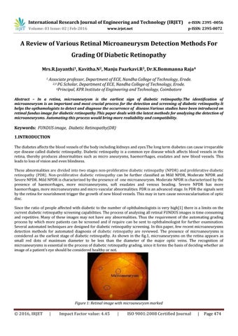
Keywords: FUNDUS image, Diabetic Retinopathy(DR) 1.INTRODUCTION The diabetes affects the blood vessels of the body including kidneys and eyes.The long term diabetes can cause irreparable eye disease called diabetic retinopathy. Diabetic retinopathy is a common eye disease which affects blood vessels in the retina, thereby produces abnormalities such as micro aneurysms, haemorrhages, exudates and new blood vessels. This leads to loss of vision and even blindness. These abnormalities are divided into two stages non-proliferative diabetic retinopathy (NPDR) and proliferative diabetic retinopathy (PDR). Non-proliferative diabetic retinopathy can be further classified as Mild NPDR, Moderate NPDR and Severe NPDR. Mild NPDR is characterized by the presence of one microaneurysm. Moderate NPDR is characterized by the presence of haemorrhages, more microaneurysms, soft exudates and venous beading. Severe NPDR has more haemorrhages, more microaneurysms and micro vascular abnormalities. PDR is an advanced stage. In PDR the signals sent by the retina for nourishment trigger the growth of new blood vessels. This may in turn cause neovascularisation of optic disc. Since the ratio of people affected with diabetic to the number of ophthalmologists is very high[1] there is a limits on the current diabetic retinopathy screening capabilities. The process of analyzing all retinal FUNDUS images is time consuming and repetitive. Many of these images may not have any abnormalities. Thus the requirement of the automating grading process by which more patients can be screened and if require can be sent to ophthalmologist for further examination. Several automated techniques are designed for diabetic retinopathy screening. In this paper, few recent microaneurysms detection methods for automated diagnosis of diabetic retinopathy are reviewed. The presence of microaneurysms is considered as the earliest stage of diabetic retinopathy. As shown in the fig.1, microaneurysms on the retina appears as small red dots of maximum diameter to be less than the diameter of the major optic veins. The recognition of microaneurysms is essential in the process of diabetic retinopathy grading, since it forms the basis of deciding whether an image of a patient’s eye should be considered healthy or not.
Figure 1: Retinal image with microaneurysm marked
© 2016, IRJET
|
Impact Factor value: 4.45
|
ISO 9001:2008 Certified Journal
|
Page 474