International Research Journal of Engineering and Technology (IRJET)
e-ISSN: 2395-0056
Volume: 03 Issue: 01 | Jan-2016
p-ISSN: 2395-0072
www.irjet.net
Survey of the Heart Wall Delineation Techniques Anjali A. Joshi1, Vitthal J. Gond2 1PG
Student, Department. Of Electronics and Telecommunication, Late G.N Sapkal College of Engineering, Nashik, Maharashtra, India 2Professor, Late G.N Sapkal College of Engineering, Nashik, Maharashtra, India
---------------------------------------------------------------------***---------------------------------------------------------------------
Abstract: Cardiac diseases are very common these
temporal resolution, excellent contrast resolution for the cardiac structures and surrounding anatomy, therefore CT scan is efficiently applied in examination of cardiovascular health. Some advantages of CT scan are as follows: 1. Less expense and wide availability 2. High spatial resolution with modern multi-slice scanners, 3. Short scan time, 4. Higher sensitivity than MR for sub-arachnoids hemorrhage, 5. Higher sensitivity in detecting intra-cranial calcifications[2] Here we have gone through five techniques. Each technique is unique. Every technique does not contain all the steps mentioned in the section 1.1.
days. An estimated 17.5 million people died due to cardiovascular diseases in 2012, representing 31% of all global deaths. Of these deaths, an estimated 7.4 million were due to coronary heart disease. Therefore early detection of cardiovascular disease is essential. Advancement in medical imaging techniques has helped in the detection of cardiovascular diseases. Use of computed tomography for cardiovascular disease detection is prevalent. Detection of heart diseases can be done by the myocardium evaluation. Myocardium can be efficiently delineated by image processing and here we have studied some techniques for heart wall delineation. Some of the techniques have used active contours, Hough transform, PCA with local shape priors for heart wall segmentation.
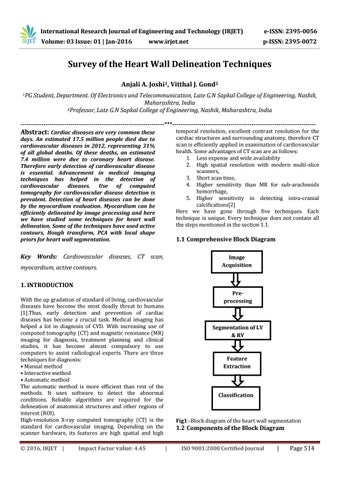
1.1 Comprehensive Block Diagram
Key Words: Cardiovascular diseases, CT scan,
Image Acquisition
myocardium, active contours. 1. INTRODUCTION
Preprocessing
With the up gradation of standard of living, cardiovascular diseases have become the most deadly threat to humans [1].Thus, early detection and prevention of cardiac diseases has become a crucial task. Medical imaging has helped a lot in diagnosis of CVD. With increasing use of computed tomography (CT) and magnetic resonance (MR) imaging for diagnosis, treatment planning and clinical studies, it has become almost compulsory to use computers to assist radiological experts. There are three techniques for diagnosis: • Manual method • Interactive method • Automatic method The automatic method is more efficient than rest of the methods. It uses software to detect the abnormal conditions. Reliable algorithms are required for the delineation of anatomical structures and other regions of interest (ROI). High-resolution X-ray computed tomography (CT) is the standard for cardiovascular imaging. Depending on the scanner hardware, its features are high spatial and high © 2016, IRJET |
Impact Factor value: 4.45
Segmentation of LV & RV
Feature Extraction
Classification
Fig1:-Block diagram of the heart wall segmentation
1.2 Components of the Block Diagram |
ISO 9001:2008 Certified Journal
|
Page 514