International Research Journal of Engineering and Technology (IRJET)
e-ISSN: 2395-0056
Volume: 10 Issue: 03 | Mar 2023
p-ISSN: 2395-0072
www.irjet.net
Deep Learning based Multi-class Brain Tumor Classification Yash A. Paddalwar1, Atharva M. Patil2, Saurav Sabu3, Harsh A. Singh4, Sheetal Gawande5 1Student, Department of Information Technology, Pillai College of Engineering, New Panvel, Maharashtra, India 2Student, Department of Information Technology, Pillai College of Engineering, New Panvel, Maharashtra, India 3Student, Department of Information Technology, Pillai College of Engineering, New Panvel, Maharashtra, India
4Student, Department of Information Technology, Pillai College of Engineering, New Panvel, Maharashtra, India 5Professor, Department of Computer Engineering, Pillai College of Engineering, New Panvel, Maharashtra, India ---------------------------------------------------------------------***---------------------------------------------------------------------
Abstract - A brain tumor is a kind of cancer that can impact
company because there is a chance that teams will need to divide the images into various categories. In such circumstances, this can be used to distinguish between the images and keep patient records secure. This could be a crucial tool that is helpful for any hospital employee because medical images are delicate and must be handled with extreme care. The objective is to identify and classify different types of tumors and most importantly to save time of doctors/patients and provide a suitable remedy at an early stage and to identify and supply good insights to doctors [20]. Examining the Brain tumor MRI images in the Healthcare industry implies the process of identifying tumors in the early stage. The framework will help in automatic detection of images containing tumors and searching for correlations between neighboring slices along with it can also do automatic detection of symmetrical axes of the image [13]. Furthermore, this framework can be used to create a full-fledged application to detect any type of Cancerous Polyps.
people, sometimes fatally or with significant quality of life impairment. Using Deep learning techniques, researchers can identify tumors and treat them more efficiently. Brain MRI pictures can be used in a variety of ways to find malignancies. Deep learning techniques have significantly outperformed the rest of these techniques. Within the framework, comparison of the models has been employed for tumor detection from brain MRI scans. Among the Convolutional Neural Network (CNN) architectures that can be employed are Custom CNN, DenseNet169, MobileNet, VGG-16, and ResNet152 models. The same hyper-parameters can be used to train these models on MR images that have undergone the same dataset and preprocessing procedures. The goal is to develop an architecture that will compare various models to classify the Brain Tumor MRI. Machine learning and deep learning algorithms can be used to directly scan and determine the presence and type of tumor. Therefore, it is useful for analyzing brain tumor detection performance using various methods. The dataset used for Brain Tumor Detection consists of approximately 5000 Brain MR Images.
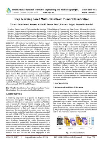
2. BACKGROUND 2.1 Convolutional Neural Network
Key Words: Classification, Neural Networks, Brain Tumor Classification, Deep Learning, Artificial Intelligence
Convolutional Neural Networks (ConvNet/CNN) is a deep learning technique that can accept input images and assign gist to various elements and objects (learnable weights and biases), and be able to distinguish between them. ConvNets, by comparison, depends upon approximately less preprocessing than other classification techniques. ConvNets can learn from filters and properties, but primitive techniques create the filters manually. To classify multigrade brain tumors, a novel convolutional neural network (CNN) is proposed [2]. Individual neurons perceive stimuli only in this restricted area of the visual field.
1.INTRODUCTION A relatively large number of people are diagnosed with secondary neuropathy. Although the exact number is unknown but this type of brain tumor is on the rise. With the use of extremely effective clinical imaging tools, early detection can always speed up the process of controlling and eliminating tumors in their early stages. A patient with the tumor may become immobile because a tumor may place pressure on the part of the brain that regulates movement of the body [6] [14] [15]. The aim of this study is to improve the detection accuracy of brain tumors on MRI picture using image processing and machine learning algorithms, as well as to develop a framework for rapidly diagnosing brain tumors from MRI images [19]. Amin et al. proposed [3] a three-step method for distinguishing between cancerous and non-cancerous brain tissue MRI. Among the steps involved are image processing, feature extraction, and image classification. This framework is useful not only for the medical staff but also for the other employees of the
© 2023, IRJET
|
Impact Factor value: 8.226
Fig - 1: CNN Architecture
|
ISO 9001:2008 Certified Journal
|
Page 778