
International Research Journal of Engineering and Technology (IRJET) e-ISSN: 2395-0056
Volume: 12 Issue: 05 | May 2025 www.irjet.net p-ISSN: 2395-0072
International Research Journal of Engineering and Technology (IRJET) e-ISSN: 2395-0056
Volume: 12 Issue: 05 | May 2025 www.irjet.net p-ISSN: 2395-0072
F. Barffuson-Dominguez1, R. Gámez-Corrales1, J. R. González-Martínez2, O. Alcantar-Jatomea3 G.T. Paredes-Quijada4
1F. Barffuson-Domínguez, Departamento de Física, Universidad de Sonora, Apdo. Postal 1626, 83000, Hermosillo, Sonora, México.
1R. Gámez-Corrales, Professor, Departamento de Física, Universidad de Sonora, Apdo. Postal 1626, 83000, Hermosillo, Sonora, México. E-mail: rogelio.gamez@unison.mx
2J.R. González-Martínez, Departamento de Investigación en Física, Universidad de Sonora, Apdo. Postal 1626, 83000, Hermosillo, Sonora, México.
3O. Alcantar-Jatomea, Departamento de Ciencias Básicas, Tecnológico Nacional de México, campus Hermosillo, 83170, Hermosillo, Sonora, México.
4G.T Paredes-Quijada, Departamento de Ciencias Químico-Biológicas, Universidad de Sonora, 83000, Hermosillo, Sonora, México. ***
Abstract – Bimetallic nanoalloys of transition metals represent a field of considerable potential, and research endeavors in this domain are of significant interest. These intriguing materials have garnered substantialinterestdueto their numerous applications as catalysts for hydrogen and Fischer-Tropsch synthesis, magnetic fluids,andmagneticdata storage. Additionally, their biomedical applications include drug delivery andcancer treatment through hyperthermia.In this pioneering study, we delve into the intricacies of the fabrication and nanostructures of CoxNi(1-x) nanoalloys. In the fabricationprocess of thesebimetallicnanoalloys,high-energy micromechanical milling was employed to produce soft transition metals. These metals were produced using powdered cobalt and nickel in varying ratios. Subsequently, a scanning electron microscope(SEM)wasemployedtoobtaina more detailed view of the nanostructures of the nanoalloys. The SEM data revealed hierarchical patterns of spherical formations with nanometric sizes down to 10 microns.
Key Words: SEM, micromechanical milling, CoxNi(1-x), nano-alloys, hierarchical structures.
In recent years, nanostructured alloys have garnered heightenedinterestduetothepotentialforprecisecontrol over their size and shape while retaining the distinct properties of each constituent metal. These properties render them suitable for diverse industrial and scientific applications. Transition metals have been the subject of numerousapplications,particularlyforbiomedicine,where bimetallic nanoalloys have been utilized in magnetic hyperthermia treatments for cancer. These nanoalloys augmentlocalheat,leadingtothedestructionofcancerous cells. The potential of bimetallic nanoalloys extends to variousfields,includingdrugdelivery,magneticresonance imaging (MRI), cell separation, biomarker detection, and bacterialinfectiontreatment.Anotherareaofapplicationis catalysis, where bimetallic nanoparticles have found
widespread use in hydrogenation reactions, including the reductionofnitrophenolsandthehydrogenationofbiomassderivedcompoundssuchaslevulinicacidandglycerol,and Fischer-Tropsch synthesis, magnetic fluids, and magnetic datastorage.Thedevelopmentofamyriadofapplicationsof thistypeofbimetallicnanomaterialcanbeattributedtoits morphology.Theseincludechainsofspheres[1],columnar structures [2], polydisperse quasi-spherical nanoalloys of various sizes [3], quasi-spherical micrometric alloys [4], starfish-type[5],nanoflakes[6],micrometricflowersformed bysequin-typestructures[7].Shell-corenanoparticles[8], Mushroom-typemicrostructures[9],nanofibers,metalalloy nanotubes, hollow spheres [10], nanocrystals [11], and nanowires[12],amongothers,havealsobeenidentified.
This study investigated the morphology of CoxNi(1-x) nanoalloys using inverted optical and scanning electron microscopy.Thehigh-energymechanicalmillingtechnique was selected for its cost-effectiveness and ease of control overthestructurestobeobtained.
2.1 Sample preparations
ThecobaltandnickelmaterialswereobtainedfromSigmaAldrichinpowderform,withapurityof99%forcobaltand 99% for nickel, and were used without prior purification treatment. The average powder size indicated by Sigma Aldrich for cobalt and nickel was less than 10 and 50 micrometers,respectively.Thenanometricparticles(alloys) were obtained by the high-energy mechanical grinding method using high-energy Fritsch Pulverisette 5 and 6 planetarymills(Figure1).Inallthesamplesinthisstudy, thecobalt-nickelratiowassystematicallyvaried,considering itsestablishedstoichiometries:CoxNi(1-x)[x=0.9,0.7,0.5,0.3, 0.1]. The grinding was performed for each sample of nanoalloynanostructuresfor24hoursat350RPMwith 6 mmand8mmdiametersteelballsinagatecontainersof65
International Research Journal of Engineering and Technology (IRJET) e-ISSN: 2395-0056
Volume: 12 Issue: 05 | May 2025 www.irjet.net p-ISSN: 2395-0072
ml capacity. The grinding was executed at a mass ratio of balls to powder mass of 20:1, with 3 grams of the sample utilizedforeachsynthesis.
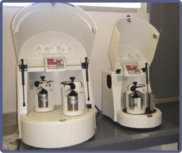
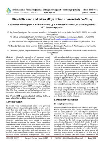
Figure 1: FritschPulverisette5and6high-energy planetarymills,eachequippedwitha65mlbowl.
OpticalmicrographswereobtainedusingaLABOMEXmodel MET 400 metallurgical optical microscopes with a 100X objective while scanning electron microscopy (SEM) micrographswereobtainedusingaJEOLmodelJSM-7800F microscope.Energydispersivespectroscopymeasurements werecarriedoutusingaBrukerXFlash6-60detector.
AsillustratedinFigure1,theFritschPulverisette6planetary mill, equipped with a 65-ml bowl functioning as a highenergyreactor,hasbeenutilizedintheexperimentalsetup. Thesehigh-energymills,whileeffectivelyreducingthesize ofthepowdercomponentsofthereactants(CoandNi),have beenshowntogeneratesufficientthermalenergytoformor aggregatetwoormoreelements,formingalloysandquasistablestructures.
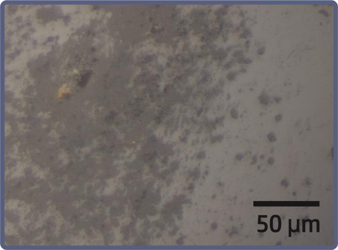
Figure 2: Amicrographwasobtainedusingoptical microscopyat200xmagnificationoftheCoxNi(1-x) alloy, withxsetat0.9.
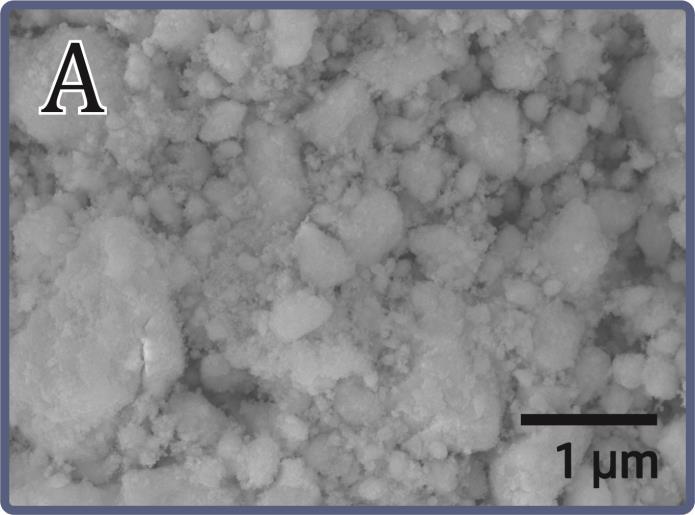
Morphological characterizations of CoxNi(1-x) nanoalloys werecarriedoutusingtwoexperimentaltechniques:optical microscopy(OM)andscanningelectronmicroscopy(SEM). The image obtained from optical microscopy reveals the presence of aggregates in a representative sample of a Co0.9Ni0.1 alloy,witha composition of 90%Co and 10% Ni (Figure 2). The sample displays polydispersity in the size distributionoftheaggregates,withsizesrangingfromafew hundrednanometerstotensofmicrons.
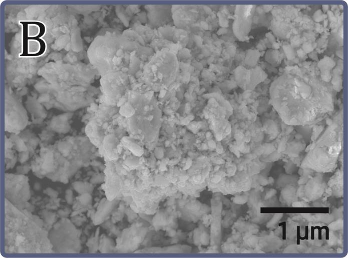
The morphology obtained in SEM is shown in Figure 3. In thisfigure,Co-Nialloyswitharatioof70:30canbeobserved (Figure 3A), and Figure 3b corresponds to the chemical composition NiCo 70:30. The following presentation provides an in-depth comparative analysis of two SEM images of Co-Ni alloys. The present study considers the micrographsidentifiedasCoNi70-30andNiCo70-30.The lower white bar of each image, corresponding to 1 µm, is usedtoestimatethedimensionandnanometerscaleinboth samples.Themicrographshowsparticleswithwell-defined contours,ideallysphericalorslightlyoval.Theuniformityin morphology suggests precise control in the synthesis process, emphasizing homogeneous nucleation and welldefinedgrowth.
International Research Journal of Engineering and Technology (IRJET) e-ISSN: 2395-0056
Volume: 12 Issue: 05 | May 2025 www.irjet.net p-ISSN: 2395-0072
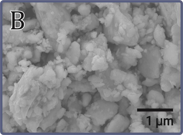
As illustrated in Figure 3A, the hierarchical structures of amorphousformandsizesofadozenmicronsareevident. Thesestructuresareconstitutedbylaminarstructureswith a thickness of several tens of nanometers and a size comparabletotheamorphousmicroparticles,asdepictedin Figure 3A, which was obtained under a magnification of 5,000X. This figure reveals the presence of amorphous microstructures,withthelargestmeasuringapproximately 10 microns. This magnification level facilitates the observationofinternaldetails.Thelamellarstructuresunder scrutiny consist of lamellar aggregates measuring several hundrednanometers.
Conversely,asdemonstratedinFigure3B(NiCo70%-30%), the image unveils a more irregular morphology, with particles displaying a propensity to form agglomerates or even branched structures. The distribution in this sample deviatesfromtheprevious oneinthatitlacksuniformity; regions exhibiting higher particle density and those characterizedbyinterparticlevoidsarediscernible.Utilizing
thesamereferenceof1µmfromthescalebar,itisestimated that the particles possess a larger average diameter or, in certaininstances,agreaterdispersionintheirdimensions, witharangeof150to400nm.Theobservedsizevariability indicateslessuniformgrowthprocessesandaheightened tendencytowardpartialcoalescence.
Observation of areas featuring augmented porosity, in conjunctionwiththeaggregationofnanoparticlesexhibiting a cushioned consistency, suggests that the growth mechanism may have been impacted by multi-centered nucleation and anisotropic growth. Evidence has demonstratedthatthismayaugmentthesizeoftheactive surfacearea.Incertainapplications,suchascatalysis,this increaseisadvantageous.
The ensuing discourse presents a thoroughgoing analysis andcomparisonofthetwoSEMmicrographsincorporated within the present document, assuming that said micrographs correspond to images of the CoNi-50 (see Figure4A)materialexhibitingvariationsinitsnanometerscale structural configuration. Figure 4A) displays a more homogeneous and compact nanometric structure. The nanoparticles under scrutiny manifest as spherical or slightlyoval-shapedentities,characterizedbywell-defined edges and a consistent distribution along the surface. The uniformity in size and compactness of the nanomaterials suggests that its growth occurred under controlled conditions that promoted the formation of a single nanocrystalline phase, with an estimated particle size ranging from 20 to 50 nanometers. The low porosity and bulk density of nanoparticles indicate a nucleation and growth process without significant coalescences, which is advantageous when uniform magnetic or electronic propertiesaredesired.
Incontrast,Figure4B displays a moreheterogeneous and branchedstructure.Agglomeratesanddendriticstructures canbeidentifiedinareaswherenanoparticlesaregrouped. These agglomerates manifest a greater dispersion in size, withestimatedsizesrangingfromapproximately40to80 nanometers,althoughsomesubstructuresappeartoexceed thesevalues.Thepresenceofregionswithobviousvoidsor interparticleporessuggeststhatgrowthwasdominatedby partial coalescence and crystal reorientation phenomena. Thesephenomenamayinfluencepropertiessuchascatalytic response or surface activity. This type of less ordered morphology could be associated with different synthetic conditions or post-synthesis treatment that favored the formationofmultiplephasesorstructuraldefects.
International Research Journal of Engineering and Technology (IRJET) e-ISSN: 2395-0056
Volume: 12 Issue: 05 | May 2025 www.irjet.net p-ISSN: 2395-0072
4. A. SEMCoNi50-50. B. SEMNiCo50-50.
The contrast between the two images indicates that the synthesis conditions or post-synthesis treatment significantlyinfluencedthenucleationandgrowthprocessof the CoNi 50%-50% material. Figure 4A which exhibits a homogeneousstructureandcompactnanoparticles,suggests controlledandpotentiallysingle-phasegrowth,aproperty that is advantageous for achieving uniform magnetic characteristics. Figure 4B demonstrates a more intricate morphologicalevolutioncharacterizedbyagglomeratesand elevatedporosity,whichmayyielddivergentfunctionalities, particularly in applications where surface-substrate interactionorcatalyticactivityarepertinent.
This work constitutes an experimental study, utilizing opticalmicroscopy(OM)andscanningelectronmicroscopy (SEM),whichwasconductedonbimetallicalloyscomprised oftransitionmetals,namelycobaltandnickel.Thesynthesis wasperformedwithmicromechanicalmillingataCox-Ni1-x ratioofx(=90,70,50,30,10)andapellet/materialratioof 20to1.Asdemonstratedbyopticalmicroscopy,thesamples
exhibited heterogeneity, accompanied by polydisperse structureswithdimensionsrangingfromtensofnanometers to several tens of micrometers. The scanning electron microscope(SEM)revealsmicroimagesofquasi-spherical nanoparticlesofbimetallicalloys,withasizerangingfrom 40to50nanometers.Thesenanoparticlesself-assembleinto lamellarstructures,witha thicknesscorrespondingtothe diameterofthe quasi-nanospheres.Duetothesubstantial amountofthermalenergyprovidedbymechanicalmilling, the lamellar structures self-assemble to form hierarchical structureswithlargersizes(approximatelytenmicronsin diameter) and amorphous shapes, different from their constituents.
[1] G.Cheng,“Fabricationandcharacterizationofironnickel chain-like fibers via simple solvothermal approach,” Mater. Res. Innov.,vol.19,no.1,pp.69–72,2015,doi:10.1179/1433075X14Y.0000000222.
[2] G.DhanalakshmiandV.Ravichandran,“Synthesisof nanocrystallinenickel-ironalloys-Anovelchemical reduction method,” Chem. Phys. Impact, vol. 6, no. March, p. 100202, 2023, doi: 10.1016/j.chphi.2023.100202.
[3] L. Li et al., “High valence metals engineering strategies of Fe/Co/Ni-based catalysts for boosted OER electrocatalysis,” J. Energy Chem., vol. 76, pp. 195–213,2023,doi:10.1016/j.jechem.2022.09.022.
[4] I. Chicinas, V. Pop, and O. Isnard, “Synthesis of the supermalloy powders by mechanical alloying,” J. Mater. Sci.,vol.39,no.16–17,pp.5305–5309,2004, doi:10.1023/B:JMSC.0000039234.58490.78.
[5] N.Nady,N.Salem,M.A.A.Mohamed,andS.H.Kandil, “Iron-nickel alloy with starfish-like shape and its unique magnetic properties: Effect of reaction volumeandmetalconcentrationonthesynthesized alloy,” Nanomaterials, vol. 11, no. 11, 2021, doi: 10.3390/nano11113034.
[6] W.Kang,“MicroforgingEffectontheMicrostructure andMagneticPropertiesofFeSiB-basedNanoflakes,” J. Mater. Sci. Technol., vol. 28, no. 4, pp. 303–307, 2012,doi:10.1016/S1005-0302(12)60058-9.
[7] L. Liu, J. Guan, W. Shi, Z. Sun, and J. Zhao, “Facile synthesisandgrowthmechanismofflowerlikeNi-Fe alloynanostructures,” J. Phys. Chem. C,vol.114,no. 32, pp. 13565–13570, 2010, doi: 10.1021/jp104212v.
[8] Y.Zhang,C.Liu,G.Fan,L.Yang,andF.Li,“Arobust core-shell nanostructured nickel-iron alloy@nitrogen-containing carbon catalyst for the
International Research Journal of Engineering and Technology (IRJET) e-ISSN: 2395-0056
Volume: 12 Issue: 05 | May 2025 www.irjet.net p-ISSN: 2395-0072
highlyefficienthydrogenationofnitroarenes,” Dalt. Trans.,vol.47,no.38,pp.13668–13679,2018,doi: 10.1039/c8dt03033b.
[9] T.Han,C.Xu,andH.Chen,“Simplesynthesisofnovel mushroom-like FeNi3 microstructures by a hydrothermalreduction,” Mater. Res. Innov.,vol.23, no. 1, pp. 39–42, 2017, doi: 10.1080/14328917.2017.1362509.
[10] G.Tong,J.Guan,Z.Xiao,F.Mou,W.Wang,andG.Yan, “Insitu generatedH2 bubble-engaged assembly:A oneapproach for shape-controlled growth of Fe nanostructures,” Chem. Mater., vol. 20, no. 10, pp. 3535–3539,2008,doi:10.1021/cm800269k.
[11] F.EbrahimiandH.Q.Li,“Structureandpropertiesof electrodepositednanocrystallineFCCNi-Fealloys,” Rev. Adv. Mater. Sci.,vol.5,no.2,pp.134–138,2003.
[12] M.Zeng,H.Yang,J.Liu,andR.Yu,“Gradientmagnetic binaryalloynanowire,” J. Appl. Phys.,vol.115,no.17, pp.113–116,2014,doi:10.1063/1.4864248.