
International Research Journal of Engineering and Technology (IRJET) e-ISSN: 2395-0056
Volume: 13 Issue: 02 | Feb 2026 www.irjet.net p-ISSN: 2395-0072
International Research Journal of Engineering and Technology (IRJET) e-ISSN: 2395-0056
Volume: 13 Issue: 02 | Feb 2026 www.irjet.net p-ISSN: 2395-0072
Mrs.D.Kavitha1, D.Chandana2, K.Ganesh3, Ch.Gyanadeep4, J.Abhishek reddy5
1Asst.Professor in Department of IT, TKR College of Engineering and Technology, Telangana, India 2345BTECH Students in Department of IT, TKR College of Engineering and Technology, Telangana, India
Abstract - Brain tumour detection is a critical task in medical diagnostics, requiring accurate and timely identification to ensure effective treatment. This project presents an automated system for detecting and segmenting brain tumours from MRI images using deep learning and image processing techniques. A pre-trained convolutional neural network model is employed to classify MRI scans as tumour-positiveortumour-negative.Tumorregionsarethen segmentedusingmorphologicaloperationsandthewatershed algorithm for precise visualization. Additionally, the system integrates a Generative AI assistant (Google Gemini via Lang Chain) to provide patients with personalized medical advice, treatmentsuggestions,andlifestyleprecautionsbasedonthe detection results. The system also includes a webbased platform for user registration, secure login, and admincontrolled account activation, ensuring controlled access.Experimental results demonstrate accurate tumour detection and effective integration of AI- driven guidance, highlighting the potential of combining deep learning and generative AI for enhanced medical diagnostics.
KEYWORDS: Brain Tumour Detection, MRI, Deep Learning, Image Segmentation, Generative AI, Google Gemini, Lang Chain, Web-Based Medical System.
Brain tumours are abnormal growths of cells within the brain that can lead to severe health complications if not detectedearly.Accuratediagnosisofbraintumoursiscrucial for effective treatment planning and improving patient survivalrates[5],[7].
Magnetic Resonance Imaging (MRI) is a widely used noninvasive imaging technique that provides high-resolution visualization of brain tissues, making it the preferred modality for brain tumour detection [2], [5]. However, manualexaminationofMRIscansistime-consuming,highly dependentonexpertradiologists,andsusceptibletohuman error,particularlyinearly-stagetumouridentification[4], [7].
Recentadvancementsindeeplearningandmedical image processinghaveenabledautomatedbraintumourdetection and segmentation, significantly improving diagnostic accuracy and efficiency [1], [6], [9]. Convolutional Neural Networks (CNNs) have demonstrated strong capability in learningcomplexspatialfeaturesfromMRIimages,allowing
accurate classification of tumour and non-tumour regions [3],[8],[10].
In addition to classification, tumour localization and segmentation play a vital role in understanding tumour shape,size,andspread.Imageprocessingtechniquessuchas morphological operations and watershed algorithms are widely used for precise tumour region segmentation and visualization[2],[9].
Furthermore, the integration of Generative Artificial Intelligence(AI)modelsenablesintelligentinteractionwith patients by providing personalized medical guidance, treatmentsuggestions,andprecautionaryrecommendations basedondetectedtumourconditions [6],[10].
By combining MRI-based tumour detection using CNNs, automated segmentation techniques, and AI-assisted recommendation systems into a web-based platform, the proposedsystemoffersacomprehensivesolutionforearly diagnosis,enhancedclinicaldecisionsupport,andimproved patientawarenesswhilemaintainingadministrativecontrol overusermanagement[1],[5],[9].
MagneticResonanceImaging(MRI)playsavitalroleinthe detectionofbraintumoursduetoitshighspatialresolution andexcellentsoft-tissuecontrast.Intheproposedsystem, MRI scans uploaded by users are first pre-processed to removenoise,normalizeintensitylevels,andenhanceimage quality.Thesepreprocessingstepsensurethattherelevant tumourfeaturesarepreservedforaccurateanalysis.
A Convolutional Neural Network (CNN) is employed to automatically analyse MRI images and classify them as tumour-positiveortumour-negative.CNNsarewell-suited for this task as they can automatically learn hierarchical features such as edges, textures, and complex tumour patterns directly from the image data. Once a tumour is detected, image segmentation techniques are applied to preciselyidentifythetumourregion.
Morphological operations combined with the watershed algorithm are used to segment the tumour area from surrounding brain tissues. This segmentation process highlights the size, shape, and location of the tumour,
International Research Journal of Engineering and Technology (IRJET) e-ISSN: 2395-0056
Volume: 13 Issue: 02 | Feb 2026 www.irjet.net p-ISSN: 2395-0072
assistinginbettervisualizationandclinicalinterpretation. Automateddetectionandsegmentationsignificantlyreduce manualeffort,minimizediagnosticerrors,andenablefaster decision-makinginmedicalworkflows.
Brain tumours are among the most life-threatening neurologicaldisorders,andtheirimpactonpatientsurvival largelydependsonearlyandaccuratediagnosis.Despitethe availability of advanced imaging techniques such as Magnetic Resonance Imaging (MRI), the conventional process of analysing brain scans is heavily dependent on expert radiologists, making it time consuming, costly, and susceptible to human error, especially in cases involving subtleorearly-stagetumours.
The growing number of MRI scans in clinical environments further increases the burden on medical professionals, often leading to delayed diagnoses. In addition,accuratelyidentifyingtheexacttumourboundaries is challenging due to variations in tumour size, shape, intensity, and location across patients. These challenges highlighttheneedforanautomated,reliable,andefficient systemcapableofdetectingandsegmentingbraintumours with high precision. Motivated by these limitations, the proposed approach leverages deep learning, image processingtechniques,and AI-drivenguidancetoassistin earlytumourdetection,improvediagnosticaccuracy,reduce clinical workload, and provide accessible, patient-centric supportthroughanintelligentweb-basedplatform.
Theproposedsystemisaweb-basedplatformdesignedto automatebraintumourdetection,segmentation,andpatient guidanceusingdeeplearningandgenerativeAI.Itaddresses the challenges of manual MRI analysis, which is timeconsuming,pronetohumanerror,andrequiresspecialized expertise.Thesystemintegratesseveralmodulestoprovide aseamlessworkflowforbothusersandadministrators. The user module allows patients to register and log in securely. Once authenticated, users can upload brain MRI scansforanalysis.Uploadedimagesarepre-processedusing grayscaleconversion,Gaussianblurring,andnoisereduction toenhancefeatureextraction.Apre-trainedConvolutional NeuralNetwork(CNN)modelthenpredictsthepresenceofa tumour,providingprobabilisticresultsfortumourdetection. To assist with visualization, the system employs image segmentation techniques, including morphological operationsandthewatershedalgorithm,tohighlighttumour regionsintheMRIscans.Thesegmentedimageshelpboth patients and healthcare professionals understand the tumour’slocationandextent. Additionally,thesystemintegratesaGenerativeAIassistant usingLargeLanguageModel(LLM)suchhasGoogleGemini via Lang Chain. Based on the detection results, the AI
generates personalized advice, recommended treatments, andlifestyleprecautionsforthepatient,enhancinginformed decision-making.
The admin module manages user accounts, allowing administrators to view registered users and activate accounts as necessary, ensuring controlled access to the system.
Overall, the proposed system provides an end-to-end solutionthatcombinesautomatedtumourdetection,visual segmentation, and AI-driven medical guidance. It aims to improve diagnostic efficiency, support early intervention, andempowerpatientswithactionableinformation,reducing the dependency on manual interpretation and enhancing overallhealthcaredelivery.
The system architecture of the proposed AI-driven brain tumour detection system is designed in a modular and layeredmannertoensureefficiency,scalability,andeaseof maintenance.ThearchitecturebeginswiththeUserInterface Layer, which provides a web-based platform for users to register,login,anduploadMRIbrainimagessecurely.This layer ensures user-friendly interaction and proper input validationbeforedata issentfor processing.OnceanMRI image is uploaded, it is forwarded to the Application ProcessingLayer,wherethecoreanalysistakesplace.The firststageinthislayerisMRIImagePreprocessing,which removes noise, normalizes intensity values, enhances contrast,andresizesimagestoastandardformatsuitablefor deeplearningmodels.Thisstepimprovesimagequalityand ensuresconsistentinputforaccurateanalysis.
Thepre-processedimagesarethenpassedtotheCNNBased Tumour Detection Module, where a trained Convolutional NeuralNetworkanalysestheMRIscantodeterminewhether abraintumourispresent.Ifatumourisdetected,theimage isfurtherprocessedbytheTumourSegmentationModule, which uses image processing techniques such as morphological operationsandthe watershedalgorithm to accuratelyextractandhighlightthetumourregion.
Followingdetectionandsegmentation,thesysteminteracts with the AI Guidance Module, which leverages a Large LanguageModel(LLM)togeneratepersonalizedguidance, precautionary advice, and general treat men related information based on the analysis results. This enhances patientawarenessand providessupportiveinsights while emphasizing that professional medical consultation is essential.
International Research Journal of Engineering and Technology (IRJET) e-ISSN: 2395-0056
Volume: 13 Issue: 02 | Feb 2026 www.irjet.net p-ISSN: 2395-0072
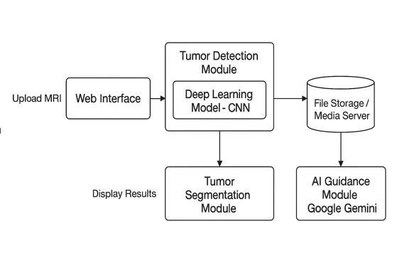
Thedataflowintheproposedsystembeginswiththeuser uploadingabrainMRIscanthroughtheweb-basedinterface. Onceuploaded,theimageisforwardedtothepreprocessing module, where noise reduction, resizing, intensity normalization,andcontrastenhancementareperformedto improve image quality and ensure consistency across different MRI inputs. The pre-processed image is then passedtothedeeplearningmodule,whereaConvolutional NeuralNetwork(CNN)analysesthescantodeterminethe presenceorabsenceofabraintumour.Ifa
Tumour is detected; the image is further processed using image segmentation techniques, including morphological operations and the watershed algorithm, to accurately extractthetumourregionfromsurroundingbraintissues. Thesegmentedoutputhighlightsthetumour’slocation,size, and shape for better visualization. Based on the detection and segmentation results, the system interacts with a Generative AI model to generate personalized guidance, precautionary advice, and general treatment related information for the user. Finally, all results, including classification outcomes, segmented images, and AIgenerated recommendations, are displayed on the user dashboard, while relevant data is securely stored in the databaseforfuturereferenceandadministrativemonitoring.
MRIimagepreprocessingisacrucialstepintheproposed system, as the quality of input images directly affects the accuracyoftumourdetectionandsegmentation.RawMRI scansoftencontainnoise,intensityvariations,andirrelevant background information that can degrade model performance. To address these issues, the uploaded MRI imagesarefirstconvertedintoa standardized formatand resizedtoafixedresolutionsuitablefortheConvolutional NeuralNetwork(CNN).Noisereductiontechniquessuchas medianfilteringorGaussianfilteringareappliedtoremove unwanted artifacts while preserving important structural details.Intensitynormalizationisthenperformedtoreduce
variations caused by different imaging conditions and scanners, ensuring uniform pixel distribution across all images. Contrast enhancement techniques, including histogramequalization,areusedtohighlighttumourregions andimprovethevisibilityofbraintissues.
ConvolutionalNeuralNetworks(CNNs)areemployedinthe proposed system to automatically detect the presence of brain tumours from pre-processed MRI images. CNNs are highly effective for medical image analysis due to their abilitytolearnspatialhierarchiesoffeaturessuchasedges, textures,andcomplextumourpatternsdirectlyfromimage data without the need for manual feature extraction. The pre-processed MRI images are fed into the CNN model as input,wheremultipleconvolutionallayersextractlow-level and high-level features relevant to tumour identification. Eachconvolutionallayerisfollowedbyactivationfunctions such as ReLU and pooling layers that reduce spatial dimensions while retaining important information. This helps in minimizing computational complexity and preventingoverfitting.Theextractedfeaturemapsarethen passed through fully connected layers, which perform classificationtodeterminewhethertheMRIscanindicatesa tumour-affectedbrainoranormalbrain
Theimplementationoftheproposedsystemintegratesdeep learning, image processing, and Generative AI within a unifiedweb-basedframework.Thesystemisdevelopedto ensureseamlessinteractionbetweenMRIimageacquisition, tumourdetection,segmentation,andpersonalizedguidance delivery. Python-based frameworks are used for model development and image processing, while the web application enables secure user interaction and result visualization. Each module is implemented independently yetworkscohesivelytoprovideaccurate,efficient,anduserfriendlybraintumouranalysis..
Project planning and requirements gathering constitute a crucial phase in the implementation of the proposed AI poweredbraintumourdetectionsystem.Duringthisstage, theprimaryobjectivesoftheprojectwereclearly defined, focusingonaccurateMRI-basedtumourdetection,precise segmentation, and AI-assisted personalized guidance throughaweb-basedplatform.Adetailedstudyofexisting braintumourdetectionmethodsandclinicalchallengeswas carried out to identify system limitations and establish realisticprojectgoals.Functionalrequirementssuchasuser registration, MRI image upload, automated tumour detection, segmentation visualization, AI-generated guidance, and administrative control were identified. In addition, non-functional requirements including system accuracy, reliability, data security, scalability, and user
International Research Journal of Engineering and Technology (IRJET) e-ISSN: 2395-0056
Volume: 13 Issue: 02 | Feb 2026 www.irjet.net p-ISSN: 2395-0072
friendly interface design were considered. Technical requirements such as dataset selection, deep learning frameworks,imageprocessingtechniques,andGenerativeAI integration were also finalized during this phase. Proper planning and systematic requirement analysis helped in designingastructuredarchitecture,reducingdevelopment risks,andensuringthatthefinalsystemmeetsbothtechnical anduserexpectationseffectively.
3.2
The system design and architecture of the proposed AI poweredbraintumourdetectionsystemarestructured to ensure modularity,scalability,andefficientdataprocessing. The architecture is divided into multiple interconnected modules, including the user interface, preprocessing unit, CNN-basedtumourdetectionmodule,segmentationmodule, AIguidanceengine,anddatabasemanagementsystem.Each module is designed to perform a specific function while seamlesslyinteractingwithothercomponents.
Theuserinterfaceallowsuserstosecurelyregister,login, and upload MRI images for analysis. Uploaded images are passedtothepreprocessingmodule,wherenoiseremoval, normalization, and enhancement are performed. The processed images are then analysed by the CNN model to detect the presence of a brain tumour. If a tumour is identified, segmentation techniques are applied to extract and highlight the tumour region. The AI guidance module generates personalized precautionary and advisory information based on the analysis results. All data and outputs are securely stored in the database, while the administrator module manages user activities, system monitoring, and access control, ensuring a reliable and efficientsystemworkflow.
Thefrontendoftheproposedsystemisdesignedtoprovidea simple, intuitive, and user-friendly interface that enables seamless interaction between users and the brain tumour detectionplatform.Thefrontendallowsuserstoregister,log insecurely,anduploadMRIbrainimagesforanalysis.Clear navigationandresponsivedesignprinciplesarefollowedto ensureaccessibilityacrossdifferentdevicesandscreensizes. The user interface displays tumour detection results, segmented MRI images, and AI-generated personalized guidance in a visually clear and understandable format. Formsandvalidationmechanismsareimplementedtoguide usersduringimageuploadandpreventincorrectinputs.The frontend also includes an administrator dashboard that enablesefficientusermanagementandsystemmonitoring. Overall, the frontend development focuses on usability, clarity, and effective visualization of medical results, ensuringasmoothandengaginguserexperience.
The backend of the proposed system is responsible for handlingcorefunctionalitiessuchasdataprocessing,model execution,user management, and secure communication between system components. The backend is developed using a server-side framework that manages user authentication,MRIimageuploads,andinteractionwiththe deeplearningandimageprocessingmodules. OnceanMRI image is received, the backend triggers the preprocessing pipeline,executestheCNN-basedtumourdetectionmodel, andappliessegmentationtechniquestogenerateanalytical results.Alluserdata,MRIimages,andprocessedoutputsare securelystoredinthedatabase,ensuringdataintegrityand privacy.
The proposed AI-driven brain tumour detection and guidancesystemwastestedusingMRIbrainscanimagesto evaluate its performance in tumour classification, tumour regionsegmentation,anduser-levelresultpresentation.The systemproducesend-to-endoutputsthroughaweb-based interface, including detection status, segmented tumour visualization,andAI-generatedprecautionaryguidance.The experimental results show that the integrated CNN model effectively identifies tumour presence, while the segmentation module successfully highlights the tumour region.Theoverallworkflowprovidesaccurateresultswith clearvisualizationanduser-friendlyinteraction.
The tumour detection module uses a pre-trained Convolutional Neural Network (CNN) to classify uploaded MRI scans as tumour-positive or tumour-negative. After successful analysis, the system displays the classification resulttotheuserthroughthedashboardinterface.Asshown inFig.2,thesystemclearlypresentsthedetectionstatusas “Tumour Detected”, enabling users to immediately understandtheoutcomeoftheMRIanalysis.Thisautomated detectionreducesdependencyonmanualinterpretationand supportsfasterdecision-making.
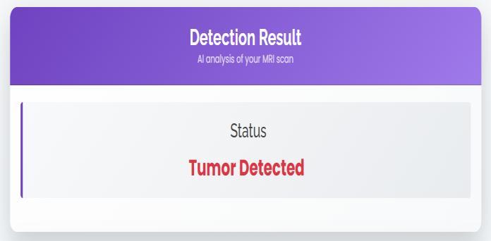
Fig. 2: CNN-based tumour detection result displayed on the web interface (Tumour Detected).
International Research Journal of Engineering and Technology (IRJET) e-ISSN: 2395-0056
Volume: 13 Issue: 02 | Feb 2026 www.irjet.net p-ISSN: 2395-0072
Aftertumourdetection,thesystemperformstumourregion segmentationtoidentifytheexactlocationandshapeofthe tumour. Morphological operations and the watershed algorithmareappliedto extractthetumourarea fromthe surrounding brain tissues. The segmentation output providesaclearvisualrepresentationoftumourboundaries, improving interpretability. As shown in Fig. 3, the system displaysboththeuploadedMRIscanandthecorresponding segmentedtumourimage,allowingusersandclinicians to observe the tumour’s position and size effectively. This segmentation stage enhances diagnostic support by providing additional visual evidence along with classification.
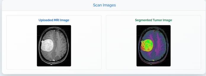
Fig. 3: Uploaded MRI image and segmented tumour region generated using morphological and watershedbased segmentation.
To improve patient awareness and provide supportive assistance,thesystemintegratesa GenerativeAIassistant usingaLargeLanguageModel(LLM).Basedonthedetection results, the AI generates personalized medical guidance, including suggested follow-up actions, precautionary measures, and lifestyle advice. As illustrated in Fig. 4, the system provides structured recommendations such as consultinganeurologist,avoidingstrenuousactivities,and monitoring symptoms. This feature enhances user understandingandensuresthatthesystemnotonlydetects tumours but also provides meaningful guidance in an accessibleformat.However,thesystemalsoemphasizesthat professional medical consultation is required for final diagnosisandtreatment.
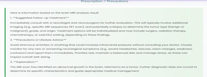
Fig. 4: AI-generated prescription, treatment suggestions, and lifestyle precautions based on MRI analysis.
Theproposedsystemdemonstratesefficientperformanceby providingresultsinacompletepipeline fromMRIupload to tumour detection, segmentation, and AI-guided recommendations withinashorttime.Thewebinterface ensures smooth interaction, clear visualization of outputs, andeasyaccessibilityforusers.Theintegrationofdetection results (Fig. 1), segmentation visualization (Fig. 2), and guidancegeneration(Fig.3)provesthatthesystemdelivers anend-to-endsolutionforbraintumouranalysis.Thismakes the platform suitable for supportive diagnostic assistance, patientawareness,andearlyinterventionworkflows.
ThisprojectsuccessfullypresentsanAI-poweredsystem forbraintumourdetection,segmentation,andpersonalized guidanceusingdeeplearningandGenerativeAItechniques. ByleveragingMRIimages,ConvolutionalNeuralNetworks effectively automate the detection of brain tumours, reducing reliance on manual analysis and minimizing diagnostic errors. The integration of image processing techniques enables accurate segmentation and clear visualization of tumour regions, aiding better clinical interpretation. Furthermore, the inclusion of AI-assisted guidance provides users with meaningful insights, precautionaryinformation,andgeneralrecommendationsin an accessible manner. The proposed web-based platform ensures ease of use, secure data handling, and efficient interactionbetweenusersandadministrators.Overall,the system demonstrates the potential of combining deep learning and AI technologies to enhance early diagnosis, improve healthcare efficiency, and support informed decision-makinginbraintumourmanagement.
The proposed AI-powered brain tumour detection and guidancesystemcanbefurtherenhancedinseveralwaysto improveaccuracy,scalability,andclinicalusefulness.Future work may include extending the system to support multiclasstumourclassificationtoidentifydifferenttumourtypes and grades. Incorporating advanced deep learning architectures such as 3D CNNs and transformer based modelscanimprovefeatureextractionfromvolumetricMRI data.Thesegmentationmodulecanberefinedusingstate-ofthe-art techniques like U-Net variants for more precise tumour boundary detection. In addition, integrating realtimeclinicaldata,radiologyreports,andpatienthistorycan enablemore personalizedandcontext-awareAIguidance. Deployment of the system in hospital environments with cloud-based infrastructure, along with mobile application support,canfurtherenhanceaccessibilityandusability.With proper clinical validation and regulatory compliance, the systemcanevolveintoareliabledecision-supporttoolfor
International Research Journal of Engineering and Technology (IRJET) e-ISSN: 2395-0056
Volume: 13 Issue: 02 | Feb 2026 www.irjet.net p-ISSN: 2395-0072
assisting medical professionals in early brain tumour diagnosisandpatientcare.
[1] R.Martínez-Del-Río-Ortegaetal.,“BrainTumour DetectionUsingMRIandCNNs,”*BigDataandCognitive Computing*,vol.8,no.9,2024.:content Reference[oaicite:0]{index=0}
[2] Z. U. Abidin et al., “Recent Deep Learning-Based Brain Tumor Segmentation Using Multi-Modal MRI,” *Front.Bioeng.Biotechnol.*,2024.
[3] M.M.Zahooretal.,“BrainTumorMRIClassification Using a Novel Deep CNN,” *Medicines*, vol. 12, no. 7, 2024.:contentReference[oaicite:5]{index=5}
[4] S.Anantharajanetal.,“MRIBrainTumorDetection UsingDeepLearningandML,”*SignalProcessing*,2024. :contentReference[oaicite:6]{index=6}
[5] F.J.Dorfneretal.,“AReviewofDeepLearningfor BrainTumorAnalysisinMRI,”*NatureReviews*,2025. :contentReference[oaicite:1]{index=1}
[6] A. Rahman et al., “Enhanced MRI Brain Tumor Detection Using Deep Learning,” *Sci. Rep.*, 2025. :contentReference[oaicite:2]{index=2}
[7] M. Ottoni et al., “Machine Learning in MRI BrainImaging:AReview,”*Diagnostics*,2025. :contentReference[oaicite:4]{index=4}
[8] A. Naeem, “Lightweight CNN for Accurate Brain Tumor Detection from MRI,” *Frontiers in Medicine*, 2025.:contentReference[oaicite:7]{index=7}
[9] S. Ganesh et al., “Brain Tumor Segmentation and Detection in MRI Using Deep Learning,” *SAGE Open Med.*,2025.:contentReference[oaicite:8]{index=8}
[10] TA Fahim et al., “Brain Tumor Detection, Classification and Segmentation by DL Models,” *Sci. Direct*,2025.:contentReference[oaicite:9]{index=9}