12 minute read
Talking with the Dead: Engaging with Human Remains in a Contemporary Medical Museum Context

from Museum Ireland, Vol. 27. Widdis, B. (Ed.). Irish Museums Association, Dublin (2021)
by irishmuseums

MUSEUM IRELAND 2020
Talking with the Dead: Engaging with Human Remains in a Contemporary Medical Museum Context
Advertisement
Evi Numen
What lessons can we learn from a 200-year-old skeleton in a closet? What can a broken wax model tell us about medical teaching? How did medical education practice evolve from body-snatching to body donation? These are some of the questions we are called upon to address, as we embark on a project to establish a medical heritage centre around a collection that spans 300 years of medical education in Ireland, and includes human remains, anatomical models, medical illustrations, and striking portraits of patients and physicians. We are challenged with striking a balance between the sensitive nature of human remains and the scientific, historical, and personal lessons we can learn from them.
To address these challenges, we must devise ways to interact with the memories of the individuals represented as remains, collectors, or creators in our collections. Doing so depends on asking the right questions and piecing together the resulting narratives. Some of these narratives are not convenient or easy to discuss, entwined as they are with attitudes and practices of a classist and colonialist past. Others complicate our understanding of human diversity, ability, and consent, and invite us to bridge the gap between specimen and visitor. However, it is our duty as custodians of medical history to give voice to the dead and tell their stories unflinchingly and honestly.
History of the Collection
Medical students today enjoy an array of remote means of learning human anatomy: they can rotate skeletons, peel back skin and muscle, and reveal structures that were only knowable to their predecessors after hours of labour over cadavers, all with a click of the mouse. They have access to digital databases that contain the culmination of centuries of collected anatomical knowledge.
Before such means were available, medical educators complemented their lectures and dissection demonstrations with anatomical atlases, drawings, and specimens. Anatomists carefully dissected and prepared each specimen to highlight a specific structure of the body in typical presentation. These masterfully created and displayed organic specimens were essential to medical teaching, and their collections reflected the standing of a medical school. The practice of collecting medical specimens spread along with the study of western medicine from Europe to Asia and the Americas. Many medical schools today still use osteological and visceral specimens to assist their students with each lesson.
The artefacts of the Old Anatomy Museum at Trinity College Dublin are the amalgam of three such collections that were used throughout the history of the School of Medicine to aid medical education, dating back to its founding in 1700. As the reputation of the school rose in the nineteenth century, so did the number of student admissions,
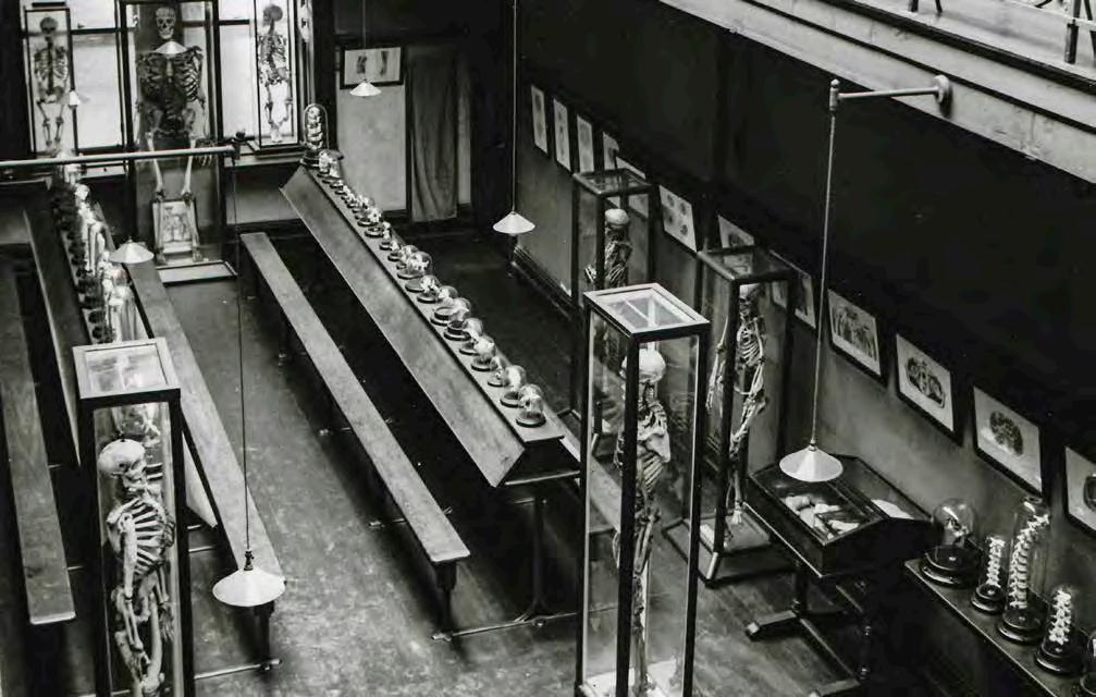
The Old Anatomy Museum when it was still in active use in 1933.
Credit: The Old Anatomy Museum, Trinity College Dublin
leading to the erection of new buildings for the Medical School in 1825, including a lecture theatre, a dissection hall, labs and classrooms, and a museum gallery and mezzanine built exclusively to house the growing medical education collection. A string of influential anatomy professors drove its curation and development efforts, and the new buildings were completed in 1876.1
In 1881, Dr Daniel John Cunningham assumed the office of Professor of Anatomy, which he occupied for twenty years. During his tenure, Cunningham dissected and prepared the potted specimens that make up the bulk of the wet specimen collection. In 1891, he established an anthropometric laboratory with the help of the Royal Irish Academy to study the Irish peoples and find ‘the origins of the race.’ Alfred Haddon and Charles Browne joined Cunningham in his travels and study of the people of the west of Ireland and the islands. The laboratory closed in 1898, but its collection remained in situ. The museum as shown in the above photo was packed away in the 1950s when Dr. Cecil Erskine became Professor of Anatomy, in favour of a more modernist museum aesthetic. The next professor of anatomy was not as interested in the collection and more changes ensued, but the museum continued to be available to students in a more diminished capacity up to 2011, when the Anatomy department moved to the new Trinity Biomedical Sciences Institute, built to mark 300 years of medical education at TCD. Following this move, Anatomy department staff began an earnest and time-consuming effort to dust it off, unpack it, catalogue, and conserve it.2
Asking the Right Questions
Why is it important to preserve and restore such a collection? As modern ideas about anatomy and medicine evolve, the study of medical history provides insight into the methodologies and theories that gave rise to contemporary discoveries, as well as the changing attitudes toward health and medicine. Human remains presented in an educational context serve as a tangible link to our technological and cultural past.
In addition, pathological specimens can teach us a great deal about the variability and vulnerability of the human condition. The Old Anatomy Collection
in particular can shed light on unfamiliar aspects of Irish history. The history of Irish medical advances is celebrated in the country’s medical societies and hospitals. Trinity College claims the legacies of physicians William Stokes, John Cheyne, William Wilde, and many others whose accomplishments have been well-studied and established.3 However, the individuals whose health and bodies made their contributions possible are largely absent from the country’s consciousness.
The 1824 catalogue of the collection states its principal function in no uncertain terms:
The Museum in the Medical School of Trinity College, Dublin, contains several Preparations, Models, and Casts, principally designed to assist the Student in acquiring a knowledge of animal structure and function, as well as of the several morbid changes to which the human frame is most subject.
From its inception and throughout its history, as is reflected by its provenance, interpretation and organisation, the collection was solely intended for use by medical educators, students,
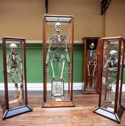
From right to left, the skeletons of an unidentified female suffering from FOP; gigantism sufferer Cornelius Magrath; centenarian James Conway; and William Clark, also an FOP sufferer.
Credit: Evi Numen, courtesy of The Old Anatomy Museum, Trinity College Dublin and researchers. Each specimen and artefact represents a diagnosis, an anatomical structure, or a pathology. Thus, we are tasked to re-interpret it from the ground up by looking beyond the medicine. Asking the right questions and following the chain of custody of each specimen, from its original owner to its placement behind glass, becomes imperative. Who did this femur belong to? Who collected it, how, and from where? For what purpose? Who studied and who learned from it? What can we learn from it today? Through our findings, we can uncover the radiating connections that culminate in the collection and display of a specimen, tracking the historical medical framework that surrounds it; we can also incorporate memorialisation practices within its interpretation to honour the individual from whom the specimen came.4
Some of the narratives we uncover in this process are bound to be steeped in controversies surrounding questionable practices that characterised facets of eighteenth and nineteenth century medicine. These include, for example, how bodies for dissection classes were procured. The increased admissions to medical schools in the 1800s meant an exponential rise in demand for cadavers for teaching. In 1791, a law was signed to allow anatomists to use the bodies of executed criminals for dissection classes. Failing to meet demand solely through this source and lacking effective preservation methods for them, anatomists resorted to grave robbers to supply the schools with recently buried cadavers. A mass pledge in our collection records how, perceiving this issue and the violence that it could incentivise, Anatomy professor Joseph Macartney (1770–1834) acquired over three hundred signatures of physicians and their relatives in support of private body donation for dissection. The Anatomy Act of 1832 cemented Macartney’s efforts.
Over two hundred years later, the body donor programme in Trinity School of Medicine is recognised for its ground-breaking approach to the practices that govern interactions between the students, the donors, and their relatives. Unlike most medical school cadaver programmes that entirely obscure the identity of a body donor, pending consent from the donor’s family, their first name may be revealed to students.
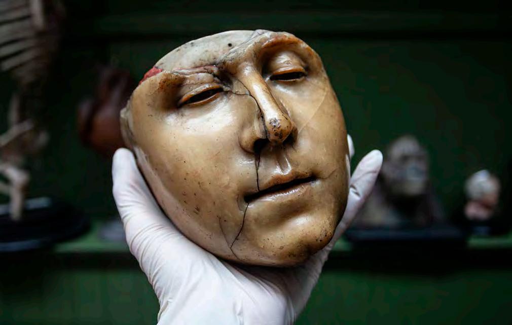
Careful cleaning of this head of a Denone wax model reveals that it was modelled on top of a human skull.
Credit: Evi Numen, courtesy of The Old Anatomy Museum, Trinity College Dublin
This practice instils respect for the individual concerned and fosters the whole patient mentality that students will be encouraged to follow in their careers.
The Named and the Unnameable
Each museum specimen is a microcosm, the axis of a story involving the multiple characters who have brought it into focus behind glass: the collector, the anatomist, the donor, and the physician, but most importantly, its original owner, the individual to whom the femur, skull, or spleen once belonged. In many cases, this last character is the one about whom we know the least: the one who remains unnameable.
This is the case with the oldest items in the collection: a handful of remnants of an extensive collection of wax models created by Denone, a Parisian anatomist and sculptor, and gifted to Trinity College Dublin circa 1739 by the Earl of Shelbourne.5 In his 1816 Catalogue of the Anatomical Wax Figures Belonging to Trinity College Dublin, John McAllister notes that,
...those who have in any degree made anatomy their study may refresh their memory, without going thro’ the fatigue of dissection, as there is scarce a part in the human body not pointed out.
Denone’s waxes served as three-dimensional mnemonic devices; life-sized illustrations sculpted in wax and laid on bone. We cannot know if the features depicted on these models / specimens / chimera are faithful to the original carriers of the skull, but we may assume that the artist, who expertly moulded wax into features, intended for anatomy students to feel that they were looking at a living individual.
Other remains belong to individuals we know fairly well. These include for example William Clarke (1677– 1738), a night watchman from Cork, whose skeleton is one of two showing the effects of called Fibrodysplasia ossificans progressiva (FOP), a very rare genetic disorder that causes skeletal muscle and connective tissue to be gradually replaced by bone.6 In 1872, his skeleton became instrumental in helping Dr Edward H. Bennet (1837– 1907) to diagnose the condition of an eleven-year-old girl who presented with bony growths,
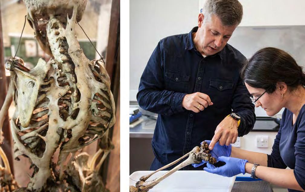
Left: Fibrodysplasia ossificans progressiva causes excess bone growth resulting in a ‘second skeleton’ as shown in this posterior view of the unidentified female FOP skeleton in the collection. Right: Endocrinology and palaeontology researchers, Adrian F. Daly and Olivia Cheronet, identify locations for sample extraction for DNA sequencing from the disarticulated lower limb of the unidentified female FOP skeleton in the collection.
Credit: Evi Numen, courtesy of The Old Anatomy Museum, Trinity College Dublin Philadelphia, has said that,
Medical heritage collections such as these ‘biomedical repositories’, if you will, can save lives. Locked within museum specimens is information that can help us inform treatments, develop cures, design vaccines. Learning about when and how these infectious disease outbreaks spread, can help us understand not only the diseases and pandemics we are dealing with now, but the future ones as well. The answers are there, it’s up to us to ask the right questions.8
…so fully developed and so well defined, that it merely required one to have seen the skeleton to recognise the nature of the cue and adjust her treatment accordingly.7
Nearly 150 years later, endocrinologist Adrian F. Daly and paleontologist Olivia Cheronet collected DNA material from Clarke’s skeleton, and that of an unidentified woman with the same condition, to contribute to research on this condition that currently plagues about 800 people worldwide.
Preserving the Past to Inform the Future
Anna N. Dhody, Co-Director and Curator of the Mütter Museum, and Director, Mütter Research Institute at The College of Physicians of In recent years, genome sequencing technology has rapidly improved, allowing for an array of incredible discoveries and insights into the past through the sampling of ancient DNA (aDNA) from historical remains found in museums.9 Today, aDNA sequencing can be achieved through minimally destructive means, such as removing dental calculus from a skull,10 a proposition that was unthinkable during the time that the Old Anatomy Museum was first populated with its silent residents. The potential benefits of such research cannot be underestimated. For example, ancient pathogen genome analysis can allow us a macro view of the evolution and epidemiology of viruses, like SARS-Cov2. By studying the patterns of epidemic and pandemic outbreak eruptions, or
Naturally, as medical heritage collection custodians, we are keen to emphasise the scientific and educational value inherent in our collections. But there is another dimension no less educational and valuable: the undeniable humanity of it. Even in the bubble of being a museum professional working with human remains, it is not easy to lose sight of how remarkable it is to hold a human skull in your (gloved) hands, or to conserve the femur of Cornelius Magrath, a 7’6’ foot skeleton with gigantism. There is a palpable reverence imbued in carrying out this work. It is this reverence and awe that we hope to inspire in visitors when, in due time, we can share the Old Anatomy collection with more than the students and researchers who currently get to see it.
Evi Numen is the Old Anatomy Museum Curator in the School of Medicine, Trinity College Dublin. This article builds on and discusses some of the ideas in her paper delivered at the February 2020 Irish Museums Association conference in Athlone.
This article has been written in collaboration with Siobhán Ward, the Museum’s Chief Technical Officer; and with Martina Hennessy, Associate Professor and Consultant for Medical Education, and Head of School Professor Michael Gill, both of the School of Medicine, Trinity College Dublin.
Notes
1. Thomas Percy Claude Kirkpatrick, History of the Medical Teaching in Trinity College, Dublin, and of the School of Physic in Ireland, 1912.
2. Davis Coakley, Medicine in Trinity College Dublin: An Illustrated History (Dublin: Trinity College Dublin, 2014).
3. Davis Coakley, Irish Masters of Medicine (Dublin: Town
House Publishers, 1992).
4. Samuel JMM Alberti and Elizabeth Hallam, Medical Museums: Past, Present, Future (London: The Royal College
of Surgeons of England, 2013).
5. George Newenham Wright, An Historical Guide to the City of Dublin, Illustrated by Engravings, and a Plan of the City, 2nd ed. (London: Baldwin, Cradock, and Joy, 1825).
6. Frederick S. Kaplan, ‘Fibrodysplasia Ossificans
Progressiva: A Historical Perspective’, Clinical Reviews in Bone and Mineral Metabolism 3, no. 3–4 (2005).
7. E.H. Bennett, ‘Extensive Osseous Depositions,
Implicating the Articulations, and Muscles. Proceedings of
the Pathological Society of Dublin, April 13, 1872’, in The Dublin Journal of Medical Science, 1872-1922 (Dublin: Fannin
& Co.), 508–13.
8. Said in conversation with the author. Used in this article
with permission by Anna N. Dhody.
9. S. Duchêne et al., ‘The Recovery, Interpretation and Use
of Ancient Pathogen Genomes’, Current Biology 30, no. 19
(2020): R1215–31.
10. N. Arning and D. Wilson, ‘The Past, Present and Future
of Ancient Bacterial DNA’, Microbial Genomics 6, no. 7 (2020).


