6 minute read
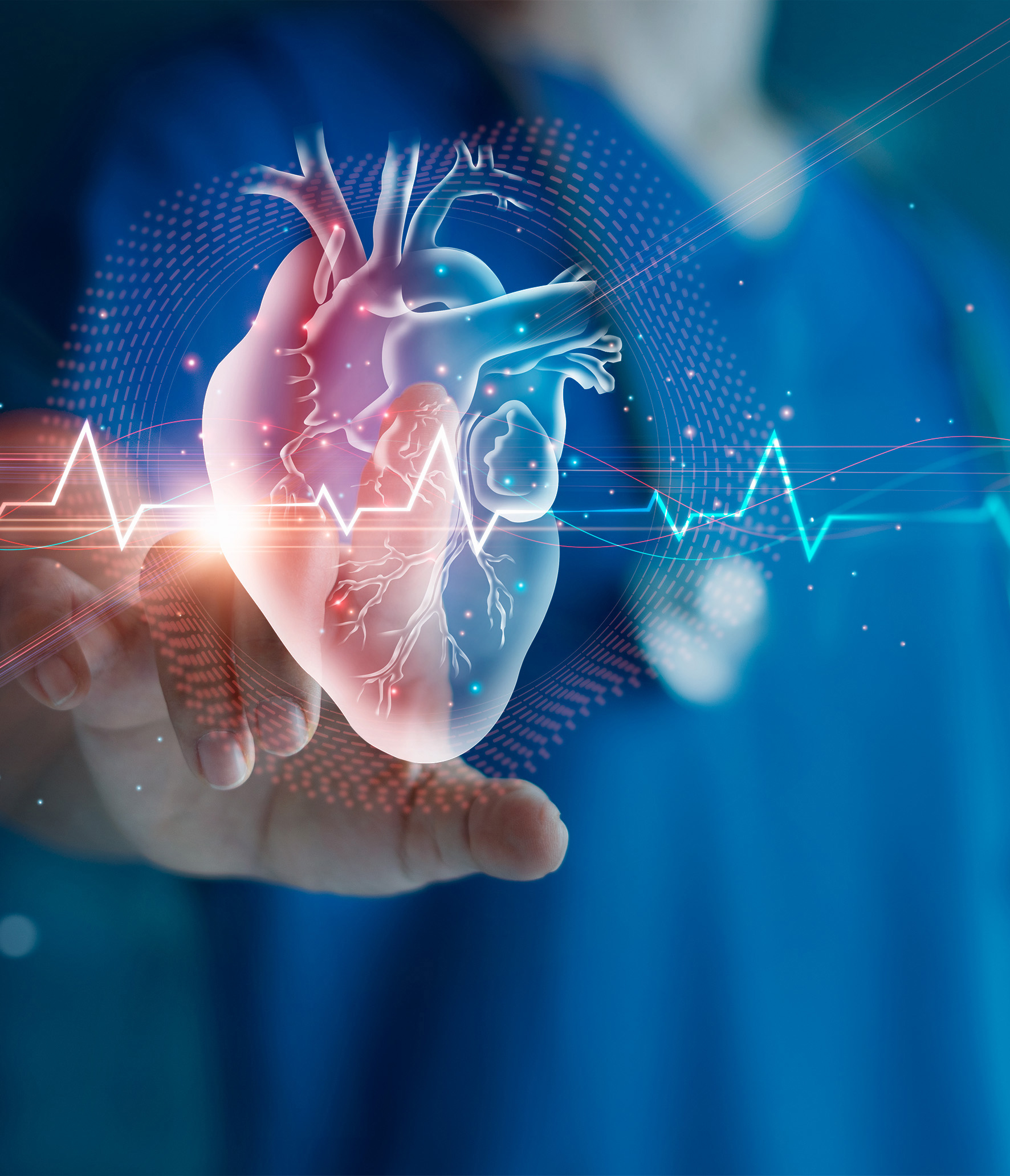
Patching up broken hearts

from Winter 2025 issue
by IN-SPHERE
Photo credit: ipobpa ( Istockphoto)
With donor hearts scarce, researchers are developing personalised, bioprinted heart patches to offer new hope for heart failure patients.
By LINDA MUSIC
The numbers are staggering. Each year, over 10,000 people in Australia will suffer a heart attack. Of those who survive, one in four will go on to develop heart failure. Their only option: a heart transplant. However, with only about 120 heart transplants conducted in Australia each year, most of these people will die. Add to this the fact that 50 per cent of heart transplant recipients die within five years, it’s clear there needs to be a better solution.
That challenge has been the focus of Associate Professor Carmine Gentile, School of Biomedical Engineering, the University of Technology Sydney (UTS), and Group Leader of Cardiovascular Regeneration at the Heart Research Institute and UTS. Over the past 20 years, he has dedicated his research to addressing this problem and now believes his team may have found a solution: a bioengineered heart patch developed from a patient’s own blood cells
Observations lead to an idea
It all began with A/Prof Gentile’s simple observation: traditional methods of studying and treating heart disease are often inadequate.
“Our research began with the recognition that current models for studying heart failure in patients, such as animal models or cell cultures in Petri dishes, fail to fully replicate the complexity of the human heart,” A/ Prof Gentile explains.
“Relying on these inadequate tools to study cardiovascular disease and treatments is a naive approach. Understanding the true development and formation of the human heart, from neonatal stages to adulthood, encompassing both healthy and diseased conditions, offers insights into effective prevention and treatment strategies.”
Leveraging Australia’s largest human heart biobank, A/Prof Gentile and his multidisciplinary team set out to uncover the molecular, cellular, and extracellular secrets of healthy versus diseased heart tissues.
Their research led them to an ambitious new solution: bioprinting “mini hearts,” tiny, functional structures combining muscle cells and blood vessel cells that mimic the real thing. These miniature replicas, laden with both muscle cells and blood vessel components, represent a paradigm shift in cardiac regeneration. Like building blocks of life, these mini hearts are bioprinted layer by layer using a specialised 3D printer that deposits bio-ink, a blend of cells and hydrogels that mimic the natural environment of heart tissue.
“In the laboratory, we analyse patient-specific MRI and CT scans and create the shape of the 3D bioprinted tissue that will be applied onto the area that has been damaged.”
This patch, custom-built for each patient, isn’t just a physical match, it’s a biological one too. Thanks to new technologies, the team can now create patient-specific stem cells using a simple blood sample. What sets these mini hearts apart is their ability to interact and synchronise, just like their full-sized counterparts. Tailored to match the anatomical and functional needs of individual patients, they hold promise as personalised patches that could restore heart function without the risks associated with traditional transplants.
"We can make the stem cells, and with the stem cells, we can make the patient-specific heart cells required for the formation of healthy muscle and blood vessels," A/Prof Gentile explains.
Then, by embedding those personalised cells into hydrogels (soft water-rich materials similar to gelatin), the team ensures that the cells survive and function properly after printing.
Crucially, the team’s mini hearts are designed to beat in synchrony with the patient’s heart, avoiding deadly arrhythmias.
“If the cells are beating at a different rate, patients will develop arrhythmias and could die,” A/Prof Gentile cautions.
This innovation could offer a lifesaving alternative for thousands who would otherwise face an agonising wait on transplant lists. It could also bypass the complications of traditional transplants, such as immune rejection and the lifelong need for immunosuppressant drugs.
The road ahead
But the road ahead is not without challenges as A/Prof Gentile and his team navigate rigorous testing phases to ensure safety and efficacy.
“We are still in the pre-clinical phase. We’ve shown that the patch can be generated according to the geometry of the patient’s heart, that it can be potentially applied onto the heart, and that it improves how the heart contracts. We are also looking at making it more durable in a way that we can test long-term effects from both efficacy and safety points of view,” A/Prof Gentile explains.
“The next challenge is to design and launch clinical trials. We need sufficient funding to support these trials, and we also need to determine where the trials will be conducted. Regulatory factors will determine whether this technology can be made available in Australia alone or internationally.”
Patients who undergo a heart transplant typically need about six months to recover. However, A/ Prof Gentile hopes that if the patch becomes the standard treatment, it could significantly shorten recovery times. This is because the flexible, elastic patch can be folded into a compact shape, inserted into the chest, and then unfolded and applied directly to the affected area of the heart using a minimally invasive procedure.
“Our dream is that the patient will be able to walk into the clinic in the morning and walk out in the afternoon with a new patch.”
It’s a bold vision: not just repairing hearts, but rebuilding them. Piece by piece, cell by cell, offering tens of thousands of people each year a chance at a new life. And with each breakthrough, that future moves a little closer to reality.
Other applications
Mini hearts in cancer research
In addition to their role in understanding heart development, mini hearts are proving invaluable in studying the long-term cardiovascular effects of cancer treatment. While advances in cancer therapies have significantly increased survival rates, many of these survivors face an elevated risk of heart disease due to the very treatments that saved their lives. One example is doxorubicin, a chemotherapy drug nicknamed the “Red Devil”—so-called for its red colour and potent cancer-fighting ability. Although effective, this drug can cause delayed cardiotoxicity, with some patients developing heart failure after treatment. However, not all patients respond to the drug in the same way, highlighting the urgent need for personalised testing tools. Mini hearts offer a promising platform for tailoring treatment strategies and long-term cardiac monitoring in cancer survivors, enabling researchers to better predict and prevent adverse cardiac outcomes.
Subjecting mini hearts to heart attacks
Beyond cancer-related research, mini hearts are also being used to model heart attacks in the lab.
A/Prof Gentile describes a project which involves creating a “heart attack in a test tube,” enabling researchers to mimic key heart attack conditions, such as fluctuating oxygen levels, to study how cardiac tissues respond to injury. This model has helped identify the damage caused by heart attacks and test potential protective therapies in a controlled, preclinical setting.
