3 minute read
International Journal for Research in Applied Science & Engineering Technology (IJRASET)


ISSN: 2321-9653; IC Value: 45.98; SJ Impact Factor: 7.538
Advertisement
Volume 11 Issue I Jan 2023- Available at www.ijraset.com
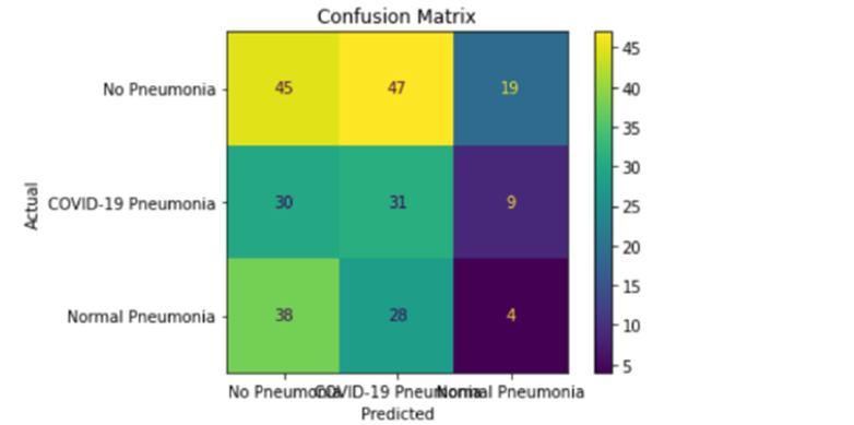
Confusion Matrix
IV. CONCLUSIONANDFUTUREWORK
The most important requirement for correctly diagnosing any type of thoracic disease is the presence of experienced radiologists. This project's main goal is to raise medical proficiency in regions with sparse radiotherapist availability. In such remote locations, our study helps with the early detection of pneumonia to avoid negative effects, including mortality. Pneumonia detection from the aforementioned dataset has not received much attention to date. The creation of algorithms in this area can greatly improve the delivery of healthcare services. Additionally, we demonstrated how hyper-parameter modification during the classification step improved model performance. Through a series of tests, we hope to provide the most effective pre-trained CNN model and classifier for use in related future research. In the near future, improved algorithms for detecting pneumonia are likely to be developed as a result of our study.
References
[1] P. M. Shah et al., "Deep GRU-CNN Model for COVID-19 Detection from Chest X-Rays Data," in IEEE Access, vol. 10, pp. 35094-35105, 2022.
[2] S. Rajaraman, S. Candemir, G. Thoma, and S. Antani, ‘‘Visualizing and explaining deep learning predictions for pneumonia detection in paediatric chest radiographs,’’ in Medical Imaging 2019: Computer-Aided Diagnosis, vol. 10950. International Society for Optics and Photonics, 2019, p. 109500S
[3] L. Qu, C. Wu, and L. Zou, ‘‘3D dense separated convolution module for volumetric medical image analysis,’’ Appl. Sci., vol. 10, no. 2, p. 485, Jan. 2020.
[4] M. F. Hashmi, S. Katiyar, A. G. Keskar, N. D. Bokde, and Z. W. Geem, ‘‘Efficient pneumonia detection in chest x-ray images using deep transfer learning,’’ Diagnostics, vol. 10, no. 6, p. 417, Jun. 2020.
[5] J. Son, J. Y. Shin, H. D. Kim, K.-H. Jung, K. H. Park, and S. J. Park, ‘‘Development and validation of deep learning models for screening multiple normal findings in retinal fundus images,’’Ophthalmology,vol.127, no. 1, pp. 85–94, Jan. 2020.
[6] Lai CC, Shi TP, Ko WC, Tang HJ, Hsueh PR. Severe acute respiratory syndrome coronavirus 2 (SARS-CoV-2) and coronavirus disease-2019 (COVID-19): The epidemic and the challenges. Int J Antimicrob Agents. 2020:105924.
[7] Z. Wang, Y. Xiao, Y. Li, J. Zhang, F. Lu, M. Hou, and X. Liu, ‘‘Automatically discriminating and localizing COVID-19 from community-acquired pneumonia on chest X-rays,’’ Pattern Recognit., vol. 110, Feb. 2021, Art. no. 107613.
[8] L. Goyal and N. Arora, ‘‘Deep transfer learning approach for detection of COVID-19 from chest X-ray images,’’ Int. J. Comput. Appl., vol. 975, p. 8887.
[9] M. Awais, M. Raza, N. Singh, K. Bashir, U. Manzoor, S. U. Islam, and J. J. P. C. Rodrigues, ‘‘LSTM based emotion detection using physiological signals: IoT framework for healthcare and distance learning in COVID-19,’’ IEEE Internet Things J., early access, Dec. 10, 2020.
[10] A. Serener and S. Serte, "Deep learning for mycoplasma pneumonia discrimination from pneumonias like COVID-19," 2020 4th International Symposium on Multidisciplinary Studies and Innovative Technologies (ISMSIT), 2020, pp. 1-5
[11] N. Hilmizen, A. Bustamam and D. Sarwinda, "The Multimodal Deep Learning for Diagnosing COVID-19 Pneumonia from Chest CT-Scan and X-Ray Images," 2020 3rd International Seminar on Research of Information Technology and Intelligent Systems (ISRITI), 2020, pp. 26-31.
[12] M. Mishra, V. Parashar and R. Shimpi, "Development and evaluation of an AI System for early detection of Covid-19 pneumonia using X-ray (Student Consortium)," 2020 IEEE Sixth International Conference on Multimedia Big Data (BigMM), 2020, pp. 292-296.
[13] Y. Wang, Y. Zhang, Q. He, H. Liao and J. Luo, "Quantitative Analysis of Pleural Line and B-Lines in Lung Ultrasound Images for Severity Assessment of COVID-19 Pneumonia," in IEEE Transactions on Ultrasonics, Ferroelectrics, and Frequency Control, vol. 69, no. 1, pp. 73-83, Jan. 2022.
[14] X. Wang et al., "Joint Learning of 3D Lesion Segmentation and Classification for Explainable COVID-19 Diagnosis," in IEEE Transactions on Medical Imaging, vol. 40, no. 9, pp. 2463-2476, Sept. 2021.
[15] M. Sevi and İ. AYDIN, "COVID-19 Detection Using Deep Learning Methods," 2020 International Conference on Data Analytics for Business and Industry: Way Towards a Sustainable Economy (ICDABI), 2020, pp. 1-6.
[16] M. Yamaç, M. Ahishali, A. Degerli, S. Kiranyaz, M. E. H. Chowdhury and M. Gabbouj, "Convolutional Sparse Support Estimator-Based COVID-19 Recognition From X-Ray Images," in IEEE Transactions on Neural Networks and Learning Systems, vol. 32, no. 5, pp. 1810-1820, May 2021.
[17] K. S. and D. Radha, "Analysis of COVID-19 and Pneumonia Detection in Chest X-Ray Images using Deep Learning," 2021 International Conference on Communication, Control and Information Sciences (ICCISc), 2021, pp. 1-6.
[18] H. N. Monday et al., "Improved Convolutional Neural Multi-Resolution Wavelet Network for COVID-19 Pneumonia Classification," 2021 4th International Conference on Pattern Recognition and Artificial Intelligence (PRAI), 2021, pp. 267-273.
[19] X. Ouyang et al., "Dual-Sampling Attention Network for Diagnosis of COVID-19 From Community Acquired Pneumonia," in IEEE Transactions on Medical Imaging, vol. 39, no. 8, pp. 2595-2605, Aug. 2020.
[20] V. K. Gupta, A. Gupta, D. Kumar and A. Sardana, "Prediction of COVID-19 confirmed, death, and cured cases in India using random forest model," in Big Data Mining and Analytics, vol. 4, no. 2, pp. 116-123, June 2021.
Author Profiles

