7 minute read
IMPLEMENTATION OF INSTINCTIVE BRAIN TUMOR SEGMENTATION AND DETECTION USING MAGNETIC RESONANCE IMAGE

from November 2022 International Journal of Innovative Technology and Creative Engineering (ISSN:2045-871

Dr. K. Selvanayaki
Assistant Professor, School of Computer Science, VET Institute of Arts and Science (Co-Edu) College, Erode, Tamil Nadu, India. {selvanayakik@vetias.ac.in}
Advertisement
Dr. K.S.Mohanasathiya
Assistant Professor, School of Computer Science, VET Institute of Arts and Science (Co-Edu) College, Erode, Tamil Nadu, India. {sathyaanandh08@gmail.com}
Dr. S. Prasath
Assistant Professor, School of Computer Science, VET Institute of Arts and Science (Co-Edu) College, Erode, Tamil Nadu, India. {softprasaths@gmail.com}
Abstract – Developments in medical imaging technologyunremittinglyrecoverthetransport of healthcare to patients. Given rising healthcarecostsandtheneedtoresponsibly steward financial resources, this paper highlights scientific, peer-reviewed studies that validate improved patient outcomes and costsavingsassociatedwithvariousimaging technologies. This review builds upon previous research conducted in past decade withanadditionalfocusoncost-effectiveness evaluations. A comprehensive search methodology was used to critically estimate publicationsfromthepast2decadeinhealth indexed in the U.S. National Library of Medicine (MEDLINE) and from leading health policyreviews.
KEYWORDS: Brain Tumor, Magnetic Resonance Image (MRI), Image Acquisition, Preprocessing, Enhancement,Segmentation,AntColonyOptimization (ACO),ParticleSwarmOptimization(PSO)
1. Introduction
Over the last decade, we can see the hasty growth of brainimagingknowledges.ithasvariety of techniques, skills and methods compare than few previous decades. MRI is the best way for brain image processing and analysis. MRI machine uses magnetic field and radio waves to harvests large amount of data and detailed exhaustive images with high level of quality. The Analysis of Brain image and segmentation from MRI dataset is deadly intricate task for specialist whohastophysicallyextractrequiredinformation. Thismanualexaminationisrepeatedly inefficient and it caused errors owing to inconsistency of trainings. At present number of originations in computer technologies to progress disease analysis and testing. Currently variety of procedures to assist clinician for MR brain image preprocessing, enhancement, segmentation, feature extraction and selection. In this paper examinedinfurthermostwidespreadapproaches usedforMRIbrainimagesegmentation.Magnetic ResonanceImaging(MRI)hasbecometheregular non-tending procedure for brain tumor diagnosis over the last few decades(2017, Subhashis Banerjee)The practice of magnetic resonance imaging (MRI) in health care and the emergence of radiology as a practice are both quite new compared with the classical specialties in medicine. We point the methods, accuracies, returns, advantages and disadvantages. We reviewthepaperwithfollowingsteps1.ImagePreprocessing and Enhancement 2.Segmentation 3. FeatureExtractionandSelection 4. Classification (K.Selvanayakietal.,)thefollowingstructureFig1 clarifies the necessary steps in tumor detection process.
Magnetic Resonance Image
MRI is used to treasure diversity of circumstances of the brain such as bleeding, abnormalities, infections, problems on vessels, etc. Mainly it shows tumor tissues on the brain images. Normally Neurologist brain tumor identification begins with Magnetic Resonance
Image. If it illustrates there is any tumor in the brain,thefurtherinvestigationlikethetypefinding, segmentinganalyzingarejustabout.Hereabrain tumor is an uncharacteristic progress of tissue in the brain or spine that can deranged the normal brainfunction. However, this type of tumor tissue basedoncellsinventionandiftheyaremalignant or benign. Anyway, brain tumor tissue segmentation and identification from the normal braintissueisthestimulatingmissionintheclinical history. In this scenario reviewed number of traditionalunearthingandbraintumorsegmenting papers from past 2010 to 2020.In conclusion, an assessment of the present innovative method for tumordetectionispresented.
2. Literature Survey
For learning of brain tumor revealing and segmentation the MRI Images is very useful in modernyears.DuetoMRIImageswecandetect thebraintumor.Fordetectionofunusualgrowthof tissuesandblocksofbloodinnervoussystemcan be seen in an MRI Images. The first step of detection of brain tumor is to check the symmetric andasymmetricShape ofbrainwhich willdefinetheabnormality.Afterthisstepthenext step is segmentation which is based on two techniques Ant Colony Optimization and Particle Swarm Optimization These two techniques are usedtodetecttumorcellsintheMRIImage.Now by this help of design we can detect the boundariesofbraintumorandcalculatetheactual area of tumor. Those optimization is usedtogive thecertaininformationlikerebuiltofmissingedges and extracting the silent edges. Accuracy and clarity in an MRI Images is dependent on each other.Saurabh Kumar et.al.,[6 ]proposed Wavelet based method is been used as a denoising. In frequency domain this method is used for de noising and preserving the actual signal. This builds the scaling coefficients freelance of the signal and therefore are often simply removed. Saurabh Kumar et.al.,[6]Support Vector Machine (SVM) approach is considered as a good candidateduetohighgeneralizationperformance, especially when the dimension of the feature space is veryhigh. Subhashis Banerjee et.al[ 10] explainedDeepConvolutionalNeuralNetworksfor classificationofbraintumorsusingmultisequence MRimages.
Swapnil R.Telrandhe, et.al [11] Proposed tumor detection inside which Segmentation separates an image into parts of regions or objects.Inthisithastosegmenttheitemfromthe background to browse the image properly and classify the content of the image strictly. During this framework, edge detection is a vital tool for imagesegmentation.Inthispapertheireffortwas madetostudytheperformanceofmostcommonly used edge detection techniques for image segmentation and additionally the comparison of these techniques was carried out with an experiment. Priya Patil et.al[5]told f-transform is used to give the certain information like rebuilt of missing edges and extracting the silent edges. Accuracy and clarity in an MRI Images is dependent on each other. The following Table1 shows the analysis report of review papers. The review said number of algorithmsandtechniques are available in the medical world for MRI brain tumor segmentation and detection but fuzzy C means occupied a special role for brain MRI segmentation and detection, this algorithm producesexcellentaccuracythanotheralgorithm.
AbdElKaderIsselmouetal.,[1]innovates number ofmethods.Thefirstmethodisimproved fuzzy c-means algorithm (IFCM), the second method is improved feed forward neural network (IFFNN), and the third method is a hybrid selforganizing map with a fuzzy k-means algorithm those methods invented for brain tumor segmentation andthosecontributedgoodfallouts for tumor detection. Selvanayaki et al.,[7,8] explained block based Ant colony optimization technique for brain tumor segmentation. Varchar et al.,[9] Md shaharier et al., [4] explained the techniqueFuccyCmeansforsegmentingidentical feature of tissues from Brain MRI. The following Table 1 shows recent review of brain tumor detection.
Table 1: Review Paper Analysis
3.ProposedSystem
certainlyrequiresalongprocessaswellas complexduetothedensityofthestructureofthe humanbrain.Obviously,theslowprocessof detectingandorderingbraintumordiseasein patientscanbasisdelayedmedicaltreatmentfor thepatient'srecovery.Forthisreason,basedon theneedformedicalinformationneededby doctorstotreatpatientsspeedilyandexactly,an imageprocessingtechniqueormethodforreading MRIimagesisestablishedbeforefewdecades. Theintentionofthedetectionmethodistoassist theradiologistindetectingtumorinmedical images.Inthispaperreviewshowsvarious techniquesormethodsthathavebeenusedto detectbraintumorsonMRIimages.Itexpectsto provideinformationondifferenttechniquesor methodsrelatedtobrainMRI.Inthispaper suggestedtoencapsulateandmatchthemethods forbraintumordetectionthroughMagnetic ResonanceImage(MRI).Inparticularnecessary stepsaredeliberateandrelated.Thispapertracks thefollowingblockdiagramFig1.
Acquirement
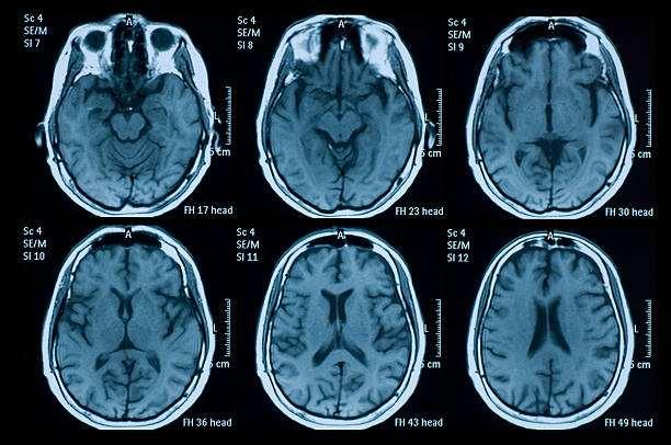
Acquirementisthefirstandforemoststep intumordetectionprocess.Itistheactionof retrievinganimagefromaMRIMachine.The detectionprocesscannotdoanyactualandformal processingwithoutanMRIimage.MRIisan especiallytechnologicalelaborationinthemedical fieldthatyieldsimageswithextraordinarytenacity tosenseandthencancategorizeillnessesthatare inventintheorgansofthesickperson’sbody.One situationispossibletoidentifyfromreadinganMRI imagewithin2.56×1011Nanoseconds.The followingviewexplainsthebrainanditsangles. ThefollowingfigureshowsthesamplebrainMRI fromRealPatientDatabase.
Fig 2: a) SingleBrain MRI b) Brain View with different angle
The intention of Magnetic acquirement varies considerably from those for water or fat, protonsinorganictissues.Thus,thesedifferences playacriticalroleintheselectionandoptimization of pulse sequences for hyperpolarized-gas applications
4. Preprocessing and Enhancement
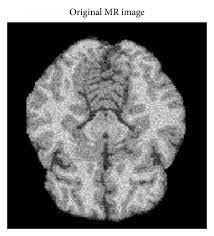
This review for pre-processing and enhancement through Magnetic Resonance Image (MRI) is aincline and erected image enhancement method and is based on the first derivative, resident data. Sonali Patil etal [2] explainedmediafilterforpreprocessinganimage enhancement. In Preprocessing and Enhancement phase, medical Image is renewed into standard format with contrast manipulation, noise reduction by background removal, edge sharpening, filtering process and removal of film artifacts. Preprocessing functions comprise those processesthatareroutinelyvitalformertothecore data study and removal of facts, and are largely convened as radiometric or regular corrections. The next one is enhancement method here the adventandbasesofimageartifactsthatcanoccur with MRI systems should be accepted and modified-raymarks, the high frequency components are removed finally unwanted skull portions on the MRI are removed. Selvanayaki etal[7] describes Median Filter can remove the noise, the high frequency components from MRI withoutdisturbingtheedges,bandwidthetcandit is used to reduce salt and pepper noise. JyotiPanwar et al [3] performed image preprocessing using image gradient method. The followingfigureshowspreprocessedImage.
3: Preprocessed MRI Image Segmentation
TheBarrierofanimagestrainsthedivision orpartingoftheimageintoprovincesofassociated attribute. The Ultimate goal in enormous quantity of image processing request is to extraction hermetic structures from the image data from which a elucidation, amplification, or imagined of the section can beprovided bythe machine. The Segmentation of brain tumor from magnetic resonance images is anvital but inefficient task performed by medical experts. The evaluation clarified numbers of procedures are available in this brain tumor detection and segmentation. ExpresslyFuzzyCMeans-Means,Supportvector Machine (SVM) approaches, Morphological Operations are used to segment tumor texture in thebrainMRI.TheaccuratesegmentationofMRI imageintodifferenttissueclasses,especiallygray matter (GM), white matter (WM) and Cerebrospinal fluid (CSF).In brief, segmentation regulates the Regions of Interest(ROIs) in an image.Thisdoesnotmeanthatthesegmentation will try to determine the type of the region, but merely determine the pixels in an image which belongtothesameitem.Thefollowingfig.4shows theSegmentedOriginalImage
The digital image processing community has developed several segmentation methods, many ofthemadhoc.Fourofthemostcommonmethods are: 1) amplitude thresholding, 2) texture segmentation3)templatematching,and4)regiongrowing segmentation. It is very important for detecting tumors, edema and necrotic tissues. These types of algorithms are used dividing the brainimagesintothreecategories(a)PixelBased (b)RegionorTextureBased(c)StructuralBased. SeveralauthorsSuggestedvariousalgorithmsfor segmentation .The segmentation is the most importantstageforanalyzingimageproperlysince it affects the accuracy of the subsequent steps. However,propersegmentationisdifficultbecause of the great verities of the lesion shapes, sizes, and colors along with different skin types and textures. In addition, some lesions have irregular boundaries and in some cases there is smooth transition between the lesion and the skin. To address this problem, several algorithms have beenproposed.Theycanbebroadlyclassifiedas thresholding, edge-based or region-based, supervised and unsupervised classification techniques Threshold segmentation, Water shed segmentation, Gradient Vector Flow (GVF) ,KmeanClustering,FuzzyC-meansClustering
5. Summary and Conclusion
In this survey paper numerous detection methods of brain tumor through MRI has been deliberate and linked for the period of recent era .This is charity to attention on the current developments of medical image processing in relationofbraintumordetectionprocess.Wehave designated several approaches in brain image processing and to discussed rations and belongings of techniques intumordetection.This paperisusedtogivemoreinformationaboutbrain tumordetectionandsegmentationmethods.Itisa breakthrough for analyzing all technologies relevanttobraintumorfromMRIinMedicalimage processing. In this paper, shows few steps about brain tumor detection process such as: The Preprocessing and Enhancement Technique Segmentation Algorithm and their performance have been studied and compared. In this paper, we have proposed different techniques to detect and segment Brain tumor from MRI images. To extractandsegmentthetumorweused different techniques such as SOM Clustering, k-mean clustering, Fuzzy C-mean technique, curvelet transform. Itcan beseen thatdetection of Brain tumor from MRI images is done by various methods, also infuture work differentautomatic methods achieve more accuracy and more efficient.
References
1. AbdElKaderIsselmou,GuizhiXu,ShuaiZhang,” ImprovedMethodsforBrainTumorDetectionand Analysis Using MR Brain Images”, Biomedical & Pharmacology Journal, Vol. 12(4), p. 1621-1631 ,December2019.
2. Selvanayaki K ,Dr Karnan M,” CAD System for Automatic Detection of Brain Tumor through Magnetic Resonance Image-A Review”,International Journal of Engineering Science and Technology”,Vol. 2(10), 5890-5901, 2010.
3. SonaliPatil1,Dr.V.R.Udupi,”PreprocessingTo BeConsideredForMRandCTImagesContaining Tumors”,OSRJournalofElectricalandElectronics
Engineering (IOSRJEEE) ISSN: 2278-1676 Volume1,Issue4(July-Aug.2012),PP54-57
4. Saurabh Kumar, Iram Abid, Shubhi Garg, Anand Kumar Singh, Vivek Jain,”Brain tumor detection using image processing method”, International Journal of Information Sciences and Application (IJISA).ISSN0974-2255,Vol.11,No.1,2019.
5. Swapnil R.Telrandhe,”segmentation methods for medical image analysis”,tesis no 1434,center for medicalimagescienceandvisualization,se-58185 linkoping,Sweden.
6. Ms.PriyaPatil,Ms.SeemaPawar,Ms.Sunayna Patil,Prof.ArjunNichal,”AReviewPaperonBrain TumorSegmentationandDetection”International JournalofInnovativeResearchinElectrical, Electronics,InstrumentationandControl Engineering”Vol.5,Issue1,January2017.
7. Md Shahariar Alam , Md Mahbubur Rahman , MohammadAmazadHossain ,MdKhairulIslam, KaziMowdudAhmed,KhandakerTakdirAhmed, Bikash Chandra Singh , Md Sipon Miah,” Automatic Human Brain Tumor Detection in MRI Image Using Template-Based K Means and Improved Fuzzy C Means Clustering Algorithm”, BigDataCoginitiveComputing,2019.
8. JyotiPanwar,Andrea S.Doria,” Magnetic ResonanceImagingDataAcquistion”,2021.
9. Vachan Vadmal, Grant Junno, Chaitra Badve, William Huang, Kristin A. Waite,Jill S. BarnholtzSloan”, MRI image analysis methods and applications: an Algorithmic perspective using brain tumors as an exemplar”,Neuro –Oncology Advances”,2020.
10.Selvanayaki K,Dr.Karnan M,”Improved ImplementationofBrainMRImageSegmentation using Meta heuristic Algorithms”,IEEE International Conference on Computational IntelligenceonComputingResearch,2012.
