What’s the Diagnosis – Case 91
1
What’s the Diagnosis – Case 91
2
What’s the Diagnosis – Case 91
3
What’s the Diagnosis – Case 91
4
What’s the Diagnosis – Case 91
5
What’s the Diagnosis – Case 91
6
What’s the Diagnosis – Case 91
7
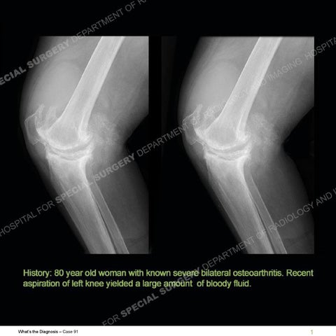
Findings As stated in the history, seen is the severe osteoarthritis of both knees. There are very large joint effusions particularly seen in the suprapatellar pouch of both knees with marked enlargement in the 9 year time interval. There is indolent bony remodeling particularly seen of the anterior aspect of the left femur. The MRI shows a markedly thickened band of tissue about the suprapatellar pouch yielding an isolated suprapatellar pouch. A marked degree of thickening is present of the joint capsule/synovial lining. A large joint effusion is present with different signal intensities. Areas of dephasing/blooming phenomenon are present on the gradient echo sequence related to hemosiderin.
What’s the Diagnosis – Case 91
8
What’s the Diagnosis – Case 91
9
What’s the Diagnosis – Case 91
10
What’s the Diagnosis – Case 91
11
What’s the Diagnosis – Case 91
12
What’s the Diagnosis – Case 91
13
Diagnosis: Complete Suprapatellar Plica There are multiple plicae of the knee that represent the remnants of embryologic structures that resorb to a great degree in most individuals. At times these plicae may become enlarged or thickened and become symptomatic. A suprapatellar plica is frequently seen on MR exams but infrequently is symptomatic. When it is symptomatic it may be related to a lack of any perforation or resorption as seen in this patient. In this case the advanced arthritis led to marked thickening of the capsule with an increased joint effusion. The difference in signal of the effusion likely relates to hemorrhagic debris layering in the joint. The imperforate nature of the suprapatellar plica made it such that there was a marked enlargement of the suprapatellar pouch. This long standing process then subsequently yielded the indolent remodeling of the anterior femur. In this example, aspiration of the knee yielded bloody fluid related to a likely hyperemic and friable synovium. The bloody fluid and enlargement of the suprapatellar pouch can be alarming and question a potential neoplasm but indeed are merely a manifestation of this uncommon etiology.
What’s the Diagnosis – Case 91
14
References Anatomy and MR imaging appearances of synovial plicae of the knee. GarcíaValtuille R, Abascal F, Cerezal L, García-Valtuille A, Pereda T, Canga A, Cruz A.Radiographics. 2002 Jul-Aug;22(4):775-84.
What’s the Diagnosis – Case 91
15