13 minute read
Clinical


Aesthetic anterior tooth restorations with nano-ceramic hybrid CAD/CAM blocks
1, 2 Prof. Dr. João Mauricio Ferraz da Silva and Danilo de Souza Andrade,
São Paulo State University, São José dos Campos, Brazil Introduction For decades now we have been observing a rise in the desire among patients for aesthetic improvements to their smiles. A multidisciplinary treatment makes it possible to achieve a harmonious and healthy smile by adapting the shape, colour, size and positioning of the teeth on the basis of facial parameters. Thanks to scientific progress overall and advances in the materials and techniques used, it is now possible to effect such changes with less invasive procedures. One such treatment technique is ceramic veneers (Belser et al., 1997; Radz, 2011; Rotoli et al., 2013).
The production of veneers begins with minimal preparation of the tooth in such a way that as much healthy dental hard tissue as possible is preserved on the one hand and changes to the shape and even small changes to the colour of the tooth are possible on the other. This novel concept was introduced under the term “minimally invasive dentistry” (Radz, 2011).
The material most commonly employed in the production of ceramic veneers is lithium disilicate-based ceramic, which displays good mechanical properties thanks to its resilience, offers excellent visual properties thanks to its ability to reproduce natural tooth characteristics well and is also bio-compatible with the neighbouring oral tissues (Chen et al., 2018; Palla et al., 2018; Zhi et al., 2016).
Composite-based blocks are also used to produce indirect restorations with the aid of CAD / CAM technology. During the production process, they are exposed to heat and pressure for polymerisation and, as a result, display superior mechanical properties in comparison with direct composite restorations (Mainjot et al., 2016). In terms of their modulus of elasticity and strength, the composite-based CAD / CAM blocks achieve values similar to those of natural tooth substance, i.e., enamel and dentine. These properties can be controlled by the percentage of resin matrix in the constituents of the blocks. In addition, this material displays greater resistance to fatigue compared with ceramics (Alamoush et al., 2018; Magne et al., 2010), and such properties make this material an excellent choice for durable, indirect restorations. Grandio blocs (VOCO GmbH, Cuxhaven, Germany) is an example of a hybrid ceramic block for CAD / CAM systems – it is a nano-ceramic hybrid material containing 86 % inorganic fillers in a polymer matrix. The composite systems for CAD / CAM are indicated for the indirect production of permanent single-tooth restorations such as inlays, onlays, full crowns and veneers.
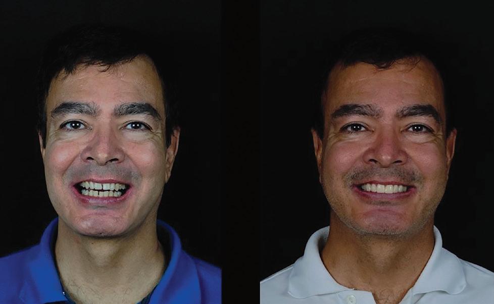
Figure 1: Portrait photo before and after treatment. Figure 2: Close-up of smile before and after treatment.
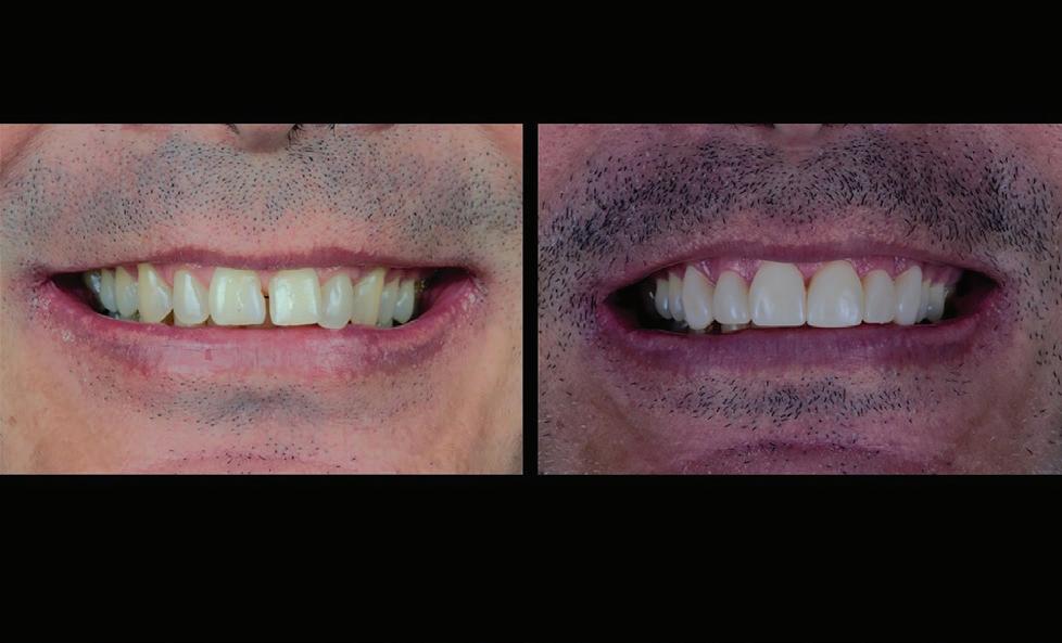
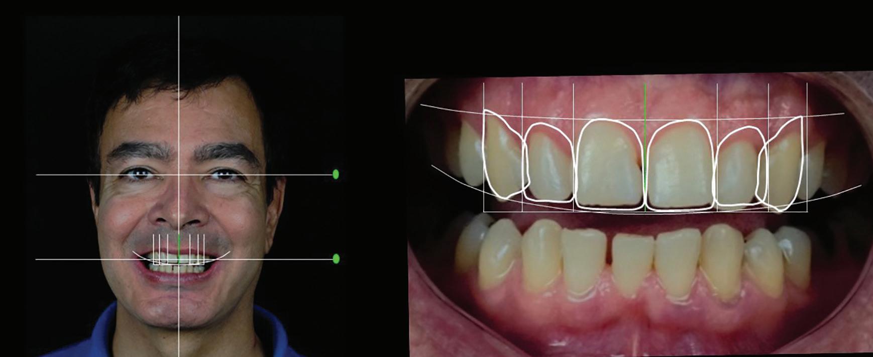
Figure 3: Digital smile design.
Alongside the mechanical advantages, the composite blocks also offer the possibility of shade adaptation directly after the milling process without any need for additional crystallisation, as is the case with ceramic blocks. This represents another advantage of this production technique (Allen et al.). A further interesting factor worthy of note when these blocks are used is that, compared with ceramics, the margins of the restoration do not display any microcracks and are more homogeneous (Tsitrou et al., 2007). The restorations produced using composite-based materials are easier to repair intraorally if and as required, if necessary by freshening up the area, silanising the restoration afterwards, applying an adhesive system and performing the repair directly with the composite (Tsitrou et al., 2010).
This clinical case thus aims to illustrate the possibility for using CAD / CAM technology and composite-based blocks for the indirect restoration of anterior teeth. Case report
A 50-year-old patient presented in the clinic of the university project Construindo Sorrisos Confiantes (Building Confident Smiles) run by the Department of Dental Materials and Prosthetics at the Institute for Science and Technology at the Federal University of São Paulo in São José dos Campos, Brazil. The patient was unhappy with his smile and particularly did not like the gaps between his teeth or their shade. In the first session, the patient’s medical history was recorded, a clinical examination performed and photographs taken for the purpose of the diagnosis and subsequent treatment planning (Fig.1, Fig. 2).
The examinations revealed that the patient was in good general health and maintained good oral hygiene without any systematic conditions which might affect and / or hinder dental treatment.
The extraoral photographs, comprising a front portrait
DA SILVA / DE SOUZA ANDRADE
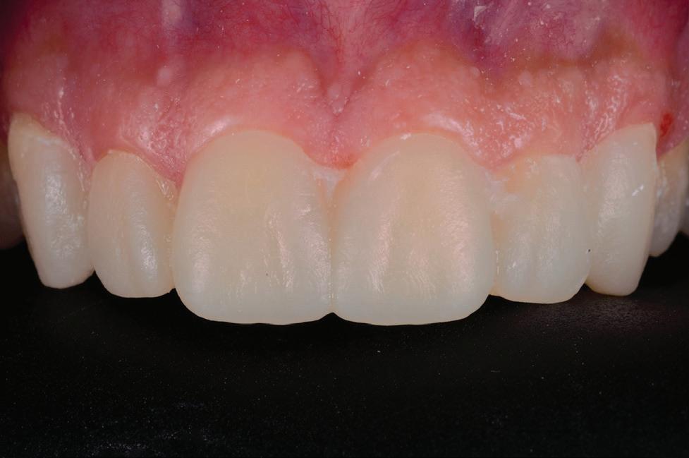
Figure 4: Mock-up. Figure 5: Preparation on the mock-up.
photograph with lip retractor and forced smile, 45º from the side and a photograph in the 12 o’clock position, and the intraoral photographs, comprising a close-up of the smile, maximal intercuspidation with lip retainers and the maxillary teeth against a black background, were taken prior to the start of treatment (Fig. 3). This was followed by 2D digital smile design (DSD) using the diagnostic photographs, based on the main problem and with the aim of resolving it. The ideal smile was constructed on the basis of the patient’s facial features and the function of the stomatognathic system. The parameters employed when designing the smile were the smile line and the position of the lips, the gingival zenith, tooth proportions, the face format and the increase in size in accordance with the principles of the RED correlations (Stanley et al., 2018). Once the final 2D smile design was complete, the data were sent along with the patient’s models to the dental laboratory for production of a diagnostic waxup for the patient.
The model with the diagnostic wax-up was then used to produce a silicone impression, which replicates the proposed new dental anatomy and allows production of a mock-up on the patient’s teeth without the need for any tooth preparation, solely with the aim of simulating the proposed aesthetic restoration for the patient. The dentist uses this simulation to analyse the aesthetics and function for the case in question and either recommends or rejects the plan. In addition, the trying-in of the mock-up is ideal for giving the patient an idea of how the proposed aesthetic restoration will look and building up his confidence in the planned treatment.
The mock-up is made using bis-acrylic, which boasts material characteristics such as low heat emission during the polymerisation reaction. It also reproduces the shape of the tooth copied using the silicone tray well and has a similar colour to natural dental tissue. The Structur 2 bis-acrylic composite (VOCO GmbH) was used. It was introduced into a silicone tray with the tip of the mixing tip always in the deepest part of the impression so as to avoid air bubbles. Once filled, the tray was inserted into the patient’s mouth and the excess material removed during the early stages of the polymerisation. The material was then allowed to cure completely. Once fully polymerised, the silicone tray was removed from the patient’s mouth and the surface of the finished mock-up wiped with a piece of gauze soaked in alcohol in order to remove any remaining monomer (Fig. 4).
Once the patient and dentist were both satisfied with the completed planning, a treatment plan for attaining the treatment goal described in the first phase was submitted to the patient. The following treatment was proposed to the patient: tooth whitening to achieve a homogeneous tooth shade and production of veneers made from hybrid blocks (Grandio blocs – VOCO GmbH) for six teeth from tooth 13 to 23 in order to redesign the shape of the teeth.
The technique proposed by Kern & Ahlers (2015) was used as a reference for the preparation of the teeth, with the mock-up serving as a guide for removal of the dental substance. A 4141 grinding bur (KG Sorensen, Cotia, Brazil), characterised by diamond-coated rings, was used to carve reference grooves into the vestibular surfaces of the respective teeth, taking into consideration the incline of the tooth in each third, and an approximately 0.5 mm thick layer of substance removed from mesial to distal. An extra-fine 3145FF diamond bur was then used to remove a second layer of substance from each vestibular surface
DA SILVA / DE SOUZA ANDRADE
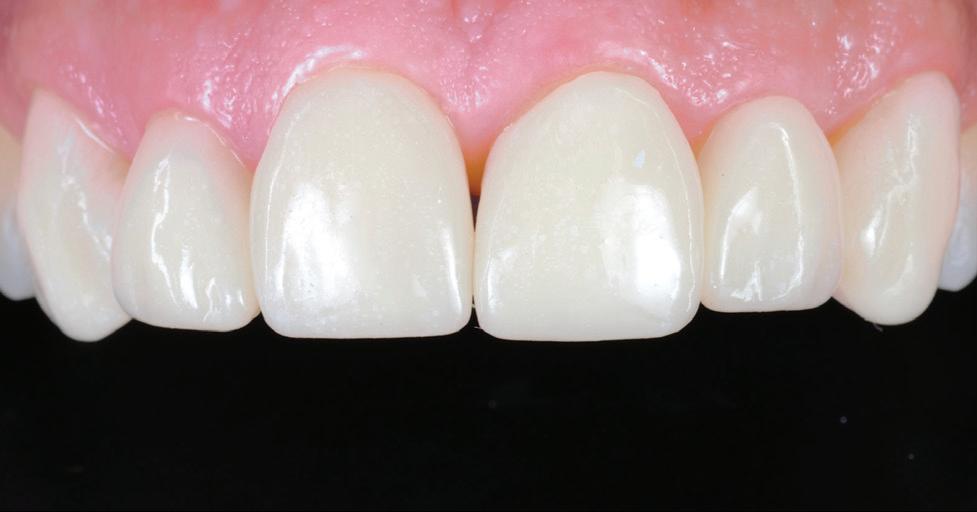
Figure 6: Dry try-in.
until the groove placed during the first step was levelled out. The preparation was completed with an extra-fine 3203FF diamond bur, with the cervical and proximal preparation margins being defined so as to achieve better marginal integrity for the respective veneers and determine the exact size of the veneers (Fig. 5).
The impression was taken in the same treatment session following the preparation and a single #000 knitted cord (Ultrapak – Ultradent Products Inc., South Jordan, USA) used for the gingival retraction in advance. The cord was soaked in a little haemostatic solution and placed into the gingival sulcus. The impression was performed in two stages with an addition-curing silicone (Virtual – Ivoclar Vivadent AG, Schaan, Liechtenstein). The DSD planning and the model were sent to the laboratory as references and for the production of the restorations.
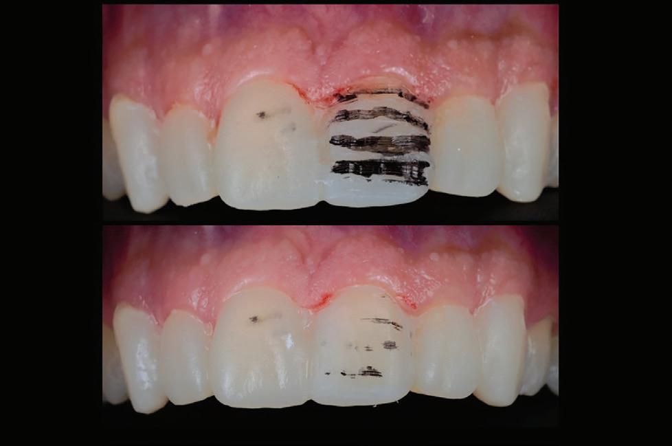
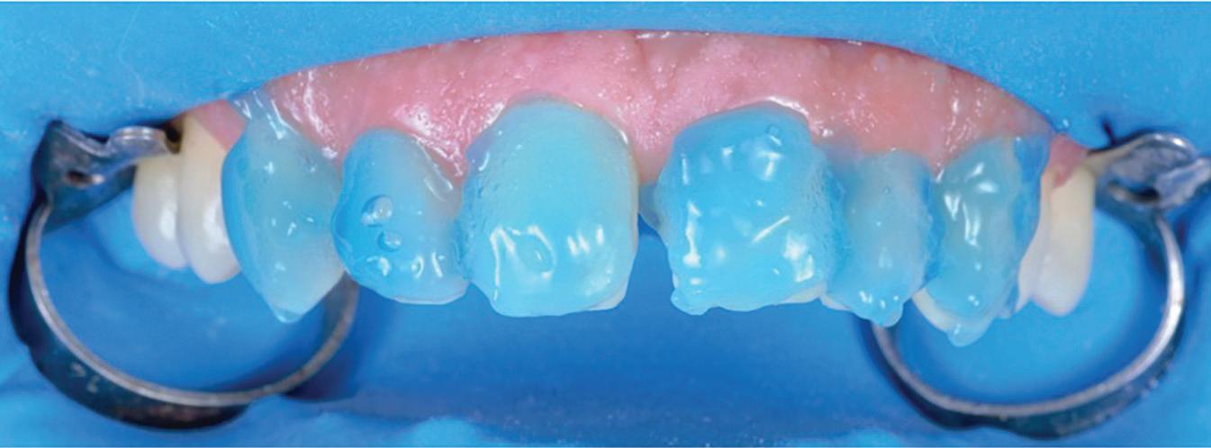
For insertion of the restorations, a dry try-in was performed in the mouth in advance in order to assess the marginal integrity and final position of the veneers and make any proximal adjustments if and as necessary (Fig. 6). The interior sides of the veneers were produced in the following way in accordance with the manufacturer’s specifications: sandblasting with aluminium oxide (25-50 µm), followed by cleaning of the pieces in an ultrasound bath for 5 minutes. The surface was then treated with the bonding agent Ceramic Bond (VOCO GmbH) (Fig. 7): an applicator brush was used to apply the agent to the interior sides of the veneers before it was allowed to work for 60 seconds and then dried quickly with compressed air. The teeth were pretreated with 37% phosphoric acid for 30 seconds as the preparation of the teeth occurred exclusively in the enamel, then rinsed thoroughly with air and water and dried with compressed air. The adhesive Futurabond U (VOCO GmbH) was applied to the tooth surface and massaged in for 20 seconds, followed by removal of the excess material with a fine suction device and light stream of air (Fig. 8).
Following the preparation of the veneers and the teeth, the next step was the final insertion of the composite. The dual-
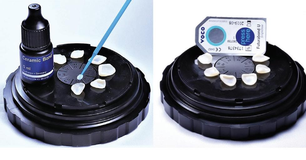
Figure 7: Application of Ceramic Bond on veneers.
DA SILVA / DE SOUZA ANDRADE
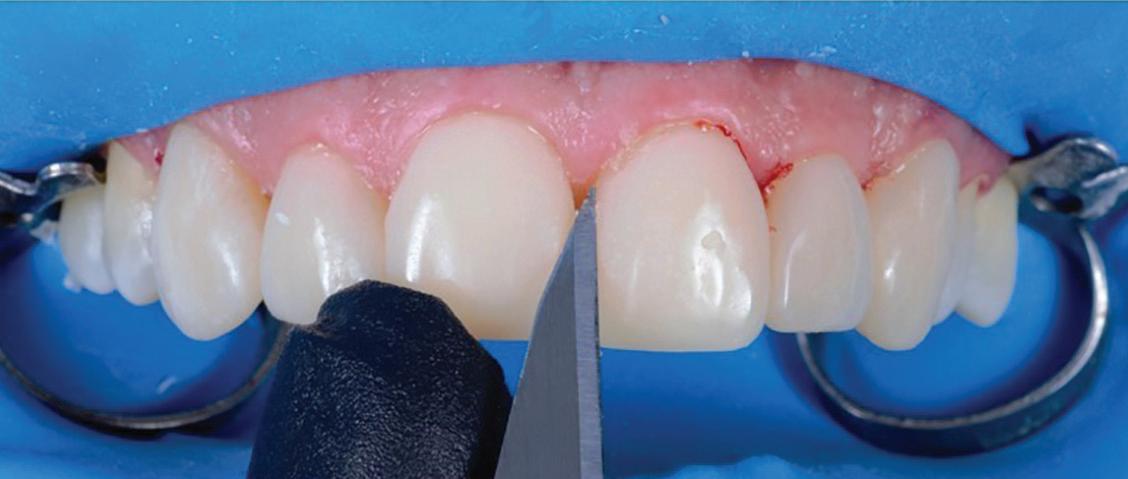
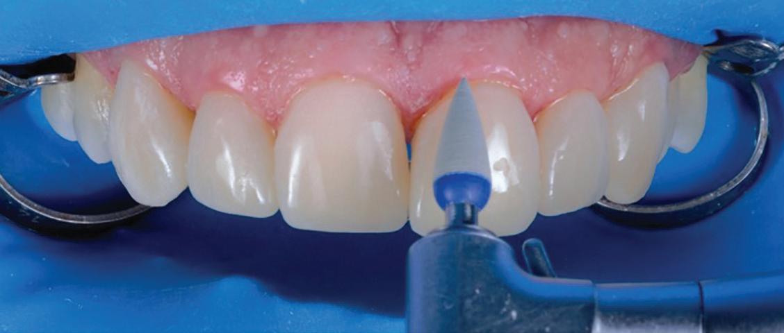
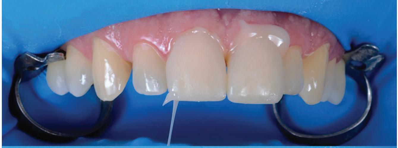
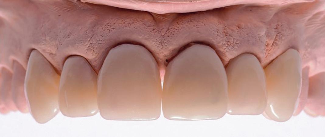
Figure 10: Removal of excess material with scalpel and polishing with the Diamanto polishers. Figure 11: The restoration on the model and in its final position in the mouth.
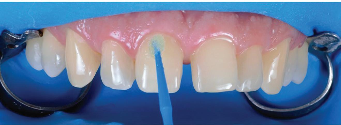
curing composite-based luting system Bifix QM (VOCO GmbH) was applied to the interior side of the veneers, the veneers brought into their final position and a check for excess material performed. Following removal of the excess material with a brush, the material was light-cured for five seconds to stabilise the veneers. With the restorations fixed in this way, the next step was to remove the excess material using dental floss on the proximal surface and a no. 11 scalpel on the cervical surface (Fig. 9).
This was followed by the final light curing for 40 seconds on each side of the veneer. Once polymerisation was complete, the remaining excess material was removed using a periodontal curette and an interproximal saw. The occlusion was then ground in on the basis of the markings using occlusal articulating film (Accufilm – Parkell Inc., Edgewood, USA) on a holder in accordance with the criteria for optimal occlusion with double-sided and even contact and clearly defined guide surfaces.
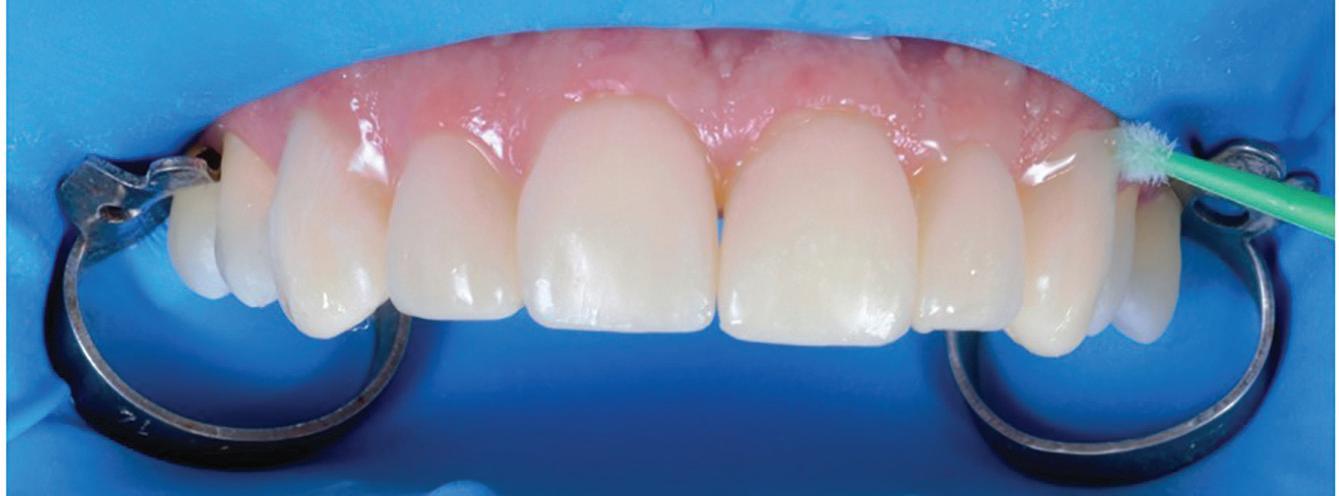
Following the grinding, the veneers were finished and polished using Diamanto diamond polishers (VOCO GmbH), giving them their final smooth and lustrous surface (Fig. 10).
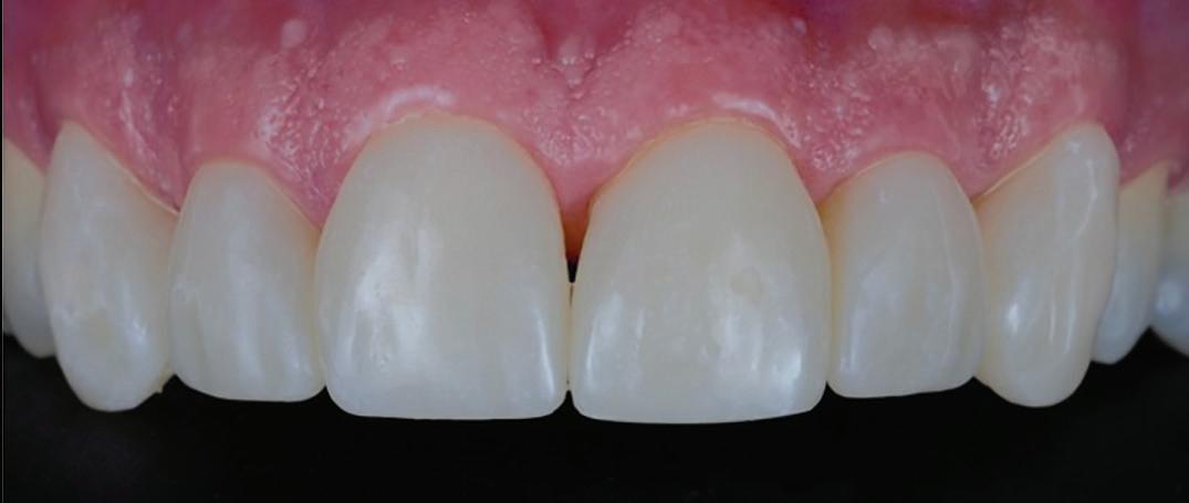
The patient was instructed in the essential care and monitoring of the restoration before being discharged with the first follow-up appointment scheduled for 48 hours later. After two follow-up sessions in which the marginal integrity, possible excess material, occlusal contact points and oral hygiene were assessed without any complaints, the patient was briefed on the importance of maintaining good oral hygiene and the requirement for a check-up every six months and then permanently discharged (Fig. 11).
Conclusion This case illustrates clearly that the CAD / CAM technique is already a clinical reality and will soon become the only practised means of producing indirect restorations. In addition, we should take the use of alternatives to ceramic materials, which have already established themselves in the scientific literature, into consideration for aesthetic restorations in the anterior region. The hybrid material described displayed good aesthetic and mechanical characteristics in this case, although further clinical studies into the longevity of the material remain necessary.
References 1. Alamoush RA, Silikas N, Salim NA, Al-Nasrawi S, Satterthwaite JD. Effect of the Composition of CAD / CAM Composite Blocks on Mechanical Properties. Biomed Res Int. 2018;2018. doi: 10.1155/2018/4893143. 2. Allen KL, Schenkel AB, Estafan D. An overview of the
DA SILVA / DE SOUZA ANDRADE
CEREC 3D CAD / CAM system. Gen Dent. ;52 (3):234–5. PMID: 15206254. 3. Belser UC, Magne P, Magne M. Ceramic laminate veneers: continuous evolution of indications. J Esthet Dent. 1997;9(4):197–207. PMID: 9468884. 4.Chen X, Zhang Y, ZhouU J, Chen C, Zhu Z, Li LEI. Effect of different surface treatments and retainer designs on the retention of posterior Pd-Ag porcelain-fused-tometal resin-bonded fixed partial dentures. Exp Ther Med. 2018;15(2):2006–14. doi: 10.3892/etm.2017.5630. 5. Kern M, Ahlers MO. Controlling the depth of ceramic veneer preparations by using a color marker in the depth grooves. J Prosthet Dent. 2015;114(6):862–4. doi: 10.1016/j. prosdent.2015.06.010. 6. Magne P, Schlichting LH, Maia HP, Baratieri LN. In vitro fatigue resistance of CAD / CAM composite resin and ceramic posterior occlusal veneers. J Prosthet Dent. 2010;104(3):149–57. doi: 10.1016/S00223913(10)60111-4. 7. Mainjot AK, Dupont NM, Oudkerk JC, Dewael TY, Sadoun MJ. From Artisanal to CAD-CAM Blocks. J Dent Res. 2016;95(5):487–95. doi: 10.1177/0022034516634286. PMID: 26933136. 8. Palla E-S, Kontonasaki E, Kantiranis N, Papadopoulou L, Zorba T, Paraskevopoulos KM, et al. Color stability of lithium disilicate ceramics after aging and immersion in common beverages. J Prosthet Dent. 2018;119(4):632–42. doi: 10.1016/j.prosdent.2017.04.031. 9. Radz GM. Minimum Thickness Anterior Porcelain Restorations. Dent Clin North Am. 2011;55(2):353–70. doi: 10.1016/j.cden.2011.01.006. PMID: 21473998. 10. Rotoli B, Lima D, Pini N, Aguiar F, Pereira G, Paulillo L. Porcelain Veneers as an Alternative for Esthetic Treatment: Clinical Report. Oper Dent. 2013;38(5):459–66. doi: 10.2341/12-382-T. PMID: 23550911. 11. Stanley M, Paz AG, Miguel I, Coachman C. Fully digital workflow, integrating dental scan, smile design and CADCAM: case report. BMC Oral Health. 2018;18(1):134. doi: 10.1186/s12903-018-0597-0. PMID: 30086753. 12. Tsitrou EA, Helvatjoglu-Antoniades M, van Noort R. A preliminary evaluation of the structural integrity and fracture mode of minimally prepared resin bonded CAD / CAM crowns. J Dent. 2010;38(1):16–22. doi: 10.1016/j. jdent.2009.07.003. PMID: 19683378. 13. Tsitrou EA, Northeast SE, van Noort R. Brittleness index of machinable dental materials and its relation to the marginal chipping factor. J Dent. 2007;35(12):897–902. doi: 10.1016/j.jdent.2007.07.002. PMID: 17977638


