50 minute read
Reproduction


Course Contents
THE SCIENTIFIC METHOD
Page 8
1. The scientific method ....................................................... 10 2. Research in the laboratory .............................................. 14 3. Research in a natural environment .............................. 18 4. Finding information ........................................................... 22 Review and practise .............................................................. 24 Glossary ...................................................................................... 26
1
The ORGANISATION OF THE HUMAN Body
Page 28
1. Levels of organisation ....................................................... 30 2. The human cell ..................................................................... 32 3. Cell differentiation .............................................................. 36 4. Human tissues ...................................................................... 37 5. Organs and systems .......................................................... 40
Research project STEP
Investigating healthy habits for the heart ...................... 42 Review and practise .............................................................. 44 Glossary ...................................................................................... 46
2
NUTRITION AND FOOD
Page 48
1. Nutrition and humans ....................................................... 50 2. Nutrients ................................................................................ .51 3. Energy supply ...................................................................... 54 4. Eating ...................................................................................... 56 5. Dietary recommendations ............................................... 58 6. Food and sustainability .................................................... 60 7. Diet and health .................................................................... 62 Research project STEP Investigating healthy habits for the heart ...................... 64 Review and practise .............................................................. 66 Glossary ........................................................................................68
3
SYSTEMS OF THE NUTRITION FUNCTION
Page 70
1. The digestive system ......................................................... 72 2. Digestion ................................................................................ 74 3. The respiratory system ..................................................... 76 4. The circulatory system ...................................................... 78 5. Blood flow .............................................................................. 82 6. The lymphatic system ....................................................... 83 7. The excretory system ......................................................... 84 8. Health and the nutrition function ................................. 86 Research project STEP Investigating healthy habits for the heart ...................... 90 Review and practise .............................................................. 92 Glossary ...................................................................................... 94
4
THE INTERACTION FUNCTION
Page 96
1.The interaction function in humans ........................... 98 2. Perception: the senses .................................................. 100 3. The sense organs and health ...................................... 104 4.Nervous coordination .................................................... 106 5. The health of the nervous system ............................. 112 6. Endocrine coordination ................................................ 114 7. The health of the endocrine system ......................... 117 8. Drugs and drug addiction ............................................ 118 9. Executing responses: the locomotor system ........ 120 10. Health and the execution response .......................... 124
Research project STEP
Investigating the human senses ....................................... 126 Review and practise ............................................................ 128 Glossary .................................................................................... 130
5
Reproduction
Page 132
1.Human reproduction ....................................................... 134 2. The reproductive system .............................................. 136 3. Human gametes ............................................................... 138 4.The ovarian and uterine cycles .................................. 140 5. Fertilisation, pregnancy and birth ............................. 142 6. Birth control and reproductive health ..................... 144 Research project STEP Investigating the human senses ....................................... 148 Review and practise ............................................................. 150 Glossary .................................................................................... 152
6
HEALTH and Illness
Page 154
8
LANDFORM MODELLING
Page 196
1. Health and illness .............................................................. 156 2. Types of illnesses .............................................................. 156 3. Transmission of infectious diseases ........................... 160 4. The immune system ......................................................... 162 5. Preventing and curing illnesses ................................... 164 6. Transplants and donation .............................................. 168 7. First aid ................................................................................. 170 Research project STEP Investigating the human senses ....................................... 172 Review and practise ............................................................ 174 Glossary .................................................................................... 176
7
DYNAMIC EARTH
Page 178
1. Landform modelling ....................................................... 198 2. Exogenous geological processes .............................. 200 3. The geological action of wild waters and torrentes ..................................................................... 202 4. The geological action of rivers ................................... 204 5. The geological action of groundwater .................... 206 6. The geological action of glaciers .............................. 208 7. The geological action of wind .................................... 210 8. The geological action of the sea ............................... 212 9. The geological action of living beings ..................... 214 Research project STEPS 2 Studying soil erosion ........................................................... 216 Review and practise ........................................................... 220 Glossary ................................................................................... 222
1. The Earth’s surface and its dynamics ....................... 180 2. The Earth’s internal energy and endogenous processes .......................................... 182 3. Magmatism and volcanoes ........................................... 186 4. Tectonic forces ................................................................... 188 5. Geological risks ................................................................. 189 Research project STEP 1 Studying soil erosion ............................................................ 190 Review and practise ............................................................ 192 Glossary .................................................................................... 194
1
The Organisation of the human BODY
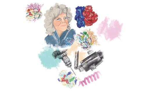
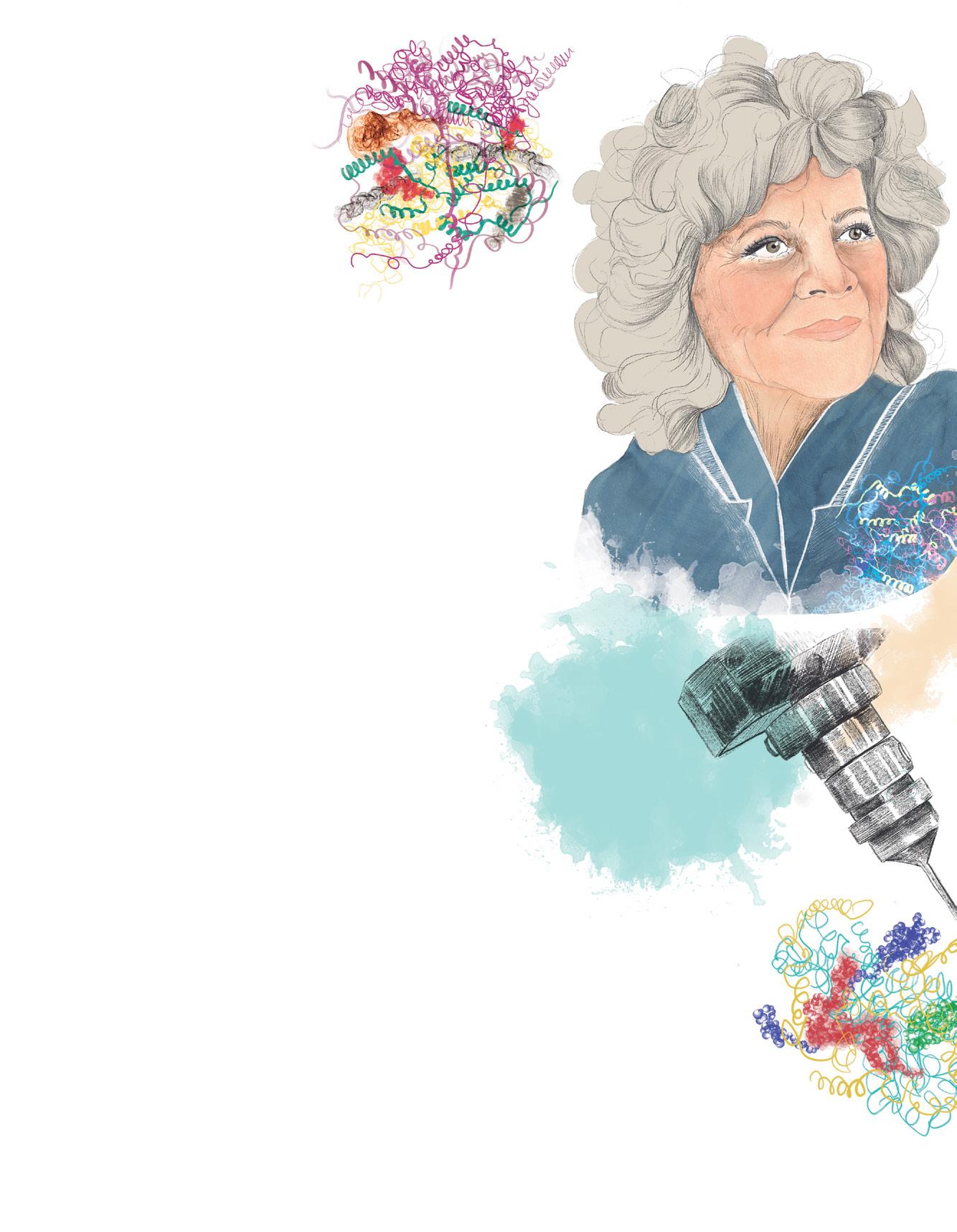
Ada Yonath. Curious by nature
To be a scientist, you must be curious. And believe me, I have always been curious! When I was five years old, I tried to find out the height of our apartment and broke my arm! My name is Ada Yonath and I was born in Jerusalem, Israel in 1939. My family never had much money, so I had to work a lot to be able to study: I gave private classes, I babysat, I cleaned the chemistry lab... It was hard, but it gave me the opportunity to learn and research in secret, so I can’t complain. Starting my career as a researcher was amazing. I was interested in something that seemed impossible: discovering what ribosomes looked like and how they worked. Ribosomes are responsible for decoding the DNA instructions in cells and using them to build proteins. But what exactly was their structure? We didn’t know. I specialise in a technique that involves creating crystals and observing them using x-rays. The problem was that the ribosome crystals we made broke extremely easily. We spent years destroying crystals until, one day, we found a way to make ribosome crystals strong enough that we could observe their structure in detail. This discovery won me the 2009 Nobel Prize in Chemistry, among other awards. While prizes don’t matter much to me, I’m really proud that my discovery can be used to attack the ribosomes of bacteria and develop new antibiotics to keep winning the fight against infections. 1 a) Why won’t Ada complain about having to work so hard when she was younger? b) What do Ribosomes do? c) Why was it difficult to observe ribosome crystals using x-rays?
Speaking
2 For Ada Yonath, curiosity is the most important quality necessary to be a scientist. She says that from a young age she was curious to explain everything she saw around her.
Discuss with a partner: a) What are the most important qualities for a scientist to have? b) Do you have any of these qualities? c) Are these qualities different from those needed in other subject areas? d) How can these qualities be inspired and encouraged within scientific education? What would you change within science classes?
3 Similar to Ada Yonath’s discovery of how to manufacture stronger ribosome crystals, there are many other inventions that have helped improve our understanding of the human body. Choose any medical invention or technique and write an imaginary interview with their inventor(s).
Include: ➜ What problem they were trying to solve ➜ How they developed their invention and how long it took ➜ What it allowed them to discover ➜ How they felt about the process
I think the most important quality of a scientist is to be patient.
We could encourage patience by doing more experiments that last a longer amount of time.

Writing An interview
LANGUAGE BANKLANGUAGE BANKLANGUAGE BANKLANGUAGE BANKLANGUAGE BANKLANGUAGE BANKLANGUAGE BANK LANGUAGE BANKLANGUAGE BANK LANGUAGE BANKLANGUAGE BANK LANGUAGE BANK 29
LEVELS OF ORGANISATION
Like other living beings, human beings are made up of cells that group together to form more complex structures, such as tissues and organs.
Biomolecules make up living beings
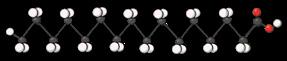
Carbohydrates
Carbohydrates are a cell’s main source of energy. They form part of some cell structures and are used to build other biomolecules. They can be complex, like cellulose, which forms the walls of plant cells, or simple, like glucose.
Lipids
Lipids are very chemically diverse. Some, like phospholipids and cholesterol, form part of cell membranes. Others, like triglycerides, provide and store energy for cells. Levels of organisation are the different degrees of complexity into which living matter is structured.
The elements in each level join together to form other more complex levels. Each new level has new characteristics and properties, making them more complex than just a collection of the elements in the previous level.
1.1 Atomic level
Atoms are the smallest units of matter that have the properties of a chemical element. Hydrogen (H), oxygen (O) and calcium (Ca) are all atoms. Atoms that form part of living matter are called bioelements.
Proteins
Proteins form part of cell structures and regulate and carry out most cell processes. For example, the haemoglobin in red blood cells transports oxygen in the blood, and membrane proteins regulate substance exchange in and out of cells.
Nucleic acids
Nucleic acids contain genetic information, which they transmit to offspring, and control cell functions. DNA (deoxyribonucleic acid) and RNA (ribonucleic acid) are nucleic acids.
Understand, think, search...
1 Give definitions of bioelement, biomolecule and chemical bond. 2 What is a polymer? Give some examples of biomolecules that are polymers.
1.2 Molecular level
Molecules are groups of atoms held together by chemical bonds. Water (H2O), oxygen (O2) and calcium carbonate (CaCO3) are all molecules. Molecules that make up living beings are called biomolecules and contain a high level of carbon. Biomolecules group together to carry out a function within a cell. They form organelles such as ribosomes and mitochondria. Biomolecules are often polymers, chains formed when molecules of the same type, called monomers, bond together. Carbohydrates or sugars, lipids, proteins and nucleic acids (like deoxyribonucleic acid or DNA) are polymers. Other substances like water and mineral salts have a simple chemical structure. They are found in both living and non-living matter.
3 Which of the following compounds are only found in living matter?
Phospholipids Glucose Iron
Sodium chloride Potassium Water
DNA Cellulose
1.3 Cellular level
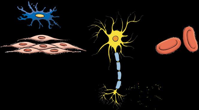
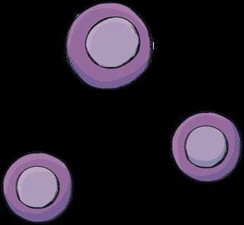
Cells are the structural and functional units of all living beings. That is, they are the smallest part capable of carrying out the three vital functions. All human cells are eukaryotic* animal cells, although they are specialised into more than 200 different types. Some examples are neurons, red blood cells, adipocytes, ova and spermatozoa.
Focus on English The word
karyotic comes from a Greek word meaning kernel or nucleus. The prefix eu- means true, while the prefix pro- means before. What do the words prokaryotic or eukaryotic mean? Which cell is more primitive?
1.4 Organism level
Tissues are groups of similar cells that specialise in the same function, and which are sometimes found along with other substances. Some examples are nervous tissue, muscle tissue and epithelial tissue. Tissues group together to form organs, such as the heart, lungs and kidneys, which carry out specific functions. In turn, organs group together in systems to perform a coordinated function. Some examples are the respiratory system, the excretory system and the nervous system. Finally, the human body is made up of a combination of organs and systems, which coordinate to carry out the three vital functions. Working with pictures
What level of organisation in the human body do the following parts or groupings belong to? a) Ovum b) Heart c) Group made up of the mouth, oesophagus, stomach, intestine, pancreas, etc. d) Protein e) Nucleus f) Carbon
Carbohydrates
Atoms
bond to form

Lipids
Proteins
Nucleic acids like Biomolecules and other substances
Water Mineral salts
which group together to form Cells
which group together to form
Tissues
which group together to form
Organs
which group together to form
Systems
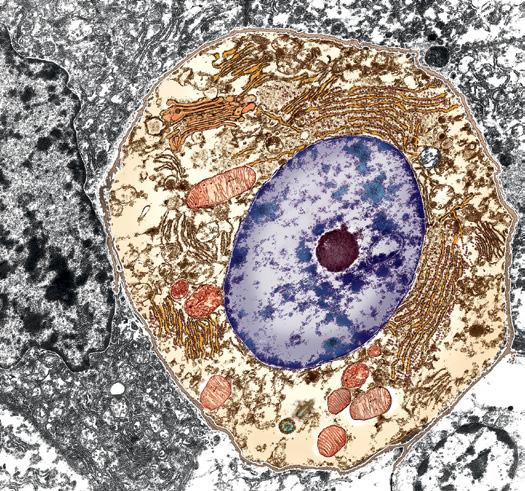
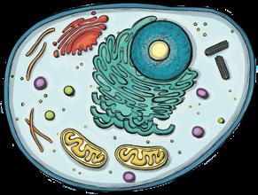
THE HUMAN CELL
Structure of a eukaryotic animal cell
Cytoplasm Plasma membrane
Nucleus
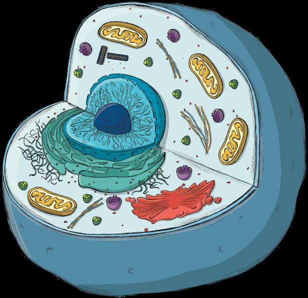
Cell organelles
Cell membrane
Carbohydrates
Interior Lipids Proteins The human being is a multicellular organism. It is made up of cells which have very different shapes and sizes. Generally, human cells have three basic parts: the plasma membrane (or cell membrane), the nucleus and the cytoplasm.
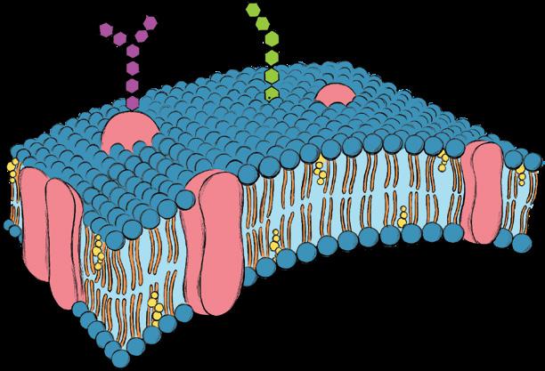
2.1 The plasma membrane
The cell membrane is a thin layer that surrounds and protects the cell and regulates exchanges of substances with the surrounding environment.
The cell membrane is made up of lipids. These form a layer, embedded with proteins, around the cell. The functions of the membrane are to: ➜ Protect the cell and separate its contents from the outside environment. ➜ Detect changes in the environment and allow the cell to respond to these changes in an appropriate way. ➜ Regulate the exchange of substances with the environment. The transfer of substances across the cell membrane takes place in different ways and depends on the size of the substance.
Small substances, such as oxygen, can cross the membrane on their own, medium-sized substances cross the membrane with the help of proteins, and large substances cannot cross the cell membrane. Instead, large substances reach the inner part of the cell by endocytosis. In this process the cell wall folds inwards to bring the substance into the cell.
Understand, think, search...
1 Give a definition of cell. 2 What are the functions of the plasma membrane? 3 Although animal cells are usually shown as being round, the cells in our bodies have a variety of different shapes. Look for pictures of different types of cell in the human body and draw them in your notebook. Label the three structures they have in common: • the plasma membrane • the nucleus • the cytoplasm.
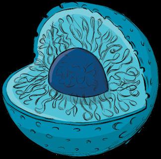
2.2 The nucleus
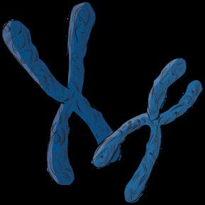
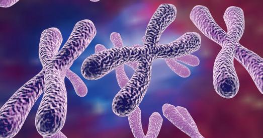
The nucleus is a large spherical structure. It contains the genetic material (DNA) which organises the cell’s activity.
The nucleus is made up of the following parts: ➜ The nuclear membrane. It contains nuclear pores which allow substances to be exchanged between the nucleus and the cytoplasm. ➜ The nucleoplasm. It is made up of a thick, aqueous liquid which contains the genetic material or DNA. ➜ The chromatin, which is the network of all the DNA fibres and proteins. When the cell divides, the chromatin fibres roll up and become compacted, forming structures called chromosomes which are visible under a microscope. ➜ The nucleolus is a spherical structure inside the nucleus where the component parts of ribosomes (a type of cell organelle) are made.
Understand, think, search...
4 anayaeducacion.es Not all cells are the same, and they do not have the same cellular organisation.
Remind yourself of the types of cellular organisation in living beings by going to ‘Prokaryotic and eukaryotic cells’ in the resource bank. What type of organisation do the following cells have? a) A protozoan d) An insect b) A fungus e) A bacterium c) An alga f) A pine tree 5 Draw a cell nucleus in your notebook, then label its parts and write what their functions are. 6 What two structural states can a nucleus be in? 7 As you already know, cells are microscopic in size. To measure them we use a unit called the micrometre or micron (μm). 1 = 0.001 mm = 0.000 001 m a) Use the bar scale to calculate the diameter of the cell in micrometres. b) Find the nucleus in this cell. What is its diameter? c) The cell in the picture is round, so we can calculate its volume like we would for a sphere. What is its volume? What is the volume of its nucleus?
The nucleus
Nucleolus
Chromatin Nuclear membrane
Nuclear pores
Chromosomes (condensed chromatin)
5 μm
2 THE HUMAN CELL
Focus on English
The phrase to be suspended in something is a technical phrase meaning that something floats within liquid or air without moving. A similar meaning for to suspend is something that is attached to a high place so that it hangs down. Alternatively to suspend can also be used to mean to stop someone from going to school or work for a short time, especially if they have broken the rules.

Working with pictures
Look at the photo of the cell. What cell structure is coloured green? Describe what you can see in the photo.
2.3 The cytoplasm
The cytoplasm is the part of the cell between the plasma membrane and the nuclear membrane.
The cytoplasm is formed from a thick, aqueous liquid called cytosol. Cytosol contains different substances and cell organelles.
Cytosol is composed of 70-80 % water and 30-20 % other substances that are dissolved or suspended* within it. These substances include proteins, carbohydrates, lipids, nucleic acids, mineral salts and ions. It is where many of the cell’s vital chemical reactions take place.
2.4 Cell organelles
There are two types of organelles: non-membranous organelles and membranous organelles.
Cell organelles are structures that are immersed in the cytoplasm and perform specific functions, like organs.
Non-membranous organelles
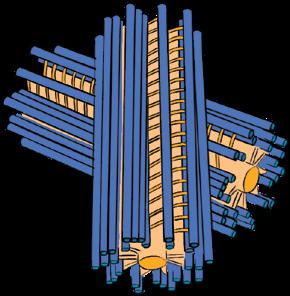
Centrioles are two fibre-based cylinders that direct the movement of the cytoskeleton and the chromosomes during cell division. Ribosomes are small particles that synthesise proteins. They can either be attached to the endoplasmic reticulum or scattered around the cytoplasm.
The cytoskeleton is a network of fibres that holds the organelles in place and gives the cell its shape. It also plays a part in cell movements (it forms cilia and flagella).
Membranous organelles
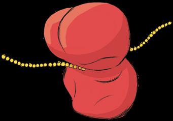
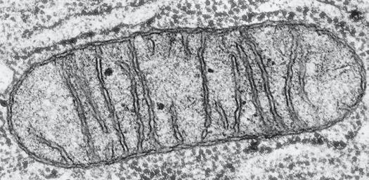
Mitochondria are oval structures with a double membrane and numerous folds in their internal membrane. They obtain energy for the cell through cellular respiration. Vesicles are small structures that transport or store substances.
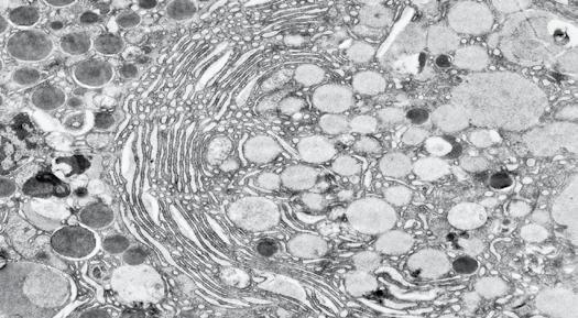
The endoplasmic reticulum (ER) is a network of tubules and sacs that synthesise substances. The rough ER (RER), which has ribosomes attached to its walls, is involved in protein synthesis. The smooth ER (SER), which does not have ribosomes, is involved in the synthesis of lipids.
The Golgi apparatus is a network of flattened sacs that modify the substances that are synthesised in the ER and transport them outside the cell via vesicles. Lysosomes are vesicles that contain digestive enzymes. They digest the particles captured by the cell.
Understand, think, search...
8 The parts add up. Each of the structures in a cell performs a specific function, and together they allow the cell to work optimally.
Create a table describing the following for each cell organelle: What is its function? What would happen if it didn’t exist? What is its relation to the other parts? To find out how to apply this skill, go to anayaeducacion.es. 9 Look closely at the micrographs on the right and answer the following questions: a) Do they show separate cells or the inside of a cell? Explain your answer. b) Use the bar scale (the black line) to calculate the size of organelle A. c) Draw a picture in your notebook showing what you think this organelle would look like in 3D. Calculate its volume.
0.5 μm
CELL DIFFERENTIATION
Focus on English
Take on is a phrasal verb which, in this text, has been used to mean to have a particular quality or appearance. However, to take something on can also mean to agree to do a new project or extra work. On the other hand, to take somebody on, could mean to employ someone. It can also mean to start a fight or compete with someone, especially if they are more powerful or stronger than you.
Understand, think, search...
1 In your own words, what is cell differentiation? 2 Are the following statements about cell differentiation true or false? a) Cells in the human body differentiate because they have different genetic material. b) All the cells that make up the human body have the same genetic material.
Cell differentiation
Differentiation is a combination of changes to the shape and structure of the cell which allow it to specialise in a specific function.
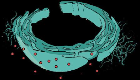
All the cells in a multicellular organism, such as a human being, are created from one single cell called a zygote, which divides to form an embryo. At first all the cells in an embryo are the same, but later they take on* different shapes and specific functions. That is, they become specialised. This process is called cell differentiation. Therefore, although all human cells share basic characteristics (for example, they have the same genetic material), they don’t all have the same shape or the same function. After differentiation has taken place in a multicellular organism, cells that look similar and that carry out the same function group together to form tissues. For example, neurons are star-shaped and have an extension filled with cytoplasm which allows the neuron to receive stimuli and to transmit electrical impulses. Neurons form nervous tissue.
Cell differentiation
Stem cell
Glial cells
Muscle fibers
Neurons Red blood cells
HUMAN TISSUES
Understand, think, search...
1 Give a definition of tissue. 2 What tissue forms part of the sweat glands?
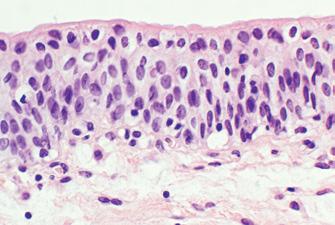
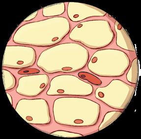
3 Look at the photo. What type of epithelial tissue can you see? A tissue is a group of cells with a similar shape and structure and which specialise in carrying out the same function.
Human tissues are classified into four basic types: epithelial, connective, muscle and nervous tissues.
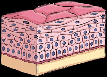
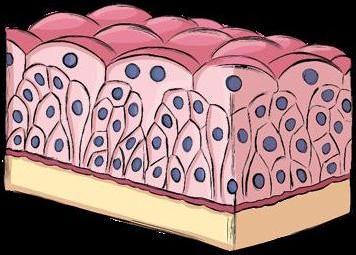
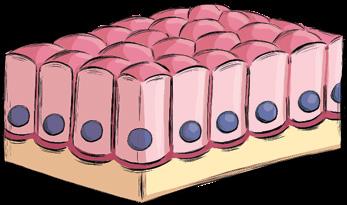
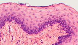
4.1 Epithelial tissue
Epithelial tissue (or epithelium) is made up of one or more layers of cells that are very close to each other. Epithelial tissue lines the organs and acts as a protective barrier. There are two types of epithelial tissue: ➜ Covering and lining epithelium. This type of epithelial tissue serves a protective function, lining both the external and the internal surfaces of organs. For example, it lines internal cavities that communicate with the outside environment. ➜ Glandular epithelium. This type of epithelial tissue is made up of cells specialised in producing substances and secreting them onto the epithelial surface. These cells can be scattered around or grouped together as glands, like the sebaceous glands, for example.
Types of epithelial tissue
Simple epithelia
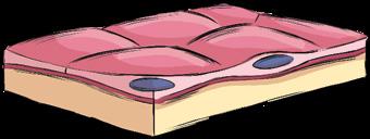
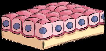
Squamous cells
Stratified epithelia Cuboidal cells Columnar cells
Glandular epithelium
Secreted substances
4 HUMAN TISSUES
Types of connective tissue
Reticular connective tissue
Fibres
Cells surrounded by lots of fibres
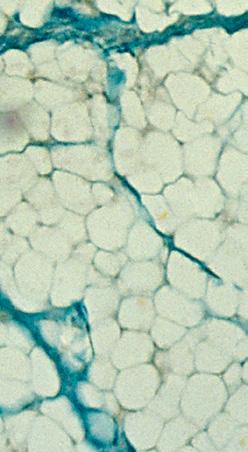
Adipose tissue
Fat cells or adipocytes with low levels of intercellular substance
Bone cells or osteocytes surrounded by a solid matrix
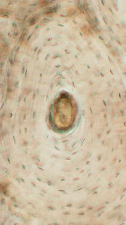
Cartilaginous tissue
Blood Solid and elastic matrix
Chondrocytes
Osseous tissue (Bone)
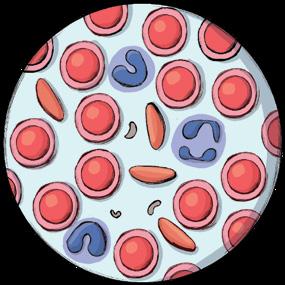
Blood plasma
White blood cells
Red blood cells
Platelets
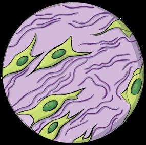
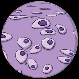
4.2 Connective tissue
Connective tissue consists of cells surrounded by fibres and an intercellular substance called a ‘matrix’.
Connective tissue supports and connects organs and other tissues in the body. There are various types: ➜ Reticular connective tissue. This tissue has an extensive gel-like matrix containing abundant fibres. It fills the space between organs, keeping them in place, and forms tendons. ➜ Adipose tissue. This tissue has a matrix containing low levels of intercellular substance. Its cells store fat as an energy reserve and for thermal insulation.
It is found under the skin. ➜ Cartilaginous tissue. Cartilage has a solid and elastic matrix. It covers the joints to prevent wear.
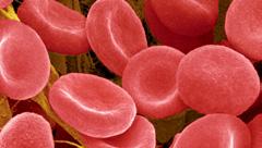
An example is ear cartilage. ➜ Osseous (bone) tissue. The matrix of this tissue is solid and hard, owing to the presence of calcium salts. It forms bones. ➜ Blood. Blood has a liquid matrix called plasma, which has blood cells suspended in it (red blood cells, white blood cells and platelets).
Understand, think, search...
4 What type of connective tissue can you see in each of the following pictures?
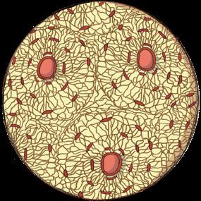
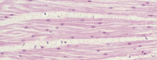
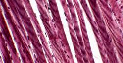
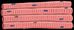
4.3 Muscle tissue
Muscle tissue is made up of elongated cells that group together to form muscle fibres and that are able to contract and relax. It is the tissue responsible for body movement.
There are three types of muscle tissue: ➜ Skeletal striated muscle tissue. This tissue forms the muscles of the locomotor system and can be moved voluntarily. The cells that make it up have several nuclei. ➜ Cardiac striated muscle tissue. This tissue forms the walls of the heart. It is similar to skeletal striated muscle tissue, but its cells only have one nucleus and it contracts involuntarily. ➜ Smooth muscle tissue. This tissue is found in the walls of various organs, like the stomach and the bladder. It contracts involuntarily. The cells that make it up only have one nucleus.
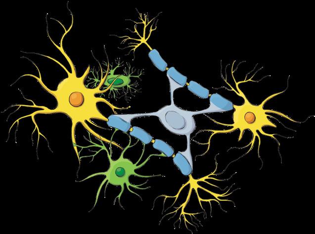
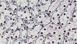
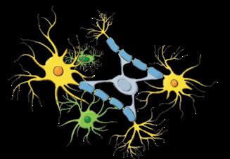
4.4 Nervous tissue
Nervous tissue is the main component of all the structures in the nervous system. It consists of neurons, which are star-shaped cells that specialise in collecting and transmitting information, and glial cells, which are responsible for neuron nutrition, defence and support.
Nervous tissue
Neuron
Glial cells
Types of muscle tissue
Skeletal striated muscle tissue
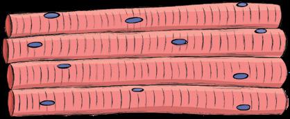
Multinucleated muscle fibre.
Cardiac striated muscle tissue
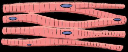
Muscle fibres that are similar to striated muscle, but with only one nucleus.
Smooth muscle tissue
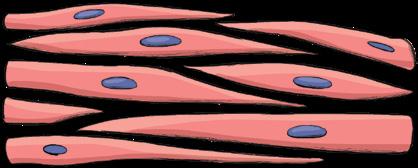
Muscle cells with only one nucleus.
Understand, think, search...
5 The mirror. Go to anayaeducacion.es to find out how to apply this thinker’s key, and draw a table comparing the different types of muscle tissue. 6 Compare the tissue shown below to the diagrams on these pages. What type of tissue is it? How do you know?
ORGANS AND SYSTEMS
Understand, think, search...
1 Puzzles. Form groups in your class and share out the topics to be researched. Each group will research a specific organ (brain, heart, stomach, small intestine, etc.) with a focus on the following: a) What is its function? b) What tissues is it made of? c) What type of cells make up these tissues?
Digestive For nutrition
Respiratory
Excretory Circulatory
Lymphatic
As we have just seen, cells of the same kind are organised into tissues. In turn, different tissues are organised into more complex structures called organs, which form coordinated systems.
5.1 Organs
The organs of the human body are made up of several tissues that work together to perform a specific function.
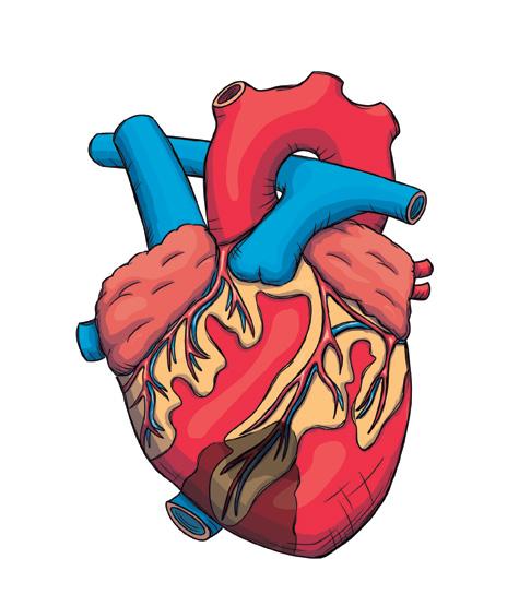
For example, the heart pumps blood, the kidneys clean blood, and the lungs exchange gases.
5.2 Systems
Systems are groups of organs that work together to perform a shared activity.
When the different systems work as a whole, the organism is able to perform the three vital functions (nutrition, interaction and reproduction).
Systems and the vital functions
For interaction For reproduction
Nervous
Muscular Skeletal
Endocrine Female
Male
5.3 The internal environment
One characteristic of multicellular organisms, like human beings, is that most of their cells are not in direct contact with the external environment. This means that they cannot exchange substances (gases, nutrients, waste, etc.) with it, as unicellular organisms do. To solve this problem, they have developed an internal environment.
The internal environment is the fluid that bathes all the organism’s cells. It is connected to the external environment by the blood. Cells take the nutrients and oxygen they need from the internal environment, and discharge the waste they produce into it.
Homeostasis
Homeostasis is all the physiological processes that keep the internal environment stable.
The organism achieves this balance thanks to its different organs and systems. This process of regulation prevents changes in the internal environment that would be fatal for cells.
External environment
Blood Food
Nutrients ORGANISM
Nutrients
Tissue cells O2
CO2
Internal environment
Waste Oxygen (O2)
Carbon dioxide (CO2)
Waste
Urine and sweat Understand, think, search...
1 Which of the following functions does the internal environment perform? a) It acts as a vehicle for substance exchange. b) It provides thermal insulation. c) It protects the cells it surrounds. 2 The What If What would happen to our bodies if the internal environment didn’t exist?
Technology CREATE The human body and its systems often offer inspiration to robotics designers. Use one of the systems of the human body as inspiration to design a plan for a sensor system.
Working with pictures
Look at the diagram showing how the internal environment works. Based on this diagram, write a description of how food, oxygen, carbon dioxide and waste are exchanged between the internal environment and the external environment.
research project Investigating healthy habits for the heart
project introduction
Leading causes of death worldwide Cardiovascular diseases
32.3 %
Cancer Respiratory diseases Diabetes Acute respiratory infections Dementia Neonatal death Diarrhoeal diseases Road accidents Liver diseases
16.3 %
6.5 %
5.8 %
4.4 %
4.4 %
3.2 %
3 %
2.5 %
2.3 %
project objective
project steps
Cardiovascular diseases, like arteriosclerosis, heart attack and stroke, are the leading cause of death in Spain and in other developed countries (see table). Knowing the annual figures for deaths and their causes is a good way to assess how effective a country’s health system is. These figures help health authorities take targeted public health measures. For example, if mortality due to heart disease and diabetes shoots up in the space of a few years, health authorities will often launch programmes aimed at encouraging healthy habits to prevent these diseases. It has been proven that the rise in cardiovascular diseases is directly linked to certain habits and lifestyles. Factors that increase the risk of developing a cardiovascular disease include obesity, smoking, a sedentary lifestyle, high levels of cholesterol in the blood, high blood pressure and diabetes. Main cardiovascular risk factors
Genetic predisposition
Age Diabetes
Alcoholism Smoking Obesity High blood pressure
Some medications Poor eating habits
A sedentary lifestyle
In this research project, you will be studying cardiovascular risk factors in people of different ages and sexes and with different lifestyles. One of the key objectives is to find out whether adopting certain healthy habits prevents cardiovascular diseases. With the information you have collected, you will prepare a presentation to report the results of your research and the conclusions you have drawn.
This project consists of three stages or steps: ➜ Step 1 (in this unit). Organising your work and searching for the information you need to start your research. ➜ Step 2 (in unit 2). Formulating a hypothesis, carrying out the research and obtaining experimental data. ➜ Step 3 (in unit 3). Analysing the results and drawing conclusions.
Finally, you will prepare a presentation that you can use to report and explain your results in class.

Organising your work
Your teacher will split the class into groups of 3 or 4. In your groups, discuss the project you are going to carry out and the methods you could use. Divide up the work among the members of your team. Share your ideas with the other groups. Discuss the number of people you are going to include in the study, the age range, what lifestyles you will look at, and so on. It is important to select a study sample that is representative of as wide a population demographic as possible. This will make your conclusions from the study more realistic.
Finding and analysing information
When searching for and selecting information for your project, make sure you use reliable sources. Consult sources in different formats (articles, infographics, graphs, etc.). Analyse information about cardiovascular risk factors, their incidence in the population and their effect on the physiological state of the circulatory system. To help you search for information, you could write a list of questions like the ones below:
✓ How does age influence the risk of developing a cardiovascular disease? What about gender? ✓ Why do excess weight and high blood pressure increase the risk of developing a cardiovascular disease? ✓ How does smoking affect the health of the cardiovascular system? ✓ How do regular physical exercise and a balanced diet help prevent cardiovascular diseases? ✓ Why is it so dangerous to have more than one of these risk factors at the same time?
Organising your ideas
1 Chain of sequences. In your notebook, copy and complete the following chain of sequences about the levels of organisation in living matter.
Find out how to use this graphic organiser at anayaeducacion.es.
Interpreting pictures
3 Copy this drawing and label the cell structures that correspond to the letters. What is the function of each structure?
I A
Organism level The human body is ?
The systems are ? such as ?
The organs are ? such as ?
The tissues are ? such as ?
Atomic level Atoms are ? such as ? Bioelements are ?
They are joined by chemical bonds.
Molecular level Molecules are ? such as ? Biomolecules are ?
They group together to perform the three
vital functions. Cellular level
Cells are ?
Human cells
are ? such as ? They group together to form organelles such as ?
Summarising
2 Write your own unit summary based on the outline below: • What is the difference between the atomic and molecular levels of organisation? Include definitions of bioelement and biomolecule. • Give a definition of cell. What higher levels are cells organised into? • What is the cell membrane composed of? What are its functions? • List the parts of a cell nucleus and explain their functions. • Write a list of the membranous organelles and the non-membranous organelles. What is the difference between them? What is the main function of each one? • Explain what cell differentiation is. • Name the different types of human tissue. What are the main characteristics of each type? • Name the different systems in the human body and the function they are involved in.
H
B C

G
F
E D
4 Look at the following pictures of tissues and cells and answer the questions:
a) What types of tissues and cells are shown? b) Link each cell to the tissue it forms part of. c) What is the function of each type of tissue? d) What characteristics of each cell type make them suitable for the function they perform in the human body? 5 What cell structure can you see in the following image? What is it composed of? What level of organisation of living matter does it correspond to?
A B
C D
Applying your knowledge
6 State what level of organisation in the human body the following correspond to: calcium; kidney; nucleus; group made up of the mouth, oesophagus, stomach, intestine, pancreas, etc.; spermatozoa; protein. 7 Copy and correct the following incorrect sentences about the cell membrane: a) The cell membrane isolates the cytoplasm from the outside. b) The plasma membrane is formed of a double layer of proteins. c) Medium-sized substances cross the cell membrane freely and without any help. d) Small substances enter the cell by endocytosis. e) The plasma membrane is not present in all cells. 8 What is the difference between: a) The nucleus and the nucleolus? b) Chromatin and chromosomes? c) The nucleolus and nucleoplasm? 9 List the names of the cell organelles in a table.
Add another column to describe the shape of each organelle, accompanied by a drawing, and another column for its function. 10 Copy the following sentences and say which tissue they refer to: a) Its matrix is liquid and is called plasma. b) It forms glands, which secrete substances. c) Its matrix is solid and elastic. We find it between the vertebrae and in the ear. d) It has a protective function in that it lines cavities. e) It contains low levels of intercellular substance and its cells store fat. f) Its cells transmit nerve impulses. g) Its cells are elongated and it is responsible for body movement. h) Its matrix is solid and contains calcium salts. 11 Why is cell differentiation important? Provide two examples.
Moving forward
12 Read the following text and answer the questions:
Invented in the 16th century, the microscope is a key tool for studying cells. As more and more powerful microscopes were developed, scientists were able to make discoveries about cellular structure. The ‘power’ of a microscope is its resolution; that is, the smallest distance by which two points can be seperated and still seen as separate points. Optical microscopes use visible light, which passes through the sample and provides an image that is magnified by a set of lenses. Their resolution is 0.2 μm. Electron microscopes don’t use light, but rather a beam of electrons. These either pass through the sample or bounce off it and are then captured by a screen, where the image appears. Their resolution is 5 μm.
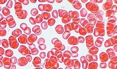
a) What type of microscope has been used to take these pictures of red blood cells? b) If the diameter of a red blood cell is approximately 5 μm, how much magnification does each microscope provide? 13 Many of the body’s cells have names related to the organ they belong to. Search for a picture of each of the following cells and indicate which organ they belong to: hepatocyte, osteocyte, myocyte, chondrocyte.
Sustainable Development Goals
14 Sustainable Development Goal 3 aims to ensure healthy lives and promote well-being for all. The UN has set numerous targets for this goal. a) In groups, find out about the targets of Goal 3 at anayaeducacion.es. Choose the target that most interests your group, and research it. b) Prepare a presentation to give to your class about the importance of your group’s chosen target and what could be done to achieve it. c) Discuss and draw conclusions about the importance of education and research in achieving this goal.
A
Atoms The smallest particles into which matter can be divided while retaining its chemical properties.
B
Biomolecules Molecules found exclusively in living beings and containing a high level of carbon.
C
Carbohydrates Biomolecules whose main function is to provide energy to cells.
Carbohydrates
Cell Structural and functional unit of a living being. That is, a cell is a unit capable of carrying out the three vital functions. Cell differentiation Process by which cells take on a shape and structure that allows them to become specialised for a particular function. Cell nucleus Cell structure surrounded by a membrane and containing the genetic material. Cell organelles Small structures in a cell that are specialised to perform different functions. Centrioles Non-membranous cell organelles made up of protein fibres that direct the cytoskeleton during cell division. Chromatin Network of all the DNA fibres and proteins found in the cell nucleus. Chromosomes Compact structures made of chromatin fibres that are visible under a microscope when the cell is about to divide. Cytoplasm Thick aqueous liquid (cytosol) that fills the inside of the cell and contains the cell organelles. Cytoskeleton Network of fibres that gives the cell its shape and enables it to move.
E
Endoplasmic reticulum (ER) Cell organelle consisting of a network of tubules and sacs extending out from the cell membrane. There are two types: the rough ER (RER), which has ribosomes attached to it and is involved in protein synthesis; and the smooth ER (SER), which does not have ribosomes attached to it and is involved in the synthesis of lipids. Eukaryotic cell A type of cell in which the genetic material is contained in a nucleus surrounded by a membrane, and which consists of many different organelles.
G
Genetic material A fibrous substance called DNA that controls cell function. Golgi apparatus Membranous organelle consisting of a network of flattened sacs that modify the substances synthesised in the ER in order to transport and secrete them.
H
Homeostasis All the processes that keep the composition and properties of an organism’s internal environment stable.
I
Internal environment Fluid that surrounds all the cells and is connected to the external environment by the blood.
L
Levels of organisation The levels of complexity into which living matter is organised. Lipids Biomolecules that perform various functions like energy storage (triglycerides) and heat insulation. Some lipids form part of the plasma membrane of cells (phospholipids and cholesterol).
Lipids
Lysosomes Membranous cell organelles responsible for digesting particles.
M
Micrometre or micron Unit of length equal to one millionth of a metre. Mitochondrion Cell organelle surrounded by a double membrane whose function is to obtain energy for the cell through cellular respiration. Monomers Simple or small molecules that form the basic unit of other, larger molecules known as polymers.
N
Nucleic acids Large biomolecules that contain genetic information, which they transmit to offspring, and that control cell functions. DNA and RNA are nucleic acids.
Nucleic acids Nucleolus Part of the cell nucleus where ribosomes are made. Nucleoplasm Part of the cell nucleus made up of a watery liquid containing the genetic material.
O
Organ Group of different tissues performing a specific function.
P
Plasma membrane Layer that surrounds the cell, protects it and regulates substance exchange with the surrounding environment. It is made up of lipids, carbohydrates and proteins. Polymers Biomolecules formed of long chains of monomers. Proteins Complex biomolecules that form part of cell structures and have various functions like regulating and executing most cell processes.
Proteins
R
Ribosomes Non-membranous cell organelles responsible for synthesising proteins.
S
System Group of organs performing a shared function.
T
Tissue Group of similar, coordinated cells that are specialised to perform the same function.
Building Blocks is an educational project of Anaya Educación for Secondary Education with the participation of: S. Clemente, A. Domínguez, A. B. Ruiz, Danny Latimer, Hannah Peat, Karen Piper, Denise Suárez, Begoña Fuente Larrazabal, Sara Gascón Martin, Paloma Rodriguez Esteban, Veronica Moro Rivera. The following people have also contributed to this book: Editorial team: Hannah Peat and the Departamento de Ciencias Naturales (Almudena Alcón, Esther Fernández, Mª Isabel García, Marco Sánchez, Mercedes Sacristán and Jorge Torres).
inclusion and anti-discrimination Advisor: Víctor Díez Mazo.
Design, technical drawings and maps: Patricia G. Serrano, Juan Carlos Quignón, Miguel A. Castillejo, Miguel A. Díaz-Rullo and Miriam Arribas. Illustrations: Sara Mateos, Celia López Bacete, Santi Martín, Alicia Posada, Mª Carmen Fuente, David Menéndez and Carlos Moreno. Layout: Mª Nieves Merino and Oscar Latorre. Translation: Montero Language Services. Proofreading: Rachel Bland (Bakun S. L.)
Graphic edition: Olga Sayans.
Photographs: Age Fotostock (ARCO/Muehlmann, K., BILDAGENTUR-ONLINE/MCPHOTO-SCHULZ, DENNIS KUNKEL MICROSCOPY, DR JUERG ALEAN, Fco. Javier Sobrino, Jennifer Waters, Jens Meyer, Leung Cho Pan, MAY FOTO, Science Photo Library, Tamara1K, ViewStock, Walt Anderson, Wellcome Images), Agencia EFE (Jesús Diges, Salvador Sas) Alamy/Cordon Press (Anne-Marie Palmer, Daria Artemenko, jeremy sutton-hibbert, Moviestore Collection Ltd), Archivo Anaya ( Alcón, A. y Santos, V.E.; CSIC/Abrisqueta, J.; Candel, C.; Canto, M.; CNB/Ruiz Castón, J.; Cosano, P.; CSIC/Abrisqueta, J.; Dpto. Histología y Anatomía Universidad de Navarra; Grajera, R. & Muñiz, E.; Gómez-Miguel, V.; Martín, J. A.; Martínez, C.; Moreno, C.; Muñoz, J.C.; Ruiz Pastor, L.; Ruiz, J.B.; Sánchez, J.; Velasco, P. - Fototeca de España; Villaboy, N.), Dreamstime (Baibaz), GettyImages (Don Mason, Hero Images, Hugh Whitaker, JacobH, Jason Edwards, Micro Discovery, Miguel Sotomayor, Milos Bicanski, Seksak Kerdkanno / EyeEm, simonkr, Slonov), iStock/Gettyimages (ajt, Akchamczuk, alekseystemmer, AlexRaths, AlinaMD, alisa24, AntonioGuillem, arachi07, areeya_ann, Banyan-RanchStudios, Cloudtail_the_Snow_Leopard, Cylonphoto, daniilphotos, dankadanka, dmbaker, Dr_ Microbe, EcoPic, Elizabeth M. Ruggiero, GlobalP, Harry Collins, hartmanc10, Helmut Feil, jez_bennett, Jillian Cooper, kappaphoto, karlumbriaco, ktsimage, lekcej, Lenorlux, Marcin Wojciechowski, mel-nik, Michelle de Villiers, mihtiander, MikeLane45, miroslav_1, MMilda, nzfhatipoglu, okugawa, panso, PLG, pum_eva, Rawpixel, ricul, seb_ra, stevelenzphoto, Tinieder, tonda, Tramper2, Usercce650b4_975, VictorTyakht, zokru), Jacques Descloitres/MODIS Rapid response Team/NASA/GSFC, Nature Picture Library/ CordonPress (Andy Rouse, Chris & Monique Fallows, David Shale, Solvin Zankl, Tony Wu), 123rf (ahasoft2000, alexmit, archnoi1, jovannig, Katarzyna Białasiewicz, Manit Larpluechai, natika, Paras Soni, Przemyslaw Koch, rido, Teodoro Ortiz Tarrascusa, Thomas Photiou, Ting Fen Zheng, vilainecrevette, Wavebreak Media Ltd, Yakov Oskanov, yurakp).and collaborators.
Special thanks to Ana Codeseda for writing the scientists’ biographies. Other thanks: to Almudena Monge for permission to use the field notebook illustation. Academic and Professional Orientation: created in conjunction with Fundación Bertelsmann. Coordinator: Juan José Juárez Calvo. Expert collaborators: Sara Lozano Santiago, Belén Pérez Castro and Pilar Vázquez Hernández.
Commitment to Sustainable Development Goals
Our publications contain carefully selected content, illustrations and language to comply with non-discrimination on the grounds of gender, culture or opinion.
Grupo Anaya considers social and environmental responsibility to be one of its fundamental values. For this reason, we are committed to: · continually improving our contents and materials related to the environment. · reducing our carbon emissions. · using natural resources responsibly. · making sure that our activity has no negative consequences for endangered forests. These commitments, among others, mean that 100% of the paper used in our books has the PEFC label.
Important information: The activities proposed in this book should be completed in a separate notebook or on sheets of paper, not in the book itself. The links to webpages which appear in this book have been checked before printing. The publisher cannot be liable for any changes or modifications which occur after the date of publication.
© GRUPO ANAYA, S.A., 2021 - C/ Juan Ignacio Luca de Tena, 15 - 28027 Madrid.
All rights reserved. No part of this publication may be reproduced, stored in a retrieval system, or transmitted, in any form or by any means, electronic, mechanical, photocopying, recording, or otherwise, without the prior permission of the publishers.
de cerca
biología y geología 3

ESO
Building Blocks
Building Blocks es un proyecto educativo de Anaya para Educación Secundaria con la participación de: CONTENIDOS: Berta Gallego (adaptación de los contenidos del libro de Biología y Geología 3.° ESO). Equipo de edición: Departamento de Ciencias Naturales (Almudena Alcón, Esther Fernández, M.ª Isabel García, Marco Sánchez, Mercedes Sacristán, Jorge Torres) y Hannah Peat. Asesoramiento para la inclusión y la no discriminación: Víctor Díez Mazo.
Diseño, gráficos y cartografía: Patricia G. Serrano, Juan Carlos Quignon, Miguel Á. Castillejo, Miguel Á. Díaz-Rullo y Miriam Arribas. Ilustración: Sara Mateos, Celia López Bacete, Santi Martín, Alicia Posada, M.ª Carmen Fuente, David Menéndez y Carlos Moreno. Maquetación: Óscar Latorre. Corrección: Encarnación Martín. Edición gráfica: Olga Sayans.
Fotografía: Age Fotostock (DENNIS KUNKEL MICROSCOPY, DR JUERG ALEAN, Fco. Javier Sobrino, Jennifer Waters, Leung Cho Pan, MAY FOTO, Science Photo Library, Tamara1K, ViewStock, Walt Anderson, Wellcome Images), Agencia EFE (Jesús Diges, Salvador Sas, Torres Molina) Alamy/Cordon Press (Daria Artemenko, Moviestore Collection Ltd), Archivo Anaya (Alcón, A. y Santos, V. E.; CSIC/Abrisqueta, J.; Candel, C.; Canto, M.; CNB/Ruiz Castón, J.; Cosano, P.; CSIC/Abrisqueta, J.; Dpto. Histología y Anatomía Universidad de Navarra; Grajera, R. & Muñiz, E.; Gómez-Miguel, V.; Martín, J. A.; Martínez, C.; Moreno, C.; Muñoz, J. C.; Ruiz Pastor, L.; Ruiz, J. B.; Sánchez, J.; Velasco, P. - Fototeca de España; Villaboy, N.), Dreamstime (Baibaz), GettyImages (Don Mason, Hugh Whitaker, JacobH, Micro Discovery, Miguel Sotomayor, Milos Bicanski, Seksak Kerdkanno / EyeEm, simonkr, Slonov), iStock/Gettyimages (AntonioGuillem, areeya_ann, Cylonphoto, dmbaker, Dr_Microbe, ktsimage, MMilda, nzfhatipoglu, panso, PLG, seb_ra ), Jacques Descloitres/MODIS Rapid response Team/NASA/GSFC, Nature Picture Library/CordonPress (Solvin Zankl), 123rf (ahasoft2000, alexmit, archnoi1, dolga-chov, jovannig, Katarzyna Białasiewicz, natika, Nelson Marques, Paras Soni, Przemyslaw Koch, rido, Thomas Photiou, Ting Fen Zheng, Wavebreak Media Ltd, yurakp).
compromiso
Nuestras publicaciones mantienen el rigor en el uso y en la selección de los contenidos, en las imágenes y en el lenguaje, para cumplir con la no discriminación por razón de género, cultura u opinión.
Grupo Anaya considera la responsabilidad social y medioambiental uno de sus valores fundamentales. Por ello, se compromete a: · mejorar nuestros resultados en materia de medio ambiente, · reducir nuestras emisiones de carbono, · hacer uso de los recursos naturales de manera responsable y · eliminar cualquier impacto negativo de nuestra actividad en los bosques en peligro. Dichos compromisos, entre otros, hacen que el 100 % del papel utilizado en nuestros libros tenga el sello PEFC.
Importante: Las actividades propuestas en este libro deben ser realizadas en cuadernos u hojas sueltas; nunca en el propio libro. Los enlaces a las páginas web que aparecen en este libro han sido revisados en la fecha de su impresión. La editorial no se hace responsable de las modificaciones o las anulaciones que se produzcan en ellos con posterioridad a dicha fecha.
© GRUPO ANAYA, S.A., 2021 - C/ Juan Ignacio Luca de Tena, 15 - 28027 Madrid.
All rights reserved. No part of this publication may be reproduced, stored in a retrieval system, or transmitted, in any form or by any means, electronic, mechanical, photocopying, recording, or otherwise, without the prior permission of the publishers.
ÍNDICE
La mETODOLOGÍA científica......................................4 1 Qué es el método científico • 2 La investigación en el laboratorio • 3 La investigación en la naturaleza • 4 Búsqueda de información
1 Los niveles de organización • 2 La célula humana • 3 Los tejidos humanos • 4 Órganos, aparatos y sistemas
1 La nutrición y los nutrientes • 2 El aporte de energía • 3 Alimentación y dieta • 4 La dieta y la salud
3. Aparatos para la función
de nutrición ...................................................................... 20 1 El aparato digestivo • 2 La digestión • 3 El aparato respiratorio • 4 El aparato circulatorio • 5 La circulación sanguínea y el sistema linfático • 6 El aparato excretor • 7 La salud y la función de nutrición
4. La función de relación............................................28 1 La relación, la percepción de estímulos • 2 La coordinación nerviosa • 3 La coordinación endocrina • 4 La ejecución de la respuesta. El aparato locomotor • 5 La función de relación y la salud
5. Aparatos para la función
de reproducción .............................................................36 1 La reproducción humana. Los gametos • 2 Los aparatos reproductores • 3 Los ciclos del ovario y del útero • 4 La fecundación, el embarazo y el parto • 5 salud y planificación reproductiva
6. Vida sana ................................................................................ 42 1 La salud y la enfermedad. Tipos de enfermedades • 2
Las enfermedades infecciosas • 3 El sistema inmunitario • 4 La prevención y la curación de enfermedades infecciosas 5 Trasplantes, donación y primeros auxilios
7. La cambiante Tierra................................................... 48 1 La superficie terrestre y sus cambios • 2 La energía interna de la Tierra y los procesos endógenos • 3 El magmatismo y los volcanes • 4 Las fuerzas tectónicas y los riesgos geológicos
8. El modelado del relieve ........................................52 1 El modelado del relieve • 2 Los procesos geológicos exógenos • 3 El modelado de las aguas de arroyada y los torrentes • 4 El modelado de los ríos • 5 El modelado de las aguas subterráneas • 6 El modelado glaciar • 7 El modelado del viento • 8 El modelado del mar
GLOSARIO ..................................................................................60
1uniDAD LA ORGANIZACIÓN DEL SER HUMANO
1 LOS NIVELES DE ORGANIZACIÓN
Los niveles de organización son cada uno de los grados de
Nivel atómico
Los átomos son las partículas más pequeñas en que se puede dividir la materia. Los que forman parte de la materia viva se denominan bioelementos.
Nivel molecular
Las moléculas son agrupaciones de átomos, unidos por enlaces químicos. Las que forman parte de los seres vivos se llaman biomoléculas y tienen un elevado contenido en carbono. Son los glúcidos o hidratos de carbono, los lípidos, las proteínas y los ácidos nucleicos. Otras sustancias, como el agua y las sales minerales, se encuentran tanto en la materia viva como en la no viva y tienen una estructura química sencilla.
Nivel celular
Las células son la parte más pequeña de los seres vivos capaz de realizar las tres funciones vitales. Todas las células humanas son eucariotas de tipo animal.
Nivel organismo
Los tejidos son asociaciones de células semejantes especializadas en la misma función. Los tejidos se agrupan formando órganos, que realizan una función concreta. A su vez, el conjunto de órganos que llevan a cabo una función común constituyen los aparatos y sistemas.
1 Escribe una definición de los siguientes conceptos: célula, biomolécula y bioelemento. 2 ¿Son el agua y las sales minerales biomoléculas? ¿Por qué? 3 Copia el dibujo y escribe en tu cuaderno en los círculos el nivel de organización que corresponda.
Los glúcidos
Son la principal fuente de energía de la célula. Forman parte de algunas estructuras celulares y sirven para construir otras biomoléculas.
Los lípidos
Son sustancias de naturaleza química muy diversa. Algunos, como los fosfolípidos y el colesterol, forman parte de las membranas celulares; otros, como los triglicéridos, son una fuente de energía y de almacenamiento energético para la célula, etc.
Las proteínas
Forman parte de las estructuras de la célula y llevan a cabo la regulación y la ejecución de la mayoría de los procesos celulares.
Los ácidos nucleicos
Contienen la información genética, que transmiten a la descendencia, y controlan las funciones celulares. Son el ADN (ácido desoxirribonucleico) y el ARN (ácido ribonucleico).
Comprende, piensa, investiga...
que se agrupan formando
que se agrupan formando
que se agrupan formando
Las células humanas tienen diferentes formas y funciones, pero todas están compuestas por membrana, citoplasma y núcleo.
La membrana plasmática
Es una fina capa que envuelve a la célula, la protege y regula el intercambio de sustancias con el medio. Está compuesta por una capa de lípidos, en la que se insertan las
El núcleo
Es una estructura grande y esférica que contiene el ma-
Se compone de membrana nuclear, nucleoplasma, nucléolo (fabrica los componentes de los ribosomas) y cromatina, formada por fibras de ADN y proteínas. Cuando la célula se divide, la cromatina se compacta y forma los cromosomas.
El citoplasma
Está formado por un líquido acuoso denominado citosol,
Comprende, piensa, investiga...
1 Lee el texto «Las funciones de la membrana» disponible en el banco de recursos de anayaeducacion.es y explica cómo atraviesan la membrana las sustancias pequeñas, medianas y grandes. 2 Copia el dibujo del núcleo y rotula sus partes.
1 2
5
3
4
3 Lee el texto «Diferenciación celular» disponible en el banco de recursos de anayaeducacion.es y escribe un texto explicando en qué consiste ese concepto.
Los orgánulos celulares
Son estructuras inmersas en el interior del citoplasma que realizan funciones específicas. Pueden ser membranosos
El retículo endoplasmático (RE) es un conjunto de canales y sáculos que sintetizan sustancias. El RE rugoso, que tiene ribosomas adheridos a su pared, participa en la síntesis de proteínas. El RE liso, sin ribosomas, interviene en la síntesis de lípidos.
Los centriolos dirigen el movimiento del citoesqueleto y de los cromosomas durante la división de la célula.
Los orgánulos no membranosos
El citoesqueleto es una red de fibras que sostiene los orgánulos y da forma a la célula. También interviene en los movimientos celulares (constituye los cilios y los flagelos).
Los ribosomas se encargan de sintetizar las proteínas. Pueden estar adheridos al retículo endoplasmático o dispersos por el citoplasma.
Las mitocondrias obtienen energía para la célula mediante el proceso de respiración celular.
Los orgánulos membranosos
El aparato de Golgi modifica las sustancias sintetizadas en el RE y las transportan en vesículas al exterior celular.
Las vesículas transportan o almacenan sustancias.
Los lisosomas son vesículas en las que se digieren partículas capturadas por la célula.
Un tejido es un grupo de células, de forma y estructura seme-
Tejidos epiteliales
Están formados por una o varias capas de células muy próximas entre sí. Recubren los órganos y actúan como barreras protectoras. • Epitelio de revestimiento. Recubre tanto las superficies externas como las internas de los órganos. • Epitelio glandular. Produce y secreta sustancias sobre la superficie epitelial. Puede formar glándulas.
Tejidos conectivos
Están formados por células que se encuentran rodeadas por fibras y una sustancia intercelular llamada matriz. Actúan de soporte y unión para los órganos y otros tejidos del cuerpo. Hay varios tipos: tejido conjuntivo, adiposo, cartilaginoso, óseo y sanguíneo.
Tejidos musculares
Están formados por células alargadas, que se agrupan en fibras musculares, capaces de contraerse y relajarse. Son responsables del movimiento del cuerpo. • Tejido muscular estriado esquelético. Está formado por los músculos del aparato locomotor. Su contracción es voluntaria y sus células tienen varios núcleos y estriaciones. • Tejido muscular estriado cardiaco. Está en las paredes del corazón.
Es similar al estriado, pero su contracción es involuntaria y sus células tienen un solo núcleo y carecen de estriaciones. • Tejido muscular liso. Se localiza en las paredes de distintos órganos.
Tejido nervioso
Es el principal componente del sistema nervioso. Está formado por las neuronas, que recogen y transmiten la información, y por las células de glía, que se encargan de su nutrición, defensa y soporte.
1 anayaeducacion.es Lee el texto «Tipos de tejidos conectivos» y haz una tabla que incluya las características de cada uno. 2 ¿A qué tipo de tejido muscular pertenecen las imágenes? Explícalo.
Tejidos epiteliales
De revestimiento Glandular
Tejidos conectivos
Conjuntivo
Cartilaginoso Adiposo
Óseo
Sanguíneo
Tejidos musculares
Estriado esquelético Estriado cardiaco
Liso
Tejido nervioso
Comprende, piensa, investiga...
A B

4 ÓRGANOS, APARATOS Y SISTEMAS
Los órganos del cuerpo humano
Están formados por varios tejidos que funcionan de manera coordinada para realizar una función concreta.
Los aparatos y sistemas
Son agrupaciones de órganos que colaboran de forma coordinada para realizar una actividad común.
Para la nutrición
Digestivo Respiratorio
Circulatorio
Excretor Linfático
Para la relación
Nervioso
Musculatura Esqueleto
Endocrino
Para la reproducción
Reproductor femenino
Reproductor masculino
Medio interno
El medio interno
Una característica de los organismos pluricelulares es que han desarrollado un medio interno, que les permite intercambiar sustancias (gases, nutrientes, desechos) con el medio externo.
El medio interno es el líquido que baña las células y que entra en contacto con el exterior a través de la sangre. Las células toman de este medio los nutrientes y el oxígeno, y expulsan los desechos.
La homeóstasis es el conjunto de procesos fisiológicos que mantienen estables las características del medio interno.
1 Busca información sobre dos aparatos o sistemas del cuerpo humano. Explica qué órganos los forman y qué funciones desempeñan. 2 ¿Qué es el medio interno? Observa la imagen superior y escribe un texto explicando cómo se realiza el intercambio de: alimentos, oxígeno, dióxido de carbono y desechos entre el medio interno y el externo.
Medio externo
Sangre Alimentos
Nutrientes
Nutrientes O2
CO2
Células de los tejidos Medio interno
Desechos
Desechos ORGANISMO
Oxígeno (O2)
Dióxido de carbono (CO2)
Orina y sudor