5 minute read
DI Europe Summer 23


Guido Gebhardt
Pioneer in Energy Saving Radiology Practice Shares Tips
Rising energy costs are a problem for radiology practices and clinics, especially since the beginning of the war in Ukraine. Prof. Hans-Martin Klein from Burbach in Germany started looking into alternative energy sources more than ten years ago. He told us more about his green practice, and how low-field MRI is experiencing a “renaissance”.

◾ Prof. Klein, you have recently become the man to watch when it comes to reducing energy costs in radiology. How did this come about?
In 2010 I built a medical center on the high Westerwald, where the wind is known to be very cold. The Energy Saving Ordinance (EnEV), which came into force in 2009, favored the use of alternative heat sources. At the time, I thought that somehow the MRI scanner could be used as an alternative energy source, because the system produces about 20 kW of waste heat 24 hours a day, seven days a week. In addition, the energy needed to cool the helium could be saved. This concept also reduced the number of requirements for the building structure, which saved additional costs.
These measures enabled us to save about 85 percent of our heating costs. We were surprised ourselves at the time.
◾ Can such heat exchangers be installed anywhere or was this something specific to your practice?
The waste heat from the superconducting MRI scanners is not something practice-specific. The problem with this interface, however, is that not all practice owners actually own the practice space, because the conversions have to be coordinated with the building heating system.
◾ Surely there are additional potential savings in radiology?
After the medical center was built, I started planning an energy-optimized practice with an open MRI. Knowing how little energy low-field
MRI scanners require, we bought an 0.35 Tesla system in 2019.
Another aspect of conservation or sustainability in radiology is, of course, helium. It is quite expensive in technical synthesis. In addition, natural helium is slowly becoming scarce. Superconducting MRI systems require up to 2,000 liters of the gas, which is in liquid form at around – 270 °C, to cool the system.
Particularly in older systems, some evaporates regularly and must be refilled. New MRI scanners get by with significantly less than 10 liters of helium. The system with the lowest helium filling is even only 0.7 l at a field strength of 0.55 Tesla.


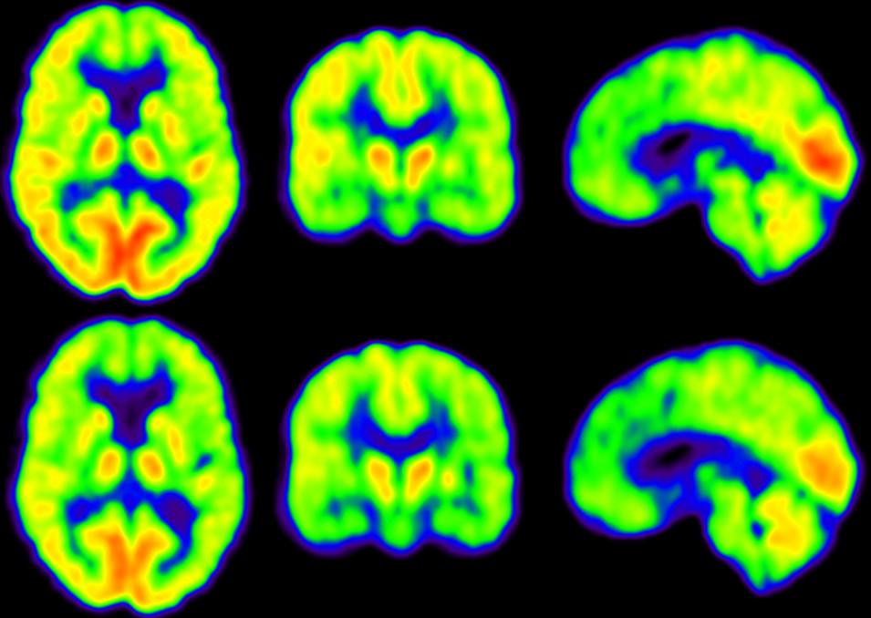
◾ Are there noticeable differences in image quality between an 0.55 Tesla low-field and a superconducting 1.5 Tesla MRI scanner?
The image quality of MRI scanners is something like this: if you double the measurement time, you get roughly identical image quality at half the field strength.
In addition to field strength, the homogeneity of the system and the gradients also play an important role in image quality. Furthermore, it is a question of how the K-space is sampled. Compressed sensing is one of the buzzwords here – along with matrix coils or concepts such as Simultaneous MultiSlice Imaging. Modern low-field MRI systems with permanent magnets already use some of these techniques. Doubling the coil elements has the same effect as doubling the field strength.
Images from low-field systems are surprisingly good. In addition, there are specific advantages of lower field strengths such as less metal artifacts, better T1 contrast, better phase separation in Dixon sequences, less dielectric effects, and less impact on implants such as VP shunts or cochlear implants.
In the past, low-field systems were always a bit frowned upon, but currently they are somehow experiencing a renaissance.
◾ To what extent does artificial intelligence play a role in MR image quality?
Image quality is essentially about the signal-to-noise ratio. So you can either increase the signal by putting more power into the field strength and the gradients, the coils and so on, or you can try to reduce noise.
If you reduce noise with classical felting, the images always lose information. Deep learning reconstruction, on the other hand, is able to reduce noise without losing information by preserving contour information.
Of course, this can only be used within certain limits, because otherwise one runs the risk of pretending an accuracy that is not there at all. But compared to the filters that were available in the past, we now have very powerful AI algorithms at our disposal. This will bring significant advantages for low-field systems.
https://www.greenscan-imaging.de/
A new approach to the Improvement of Energy Efficiency in Radiology Practices, Prof. Hans-Martin Klein, Verlag, https://www.thieme-connect.com/products/ejournals/abstract/10.1055/a-1123-7944