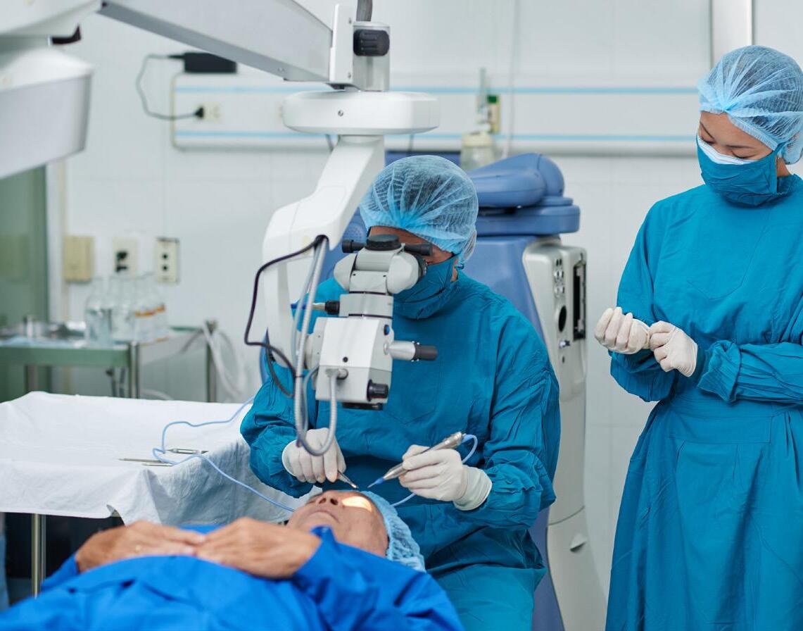
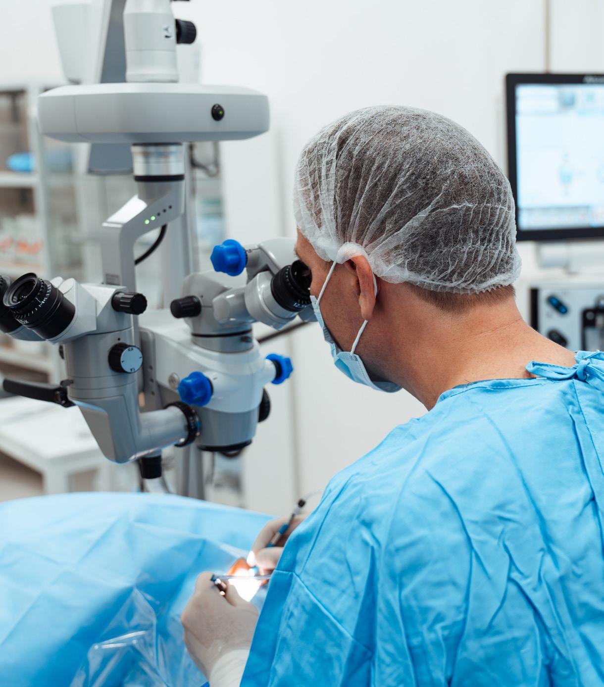
A holistic approach to health can magnify benefits of cataract surgery.
ALSO IN THIS ISSUE
Optimising Cataract Surgery
Oliver Findl reviews the ESCRS Guideline for Cataract Surgery and discusses how evidence-based research can improve clinical outcomes.
Orbital Inflammation: Important Associations
Complex clinical presentations require a straightforward approach.
Optimising Innovation through AI Collaboration
Beyond chat apps, specialised prompt patterns can help develop solutions.
https://congress.escrs.org/
Contents
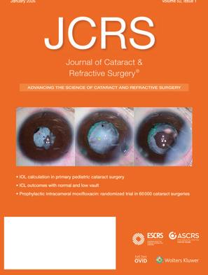
10 Cover
Focusing on the Patient with Dementia A holistic approach to health can magnify benefits of cataract surgery.
Preventing Corneal Haze with Losartan
Lycia Pedral Sampaio MD, PhD
Pushing Envelopes with
Victor L Perez Quiñones MD
Mona Harissi-Dagher MD, FRCSC
Orbital Inflammation: Important Associations
Endothelial Disease
Lamis Baydoun MD, PhD
Risk of Rhegmatogenous Retinal Detachment
Birgitte Romme Nielsen MD, PhD
Tackling the Treatment Burden in Retinal Diseases
Aude Couturier MD, PhD
Subretinal Drug Delivery Eye Drops Reimagined
Greener Cataract Surgery: Less Waste, Lower Costs
Finding Happiness at Work
Publisher Tom Ogilvie-Graham
Executive Editor
Stuart Hales
Editor-In-Chief
Sean Henahan
Senior Content Editor
Kelsey Ingram
roo.khan@wearemci.com
is registered with the European Union Intellectual Property Office and the US Patent and
Published by the European Society of Cataract and Refractive Surgeons, Suite 7–9 The Hop Exchange, 24 Southwark Street, London, SE1 1TY, UK. No part of this publication may be reproduced without the permission of the executive editor. Letters to the editor and other unsolicited contributions are assumed intended for this publication and are subject to editorial review
is not responsible for statements made by any contributor. These contributions are presented for review and comment and not as a statement on the standard of care. Although all advertising material is expected to conform to ethical medical standards, acceptance does not imply endorsement by ESCRS EuroTimes. ISSN 1393-8983 32 30 22
Learn more about EuroTimes or connect with ESCRS at ESCRS.org
Fix the Roof While the Sun is Shining!
There is a huge unmet need in providing cataract surgery to people living with dementia. We know this thanks to a landmark study published in 2016 called the Provide Study.
Undertaken by the College of Optometrists in the UK, the study demonstrated that people living with dementia are often underserved by healthcare providers, from lacking spectacles to not being considered for cataract surgery.
As in the general population, cataract surgery can reduce the risk of falls and considerably improve the lives of people with dementia as well as their caretakers.We also now know that early intervention with cataract surgery protects against the progression of dementia. Indeed, the World Health Organization recently declared vision a modifiable risk factor for dementia.
When you see a patient who has both cataracts and dementia, you can’t assume they will come back in six months due to the probable progression of their dementia. When you explain this to the family members, they will say, “Mum? You know what, you’re gonna get your eyes fixed next week—we’re going to get this sorted”.
Therefore, my approach is to act as soon as possible. If they are still driving, I would aim for emmetropia. If they are not driving, I aim for mild myopia, -1.0 D, so their arms’ length view is in focus: they can see their hands, the TV, and stairs. Ideally, I would use standard monofocal IOLs with same-day bilateral dropless surgery with intracameral anaesthesia.
I would generally not consider using simultaneous vision lenses in people with dementia. These lenses require a significant amount of neuroadaptation over time, and we are talking about a group who are progressively losing their brain function.
EDITORIAL BOARD
Adi Abulafia (Israel)
Bruce Allan (UK)
Noel Alpins (Australia)
Juan Alvarez de Toledo (Spain)
Gerd Auffarth (Germany)
Başak Bostanci (Turkey)
John Chang (Hong Kong SAR, China)
Béatrice Cochener-Lamard (France)
Burkhard Dick (Germany)
Mor Dickman (The Netherlands)
Joaquín Fernández (Spain)
Oliver Findl (Austria)
Nicole Fram (US)
Sri Ganesh (India)
Christina Grupcheva (Bulgaria)
Farhad Hafezi (Switzerland)
Nino Hirnschall (Austria)
Soosan Jacob (India)
Jack Kane (Australia)
Yao Ke (China)
It is important to note that having a diagnosis of dementia doesn’t mean a person lacks the capacity to make decisions. I can usually get somebody living with dementia to cooperate for 20 minutes because I do both eyes at the same time. After treatment they can read, feed themselves, and maintain independence.
How do we move the agenda forward? We need a multilayered approach from the ground up. We need to get the message to people living with dementia that vision is a modifiable risk factor. We need to encourage annual vision check-ups. We want to encourage ground-level optometrists to refer patients and surgeons to intervene early. Further up the ladder, we need to convince hospital administrators and broader healthcare systems of the value of early intervention. At the higher levels of budgeting and policy, we can emphasise the benefits of fall prevention. This is a very, very big-ticket item amongst governments and payors.
I would also like to reach out to young ophthalmologists. If you are looking for a research area, the field of dementia and vision is there for you. There is a lot of opportunity to create a name for yourself academically in what is currently not a crowded field.
Paul Ursell MBBS, MD, FRCOpth Consultant Ophthalmologist in Wimbledon, Honorary Professor, University of Bradford, UK, and immediate past-president of the United Kingdom & Ireland Society of Cataract & Refractive Surgeons.
Mika Kotimäki (Finland)
Georgios Kymionis (Greece)
David Lockington (UK)
Artemis Matsou (Greece)
Cyres Mehta (India)
Jod Mehta (Singapore)
Sorcha Ní Dhubhghaill (Belgium)
Rudy Nuijts (The Netherlands)
Catarina Pedrosa (Portugal)
Konrad Pesudovs (Australia)
Nic Reus (The Netherlands)
Filomena Ribeiro (Portugal)
Andreia Rosa (Portugal)
Giacomo Savini (Italy)
Julie Schallhorn (US)
Sathish Srinivasan (UK)
Paola Vinciguerra (Italy)
Shin Yamane (Japan)
Ron Yeoh (Singapore)
Mihail Zemba (Romania)
Thomas Kohnen José Güell
Paul Rosen
Drive Your Career Forward
Want to improve your clinical skills, stay on top of developments in the field, and have an active voice in the evolution of cataract and refractive surgery?
As an ophthalmologist in training, all these things (and much more) are within your grasp—and at no cost to you— by joining ESCRS!
Since 1991, ESCRS has promoted education in the field of implant and refractive surgery and supported research into the practice of intraocular lens implantation and refractive surgery. With more than 7,000 members across Europe and the world, ESCRS wields a powerful voice in the global discourse about ophthalmology and provides members with a rich and diverse network of professional connections.
Drive ophthalmology—and your career—forward. Take advantage of free ESCRS trainee membership today!
ESCRS YO membership has given me a strong international community, real integration across cultures and languages, and meaningful connections that extend far beyond meetings. It has opened opportunities to present on stage, build confidence, and grow as a leader within ESCRS and the wider ophthalmology community.
—LAURA MAUBON
As a young ophthalmologist, ESCRS membership opened doors to high-quality education, mentorship, and opportunities that have shaped my early career. Being an ESCRS YO member has connected me with an inspiring global community and given me access to resources that continually improve my clinical practice.
—VALENTIN HOOIJER
Much More Than a President
Filomena Ribeiro’s leadership of ESCRS made our Society ‘more human.’
Filomena Ribeiro has undoubtedly been much more than a president of the European Society of Cataract and Refractive Surgeons. She has been a reference, a guide, and an inspiration to all of us who have had the privilege of working alongside her.
Her presidency was defined by a clear vision and, above all, a deep conviction in the values that should underpin our Society. Among them, her strong commitment to diversity and inclusion stands out. By declaring 2024 the ‘Year of Diversity’ and promoting concrete initiatives such as mentoring programmes and member surveys, she not only drove structural change but also conveyed a powerful message: European ophthalmology can only truly
progress if all voices are given space and recognition.
Under her leadership, ESCRS took important steps to strengthen its international presence. The affiliation with BRASCRS (Brazilian Association of Cataract and Refractive Surgery) is a clear example of her ability to build bridges and foster a more connected global community. This outward-looking vision has been reflected in multiple initiatives that have enhanced the Society’s relevance beyond Europe.
Education was another cornerstone of her presidency. Filomena played a decisive role in advancing the Simulation Training Lab, establishing it as a central component of the ESCRS Congress and as a key tool for training
the next generation of surgeons. Thanks to her leadership, surgical education has gained in quality, structure, and longterm vision.
However, her impact goes beyond major initiatives. She also transformed the Congress experience, making it more engaging, interactive, and human. Initiatives such as the Arena, World Café discussions, and guided exhibition tours have helped position ESCRS not only as a place for learning but also as a space for connection and belonging.
Her sensitivity towards people was also reflected in projects such as BoSS (Building Our Sustainable Society), aimed at building a more inclusive and accessible Society, as well as in initiatives that preserve and celebrate our history, such as the IOL Museum and the Heritage Project. Likewise, her involvement in charitable work, including her visit to Mozambique, highlighted a genuine commitment to improving access to eye care globally.
Those of us who have worked closely with Filomena have also witnessed a leadership style grounded in common sense, scientific rigour, and strong ethical principles. She has shown that it is possible to move forward with ambition without ever losing coherence, integrity, and, ultimately, the humanity that gives meaning to everything we do.
Above all, she has reminded us of something essential: that behind every project and every strategy, there are people. Her ability to combine determination with empathy, vision with closeness, has left a lasting impression on all of us.
For all these reasons, Filomena deserves not only our recognition but something far deeper and more sincere: our affection, our admiration, and a gratitude that will endure well beyond her presidency.
Her presidency leaves ESCRS stronger, more open, and, above all, more human.
Joaquín
Fernández Pérez MD, PhD
ESCRS Welcomes New Members to Council of Management
The new year brought some new faces to the ESCRS Council of Management, with turnover in the ranks of trustees, council members, and co-opted council members.
Rebecca Emerson was named a new trustee, replacing Paul Rosen. Until 2024, Rebecca was a senior partner and the UK and Ireland head at Oliver Wyman, a global management consultancy; in 2025 she joined JP Morgan Markets Ltd, UK, as an independent non-executive director and Risk Committee chair. She earned a doctorate in economics from the University of Oxford and a master’s in economics from the University of California and is highly skilled in financial services consulting, formerly advising large banks and market infrastructure players.
During her career, Rebecca has demonstrated a genuine interest in healthcare provision and innovation. She has invested in healthcare start-ups and funded a post-doctoral three-year fellowship at Oxford (with the proviso
that the department support the programme with lab space).
“The opportunity to be a non-medical trustee at the ESCRS is of particular interest since I am the proud owner of extremely myopic eyes and have benefited from previous research and innovation (and expect that I will continue to do so),” she wrote in her letter of application to become a trustee.
Rebecca replaced Paul Rosen (UK), who served as president of ESCRS in 2008–2009 and was named a trustee in 2010. Paul has been active in ESCRS for roughly 30 years and currently chairs the Leadership, Business & Innovation Committee. He also serves on the Education and Publications Committees and as a medical editor of EuroTimes. Thomas Kohnen (Germany), who was elected to the council in 2021, was re-elected to serve another four-year term. He is the European editor-in-chief of the Journal of Cataract & Refractive Surgery, a medical editor of EuroTimes, and chair of the ESCRS Publications Committee. He also serves on the Education
ESCRS iLearn is an online learning platform, free for ESCRS members.
Visit elearning.escrs.org to access over 30 hours of interactive, assessed, and accredited e-learning content, including surgical videos, diagrams, animations, quizzes, and forums.
Committee and the ESCRS Functional Vision Working Group.
Christina Grupcheva (Bulgaria), who was serving as a co-opted Council member, was elected to the Council. She is a member of the Research, Publications, and Leadership, Business & Innovation Committees. George Kymionis (Greece) also was elected to the Council; he serves on the Cornea and Research Committees. Stepping down from the Council was Nino Hirnschall (Austria).
Finally, Riku Arvola (Finland) and Mirko Jankov (Serbia) were appointed co-opted Council members, replacing Mika Kotimäki (Finland) and Dimitrios Mikropoulos (Greece).
Elected council members serve fouryear terms. They can run for re-election to one additional four-year term. Co-opted council members serve two years; their term can be extended for another two years.
For more information about the Council, scan the QR code.
We all understand that our main task is to save the vision of our patients.
GLAUCOMA TREATMENT IN A WAR ZONE
War in Ukraine has forced the country’s doctors to improvise and overcome remarkable challenges.
ANDREW SWEENEY REPORTS
Four years after Russia’s full-scale invasion of Ukraine, the conflict there continues to make headlines. Hundreds of thousands of people have been killed and millions more displaced, and the world has seen it all unfold in real time.
Ever since the first Russian troops crossed the border, ophthalmologists from across Europe and elsewhere have done their utmost to support their Ukrainian colleagues and have learned much in return. Experience in treating ocular trauma has been particularly valuable—especially insights from glaucoma specialists who have kept working despite the war.
“We all understand that our main task is to save the vision of our patients, to save their vision at the early stages if they have glaucoma. We must use effective drops and laser treatment, regularly monitor them, and move to invasive surgery if required,” said Volodymyr Melnyk MD, PhD.
“We need compliance for that, for good feedback from the patient. Do we have a problem with that in Ukraine? Yes. Has the war made this situation worse? Unfortunately, yes.”
Fewer patients are visiting their doctors
Early diagnosis is always the key in glaucoma treatment. While ophthalmologists can identify it, the patient must be willing and able to visit the clinic in the first place, which they might not do if they do not have symptoms.
Unfortunately, fewer patients in Ukraine are getting to their doctor’s clinic, either due to conflict conditions or to prioritising other matters over their health. Dr Melnyk carried out studies on patients undergoing late-stage glaucoma surgery in 2020–2022 and 2022–2024 to understand the delays in receiving treatment.
Between 2020 and 2022, 32% of patients polled were not aware of glaucoma, which increased to 42% in the second study. Also, those patients who couldn’t receive earlier-stage treatment due to logistical or equipment constraints rose from 28% to 32% in the same period.
Dr Melnyk also commissioned a study into patient behaviour, surveying 1,200 ophthalmologists across Ukraine. They reported the number of patients arriving late was 75%, 84% of patients attend their appointments irregularly, and 63% of patients do not follow their doctors’ recommendations properly.
“Many of these patients live in temporarily occupied areas or places in active war zones,” Dr Melnyk said. “This means there’s no ophthalmic care, no examinations, and no regular eye drops.”
Delays cost patients their sight
These problems can be exemplified in case studies, like that of a patient born in 1956 who had advanced, operated, non-compensated glaucoma and pseudophakia. This individual was scheduled to undergo a trabeculectomy procedure, but after the war began in 2022 the procedure became impossible, and they could only be seen halfway through 2024.
By this time, the diagnosis had worsened to terminal glaucoma, with 46 mmHg of intraocular pressure (IOP) in one eye. Dr Melnyk and his team were finally able to perform a trabeculectomy and reduce the IOP to 16 mmHg, but unfortunately, the patient’s sight could not be saved.
“This is just one example of the situations we face in our country. The other problem is that we don’t have access to techniques like microinvasive glaucoma surgery (MIGS) and others because we don’t have distribution,” Dr Melnyk said.
“In Ukraine, we also don’t have access to legal distribution of valves, including Ahmed valves. However, we do have friends like the ESCRS who buy valves for us, which can be applied in treating injured soldiers.”
Unity creates lasting power
The effect of Ahmed valves on serious cases was demonstrated by another case study, this time involving a wounded soldier. Vision in the right eye was recorded at 2/100 with an IOP of 32 mmHg while the left measured 8/100 and 44 mmHg. The patient was implanted with an Ahmed valve to reduce this pressure.
The result was a significant reduction in IOP in both eyes, falling to 16 mmHg in the right eye and 19 mmHg in the left. While this result was made possible thanks to the Ahmed valve, Dr Melnyk relies on implanting a tube with small holes when circumstances dictate, which he reports to be nearly as effective in reducing IOP.
Much of Ukrainian ophthalmology is now forced to improvise like this due to wartime disruption, such as legal issues affecting distribution, among other factors. This uncertainty, Dr Melnyk explained, makes the support of organisations such as the ESCRS crucial in securing the best outcomes for patients in Ukraine.
“We need to focus on people at risk of glaucoma, and we also need to focus on developing portable diagnostic machines based on AI. It’s also important to use different types and methods of glaucoma surgery,” Dr Melnyk said.
“We need to create new glaucoma surgical techniques that could make surgery easier, less expensive, and less likely to be affected by distribution issues. Cooperation and collaboration; together we are powerful.”
Dr Melnyk presented at the 2025 Society of Ukrainian Ophthalmic Surgeons Congress in Kyiv.
Volodymyr Melnyk MD, PhD is the Head of the Society of Ukrainian Ophthalmic Surgeons. suo.org.ua@gmail.com
Dementia Focusing on the Patient with
A holistic approach to health can magnify benefits of cataract surgery.
BY ANDREW SWEENEY
atients often struggle to accurately describe their symptoms at the best of times. So, how can surgeons best manage when patients present with diminished mental function? How can ophthalmologists help as rates of dementia rise?
Cathleen M McCabe MD works with patients living with dementia, many of them in the advanced stages of the disease. With cataract surgery being associated with a 25–30% reduced risk of dementia, she notes that ophthalmologists can play a major role in mitigating the disease’s impact.
“Visual restoration can help support good brain health. We need to assess patients when they present with a cataract. Are they able to participate in decision making?” Dr McCabe said.
“Patients can be very anxious. I like to reassure them that they’re the boss of their eyeball,” she continued. “You should simplify the information you provide depending on the degree of cognitive
Two types of characters with cataracts
Cataract patients affected by dementia can be split into two groups, according to Dr McCabe. The first, with early cognitive impairment, may not want to admit to a loss of vision because they fear losing their independence, and family members may prioritise other health issues.
The second group, affected by advanced cognitive impairment, may not be able to communicate loss of function at all. They present unique challenges, especially when it comes to intraocular lenses (IOLs). This means ophthalmologists must rely more on caregivers and family members.
“Patients with cognitive impairment oftentimes have a more restricted environment. They interact more with the intermediate environment than at distance. Often enough, the only time they need to wear glasses is when they’re seeing distance,” Dr McCabe said.
As such, Dr McCabe recommends using monofocal IOLs rather than simultaneous vision IOLs (SVLs), as the latter require more onerous adaptation. She also advises doctors to target emmetropia or mild myopia based on need, and in the case of the latter condition to consider nondiffractive SVLs along with monofocal.
Dementia is not a normal part of ageing
The mistake clinicians and people in general often make is assuming dementia is a normal part of ageing. It is not—it is just more likely to occur with ageing, much like declining vision.
Renate Claasen MD, a consultant in medicine for the elderly, points out that roughly a third of people living with dementia have a visual impairment, and 50% of people with dementia living in care homes have a visual impairment.
“Dementia can lead to social isolation, depression, and physical inactivity, the same as cataracts,” Dr Claasen said. “You can imagine how having both conditions could interact. Dementia isn’t a normal part of ageing, and it needs to be addressed.”
Dementia disrupts the visual signals the brain receives as its
Dementia can lead to social isolation, depression, and physical inactivity, the same as cataracts.
ability to process information decreases. With less processable stimuli, patients can start to see faces where none exist, their episodic memory based on vision becomes compromised, and they become significantly more likely to hallucinate.
The impact of these hallucinations in dementia patients is stark: Dr Claasen reported that hallucinations are present in 23% of cases of Alzheimer’s dementia, in 50% of patients affected by Parkinson’s disease dementia, and a staggering 80% of patients suffering from Lewy body dementia.
“We know that people living with dementia have an eightfold higher risk of falls and have a higher risk of injuries due to these falls. If falls result in a hip fracture, people living with dementia do way worse than people without dementia; they’re less likely to regain their walking ability and have a 67% higher risk of mortality,” Dr Claasen said.
“Cataract surgery reduces the risk of falls in older patients by approximately a third. We can’t always treat cognitive problems, but we can try to improve visual ability.”
Peaceful patient, peaceful practice
When it comes to operating on patients living with dementia to improve their vision, Dr McCabe favours minimising sedation use when possible, as this will reduce the risk of delirium. A topical or local anaesthetic should always be preferred, as this reduces the risk of postoperative cognitive dysfunction (POCD).
However, there will be patients for whom a local anaesthetic will not be possible due to the nature of their dementia. Dr McCabe recommended that if the patient exhibits severe dementia and/or agitation, the doctor should consider a general anaesthetic and immediate sequential bilateral cataract surgery (ISBCS).
“With ISBCS, there are fewer visits, a single anaesthesia exposure, and less need for the patient to adapt to the difference between the two eyes. This means there’s less concern for the patient when they’re wondering why one eye is functioning differently from the other,” Dr McCabe said.
“Ensuring a calm environment and providing verbal reassurance to the patient will ensure efficient surgery. I always have someone holding the patient’s hand.”
Issues with compliance also affect the medication stage. Dr McCabe believes it’s best to use tapered treatment regimes and consider using combination drops, and said that ‘dropless drops’—such as subconjunctival triamcinolone, intracameral antibiotics, and phenylephrine/ketorolac infusions—achieve the best results.
“A recent study found that inflammation and complications such as intraocular pressure spikes, changes in macular thickness, or risk of cystoid macular oedema and pain and discomfort were not statistically significantly different for a topical versus a droplet regimen. Patients uniformly liked this regimen better,” Dr McCabe said.
Health is holistic
Throughout it all, one must remember that patients don’t just have dementia, they are living with it. As such, a holistic approach is required, one that incorporates a fundamental understanding of what it’s like to live with the disease.
Dr McCabe is laser-focused on highlighting the importance of tailoring one’s schedule to the needs of patients and their caregivers. Keep things simple with monofocals and droplets, and support and always listen to caregivers.
“You must think about safety with anaesthesia—and eye protection. As for postoperative care, it’s essential to involve the caregivers and family too if you can,” Dr McCabe said. “Then you can consider the visual needs of the patient.”
Dr Claasen has long called for doctors across the medical spectrum to collaborate more closely in treating dementia, and she believes cataract surgery represents an excellent opportunity for greater cooperation.
“It’s good to realise that dementia and visual impairment often coexist and worsen each other’s impact. It’s essential to have a multidisciplinary approach, as this obviously impacts the day-to-day life of patients and their families,” Dr Claasen said.
Drs McCabe and Claasen presented at the 2025 ESCRS Annual Congress in Copenhagen.
Cathleen M McCabe MD is the Chief Medical Officer at Eye Health America, Florida, US. cmccabe13@hotmail.com
Renate Claasen MD is a consultant in geriatric medicine at Cambridge University Hospital, UK. renate.claasen@nhs.net
Lenticule Extraction Procedures Show Safety and Efficacy
Ten years of SMILE follow-up and new procedures show good safety outcomes for refractive patients.
LAURA GASPARI REPORTS
Small incision lenticule extraction (SMILE) has a decade of follow-up data demonstrating its stability. New platforms for lenticule extraction show promising results and good outcomes when considering dry eye and corneal biomechanics, according to Pooja Khamar PhD.
“Right now, we are in the era of lenticule extraction, with an increasing number of procedures,” she reported. “We have a 4% compound annual rate increase in global demand from 2019 to 2025. From the beginning (2007), more than ten million lenticule procedures have been performed across all the platforms that we have.”
New platforms have emerged over the years, and each technology differs in terms of laser cut time, patient interface, and lenticule geometry, which affects healing, visual quality, and nerve regeneration. More features have been added to the lenticule extraction procedure to improve surgery performance, such as cyclotorsion compensation systems to reduce cylindrical errors.
The newer platforms, even with just 6 to 12 months of data, show faster recovery and good quantity and quality of vision. Safety and efficacy profiles are good, and when it comes to aberrations, there is a lower induction of higher-order aberrations comparable to LASIK and femtoLASIK, according to a meta-analysis.1 Lenticule extraction procedures seem to preserve better corneal strength compared to LASIK due to the flapless architecture, giving better structural preservation with less reduction in biomechanics and less weakening. This is proven to lower the long-term ectasia risk. However, as Dr
Khamar highlighted, these findings still need to be proved in multicentre trials.
Moreover, nerve-sparing procedures like SMILE, because of their smaller cuts, preserve nerves, affecting wound healing and nociception-related symptoms. Proteomic analysis shows lower stress and better extracellular matrix remodelling in SMILE compared to LASIK.2
For these reasons, when it comes to dry eye, SMILE has been shown to have fewer early symptoms than LASIK, with smaller reductions in tear breakup time, lower ocular surface disease index scores, better sensitivity and corneal nerve preservation, and a faster recovery. However, even in SMILE, nerves do not fully normalise by 5.5 years, so there is a longterm limitation in evaluating the healing process.
Nerve regeneration is important, Dr Khamar warned, because it affects epithelial healing and remodelling and is related to nociception-related symptoms, which can worsen quality of vision. So, in patients with dry eyes, Dr Khamar suggested pretreating them and planning a SMILE at a deeper cap to preserve the nerve.
Finally, ectasia is rare but not negligible in any lenticule extraction procedure. Suitable selection criteria should be crucial and, like those of LASIK, supported by a good multimodal screening with tomography, percentage tissue altered, and residual stromal thickness to exclude suspicious cases. A KLEx Xtra procedure with cross-linking using 0.2% riboflavin drops, three minutes of soak time, and UVA irradiation at 45 mW for 90 seconds can be performed selectively in patients with risk of ectasia.
As enhancement strategies, it is also possible to have a photorefractive keratectomy, convert it to a SMILE, or do a re-SMILE.
“For these patients, counselling does hold an important dimension when it comes to lenticule extraction. We should not give any wrong hopes but just tell them the right things about what they have to expect post-surgery,” she concluded.
Dr Khamar spoke at the Global Refractive Summit during the 2025 ESCRS Annual Congress in Copenhagen.
For citation notes, see page 38.
Pooja Khamar MS, FCRS (NN), FWCRS, PhD is a Consultant and Lead Trainer, Clinician and Translational Scientist, Cataract, Cornea, and Refractive Services, Narayana Nethralaya, Bengaluru, India. dr.poojakhamar@gmail.com
Cataract Surgery Grey Areas
Cataract surgery recommendations continue to evolve in the quest for optimal outcomes.
ROIBEARD O’HÉINEACHÁIN REPORTS
The ESCRS Cataract Guideline is a significant step in integrating clinical evidence with cataract surgery practice, but further research is still needed to address outstanding issues, said Oliver Findl MD.
“There is a need for good randomised controlled trials and meta-analyses to answer some questions that currently have quite a low rate of evidence,” he said.
Fine-tuning presbyopic IOL definitions
To gather this strong data, the ESCRS Functional Vision Working Group has introduced a new functional classification system for presbyopia-correcting IOLs, also called simultaneous vision IOLs (SVLs). Using outcome data from scientific literature on SVLs, the group performed a cluster analysis to classify SVLs into two main categories: partial range of field (ROF) and full ROF IOLs. Partial ROF is further divided into narrow range (standard monofocal IOLs), enhanced ROF (monofocal-plus IOLs), and extended ROF (EDOF IOLs). Full ROF IOLs are divided based on transition into continuous, smooth, and steep.
“The hope is that this is an evidence-based, clinically relevant system derived from statistical analysis, with mutually exclusive groups and subgroups that represent the progression from narrow to full range of field,” Professor Findl explained. “The roadmap also includes endpoints apart from defocus curve, including contrast sensitivity and dysphotopsia, which is much more challenging because everybody measures them slightly differently.”
Threshold of astigmatism, FED, and remote care
Prof Findl noted the threshold of astigmatism at which toric IOLs are beneficial remains to be clearly defined. Studies using anterior keratometry in cataract patients with regular corneal astigmatism show that toric IOLs are most effective in eyes with 2.0 D or more, while evidence for benefits in cases with 1.0 D is limited.
The ESCRS is working to close this gap in evidence by conducting a toric IOL study. The multicentre randomised controlled trial is recruiting cataract patients with 0.75 D to 1.50 D of astigmatism to undergo bilateral implantation with either toric IOLs or non-toric IOLs. Visual acuity will be assessed 12 months postoperatively.
Prof Björn Bachmann is working in Germany to address the best approach to use in cataract patients with Fuchs’ endothelial dystrophy (FED). He has initiated an ESCRS-sponsored randomised multicentre trial that will include patients with cataract and FED randomised to either cataract surgery without DMEK or triple DMEK; visual acuity will be assessed at 22 weeks.
Remote postoperative care, also known as telemedicine, has become more important as physicians contend with an increasing caseload. However, the efficiency it provides
cannot come at the cost of quality of care. The ESCRS Cataract Guideline advises that remote care may serve as an alternative to short-term postoperative clinical examinations, adding patients should first be screened for risk of adverse events and the approach should be limited to uncomplicated cases.
Anti-inflammatory prophylaxis
Another area the literature does not make clear, said Prof Findl, concerns the best approach to postoperative anti-inflammatory prevention for patients who have difficulty using eye drops or adhering to their regimen. However, the ESCRS EPICAT study by Prof Rudy Nuijts and his team in Maastricht, the Netherlands, has provided some valuable new data.
The randomised controlled trial assigned 626 cataract patients into a control group (topical bromfenac 0.09% and dexamethasone) and 3 dropless groups—subconjunctival triamcinolone, intracameral ketorolac tromethamine, and combined subconjunctival triamcinolone plus intracameral ketorolac—all administered at the end of surgery. Patients who received only intracameral ketorolac without drops had significantly greater central retinal thickness than those in the other groups.
“It is quite clear that triamcinolone is effective at preventing CME in those patients where you don’t want to give, or cannot give, drops after surgery. However, cost-effectiveness was not found,” Prof Findl said.
Complicated corneas and unusual eyes
Management of cataract patients with keratoconus and other corneal abnormalities has become more standardised as clinical data and research advances, Prof Findl said. In eyes with keratoconus, the guidelines recommend corneal stabilisation in progressing cases and advise that the most accurate IOL power calculations incorporate both anterior and posterior corneal parameters. The online ESCRS IOL Calculator provides an option for keratoconus as well as for short eyes and eyes that have undergone LASIK or PRK, allowing access to every modern formula. The ASCRS also has a post-refractive surgery IOL calculator, which research has shown to be effective.
The compete ESCRS Cataract Guideline is available at https://www.escrs.org/media/ooama3sf/fullextended-guideline-24092025.pdf
Prof Findl made his presentation at the 2025 ESCRS Annual Congress in Copenhagen.
Oliver Findl MD, MBA, FEBO is Chief of the Department of Ophthalmology at the Vienna Hanusch Hospital, Vienna, Austria. oliver@findl.at
Managing Visual Complications After Refractive Surgery
Epithelial mapping crucial for treatment success in correcting visual disturbances after corneal refractive surgery.
ROIBEARD O’HÉINEACHÁIN REPORTS
In eyes with visual complications following corneal refractive procedures, accurate diagnostics are essential for effective treatment. Alongside aberrometry, topography, OCT, and assessments of visual quality, epithelial mapping is vital for choosing the best approach, said Dan Z Reinstein MD.
Studies using high-frequency ultrasound have shown that changes in the stromal surface alter the epithelial pattern— thickening in depressions and thinning over peaks—in proportion to the magnitude and curvature rate of the stroma’s change, he noted.
“In eyes with irregular astigmatism following corneal refractive surgery, there is typically an irregular epithelium. An irregular topography is by definition an indication the epithelium has compensated to its maximum extent, although not enough to compensate for the stromal change,” he added. However, topography can be misleading, and post-refractive surgery eyes can have identical topographic patterns but completely different stromal surfaces. Unless there is a close correlation between the epithelial map and the stromal surface, neither topography-guided nor wavefront-guided treatments will resolve postoperative visual disturbances and may even make them worse.
If corneal irregularity is mainly due to its topographic profile and the epithelium remains relatively uniform—as seen in corneas with small optical zones and decentrations—then a topography-guided treatment may be appropriate. However, if the cornea is highly irregular due to localised epithelial defects, a topography-guided treatment should be avoided. In these cases, transepithelial phototherapeutic keratectomy (PTK) should first be used to regularise the surface and promote a more uniform epithelium, after which a topography-guided treatment can be considered, Dr Reinstein said.
He described a patient with 20/20 vision who reported ghosting after LASIK for low hyperopia. The surgeon diagnosed decentration and performed a topography-guided treatment, but symptoms worsened. A later wavefront-guided procedure led to new issues, including starbursts, headaches, and double vision.
Ultrasound biomicroscopy demonstrated the stromal surface and epithelium were compensating for a short flap. During initial ablation, insufficient protection of the nasal hinge led to the collapse of Bowman’s membrane and consequently created a crevice-and-bump pattern in epithelial regrowth. Dr Reinstein therefore decided to perform transepithelial PTK on the affected eye. To optimise tissue preservation and procedural accuracy, he used simulation software developed in collaboration with Zeiss to anticipate the postoperative stromal profile,
together with his intraoperative method for gauging ablation depth and setting the ablation rate.
After transepithelial PTK treatment, the surface irregularity was corrected, resulting in a regular topography and epithelial pattern similar to normal post-hyperopic ablation, along with improved visual symptoms.
In eyes with decentred ablations and small optical zones, topography-guided treatments are more effective than wavefront-guided treatments in reducing spherical aberration. That may be in part because topography-guided treatments are cantered on the corneal vertex, whereas wavefront-guided treatments are cantered on the pupil. If there is zero angle kappa, the corneal vertex and the pupil centre will be the same. However, 30% of myopes, hyperopes, and ametropes have more than 0.2 millimetres of angle kappa. In such cases, pupil-centred ablations will be decentred.
Topography-guided ablations using the corneal vertex are therefore more effective in expanding optical zones and ablation recentrations. However, patients should be advised that in about 15–20% of cases, a second procedure may be necessary to fine-tune the refraction.
Dr Reinstein made his presentation at the 2025 ESCRS Annual Congress in Copenhagen.
Dan Z Reinstein MD, MA (Cantab), FRCOphth, FRCSC, FEBO is Founder and Medical Director of the London Vision Clinic, London, UK. dzr@londonvisionclinic.com
Keep Up with the Boom
Organisation, collaboration, and tempo are key to managing the growing challenge of ageing boomers.
The baby boomer generation will have a massive impact on future ophthalmology, and some strategies must be put in place to handle all the new patients who will need care, according to Mats Lundström MD, PhD.
The term ‘baby boomer generation’ refers to people born between 1946 and 1964. This year, the youngest members of this generation are turning 62, while the oldest are turning 80. According to Eurostat data shown by Professor Lundström, there will be more and more people over the age of 80 in the future, and baby boomers will be a significant percentage of them. To put this in visual perspective, the increase in the elderly creates more of a pentagonal shape than the expected pyramid of a steady generational growth. This phenomenon already has had, and will continue to have, a significant impact on healthcare and, of course, ophthalmology, he stressed.
“We have three frequent ophthalmic diseases that are age-related: cataract, dry and wet macular degeneration, and glaucoma,” Prof Lundström said. “The number of cases we will treat will expand over the years, so this will be a challenge for ophthalmology to take care of.”
Ageing, however, is not the only factor in play. The development of treatments for age-related ophthalmic diseases, as well as the expansion of new surgical technologies and capacities and greater awareness of these treatments and technologies, has contributed to an increase in the burden of ophthalmic management of the growing number of cases, said Prof Lundström, citing data from the Swedish National Cataract Registry.1
So, how are doctors to navigate this impact? All areas of ophthalmology must be prepared for the upcoming challenge, Prof Lundström emphasised. Increasing medical personnel, involving primary care physicians, better managing ophthalmic practices, and increasing the tempo of treatment will help address the challenges of growing demand. Increasing the
number of personnel should involve not only ophthalmologists but ophthalmic nurses, scrub nurses, assistant nurses, optometrists, and opticians, he specified.
Cooperation with primary care should be strengthened with repeated education activities, possibilities for auscultation, and making agreements on a local level to manage ophthalmic cases without always referring all of them to eye clinics, he added.
Using Swedish National Cataract Registry data as an example, Prof Lundström showed a shift from community-based to private cataract surgeries, indicating a successful change that leads to optimisation. He emphasised that private clinics, which do not need all of the resources of community-based clinics, could focus on specific procedures like cataract surgery. Especially in areas with limited ophthalmic services, a change of tempo in treatment featuring quick decision making and action can lead to more effective case management, he added.
What comes next, when the impact of the baby boom generation has passed? Will there be a problem of overstaffing? According to Dr Lundström, this concern does not take into account the whole picture.
“I do not think we will have too many employees, because there will be ongoing development within ophthalmology, with new treatments and new challenges,” he concluded.
Prof Lundström spoke at the 2025 ESCRS Annual Congress in Copenhagen.
For citation notes, see page 38.
Mats Lundström MD, PhD is Professor Emeritus at Lund University, Sweden. mats.lundstrom@med.lu.se
TIMOTHY NORRIS REPORTS
Training the Future of Cataract Surgery
Simulation models and methods improve trainees’ skills.
TIMOTHY NORRIS REPORTS
By 2050, global blindness is expected to triple. Will there be enough skilled ophthalmologists to meet that challenge?
A recent paper published in Ophthalmology underlined a growing mismatch between the supply and demand of ophthalmologists in the US.1 Moreover, ophthalmologists have an unequal distribution globally, with a higher density in developed countries compared to developing regions, as well as a difference between urban and rural regions. The World Health Organization’s latest report presented a global eye care target for 2030, aiming to achieve a 30% increase in effective cataract surgery capacity.
According to Ann Sofia Thomsen MD, PhD, meeting this goal will require focusing on three key areas for building capacity: people, skills, and systems.
“We would have to expand our workforce, train the new generation in an effective way using available technologies, and strengthen structures such as standardised international curricula and the integration of telemedicine, AI, and digital learning platforms,” she said.
Regarding skills, she underlined that what was once the realm of opinion has now entered an evidence-based era.
Knowledge of how to train cataract surgeons more effectively is already available, including different simulation models. Yet choosing between these models can be overwhelming, as the most expensive equipment does not always prove to be the most efficient or appropriate, she emphasised.
Dr Thomsen believes virtual reality simulation training can have a significant impact on patients’ safety and surgical outcomes, citing recent literature that shows a significant decrease in PCR rates after virtual reality training.2 Furthermore, combining several simulation models such as virtual reality and wet labs can have a positive effect on surgical outcomes.3
The cost effectiveness of these measures strongly depends on the willingness to pay per outcome variable. For example, adding a synthetic eye model to a wet lab could improve cost effectiveness. Investing more in the combination of virtual reality and wet lab training could be worthwhile, depending on the number of trainees.
Self-directed training is not effective, showing varying training times irrespective of the initial skill level. Moreover, recent research showed pretrained trainees did not transfer basic skills to procedure-specific skills any better than those who received no pretraining.4 Dr Thomsen emphasised the training programmes should be specific to the procedure of interest.
Proficiency-based training has a major impact on efficiency, showing high variability amongst trainees at the beginning, becoming more consistent over time.
To ensure there will be enough skilled cataract surgeons, Dr Thomsen stressed that ophthalmic training programmes should use context-driven simulation models and said adding an early introduction to microsurgery, procedure-specific mandatory training, and competency-based training will lead to surgeons delivering better quality of care.
“I think we all should ask ourselves whether we would board a flight with a pilot who had trained a fixed amount of hours in a simulator without being tested, or, rather, with a pilot who had passed a competency-based test,” Dr Thomsen concluded.
Dr Thomsen spoke at the 2025 ESCRS Annual Congress in Copenhagen.
For citation notes, see page 38.
Ann Sofia Thomsen MD, PhD, FEBO is Research Associate Professor of the Department of Ophthalmology and Copenhagen Academy for Medical Education and Simulation, Denmark.
Cataract Under a Corneal Graft
Mitigating the impact of post-graft surgery comes down to communication and the right techniques.
ANDREW SWEENEY REPORTS
Cataract surgery inevitably raises complications, but performing the surgery under a corneal graft raises further issues. How can ophthalmologists plan surgery to ensure the best results for such patients?
In a recent presentation, Vito Romano MD shared his approach, which he divided into three sections: preoperative considerations, intraoperative management, and postoperative and refractive management.
Dr Romano first shared that communication is crucially important during the preoperative stage, as the patient needs to understand the higher risk of refractive surprise for cataract patients under a corneal graft. Irregular cornea and progressive conditions such as wound dehiscence may occur, so emphasising the risk of graft failure is just as important.
With visual prognosis limited, Dr Romano noted the importance of considering as many factors as possible to boost patient outcomes. He recommended ensuring the ocular surface is optimised and considering the use of a toric intraocular lens (IOL) and raytracing biometry. If patients wear contact lenses, these also need to be removed in advance: three days prior for soft lenses, one week for toric, and one month for gas-permeable lenses.
Using raytracing biometry will help you mitigate posterior astigmatism.
the anterior chamber.” He advocated for clinicians to actively change IOP settings according to tip type and torsional power.
“Sometimes there’s posterior astigmatism, especially due to mushroom keratoplasty. Using raytracing biometry will help you [to mitigate this],” Dr Romano said.
For the intraoperative stage, Dr Romano stressed the importance of deciding on a clear corneal or scleral incision. He also advised removing any residual suture before surgery, as this helps stabilise the graft’s curvature, adding that scleral incisions are more likely to ensure endothelial integrity.
To protect the endothelium, Dr Romano recommended the ‘soft-shell technique,’ which uses dispersive and cohesive viscoelastic agents to create a protective coating over the endothelium while maintaining surgical space.
“After you carry out Descemet membrane endothelial keratoplasty (DMEK), you use the soft-shell technique,” he said. “This helps you to coat the endothelium and reduce the risk of needing another graft.”
Dr Romano also strongly recommended that surgeons ensure low intraocular pressure (IOP) during phacoemulsification, as using a machine with “20 mmHg can destabilise
Finally, if refractive surprise does occur, Dr Romano suggested using spectacles and specialist contact lenses, but cases involving significant error can be addressed surgically. If laser surgery isn’t sufficient, he recommended an IOL exchange, which can be carried out using one scleral tunnel of 4–6 mm.
“Normally you can do this after 12 weeks, and it should be easy to carry out,” Dr Romano explained. “After you remove the old lens, you inject viscoelastic fluid before putting the new lens in to avoid tagging the posterior segment; then all you need to do is remove the old IOL and protect the endothelium.”
Dr Romano presented at the 2025 ESCRS Annual Congress in Copenhagen.
Vito Romano MD, FRCOphth is an Associate Professor of Ophthalmology at the University of Brescia, Italy. vito.romano@unibs.it
Dropless Cataract Surgery: A New Standard of Care?
UKISCRS guidelines, EPICAT, and the evolution of postoperative management.
LAURA GASPARI REPORTS
The newly published guidelines on dropless cataract surgery (DCS) from UKISCRS (United Kingdom & Ireland Society of Cataract & Refractive Surgeons), in parallel with the EPICAT (Effectiveness of Periocular drug Injection in CATaract surgery) study by the ESCRS, pave the way for a new option in perioperative and postoperative management for cataract surgery that is both regulated and safe for patients.
Dropless cataract surgery has emerged as a significant evolution in patient management in cataract surgery, and it is one of the hottest topics at conferences, generating considerable debate. Despite the availability of scientific evidence and major ongoing studies with extremely interesting findings (such as EPICAT), no one had developed guidelines that could clearly document its safety and effectiveness.
One of the stumbling blocks to doing dropless cataract surgery was the fact that there were no guidelines and nobody was quite sure what they should be doing.
“One of the stumbling blocks to doing DCS was the fact that there were no guidelines and nobody was quite sure what they should be doing,” said Bita Manzouri MBBS, PhD. Everything started with a survey among UKISCRS members last summer, which was published in Eye in December 2025.1
“We had 73 responses from the National Healthcare Service, the private sector, and the independent sector, and we looked at what they usually do with antibiotics and steroids at the time of cataract surgery,” explained David Lockington MB BCh, PhD.
The results showed that the dropless approach was more popular than previously thought, leading to a process of six-plus months of reviewing the literature and writing evidence-based and easy-to-read guidelines, which were ultimately published and are available on the UKISCRS website.
More compliance, less burden
The greatest strength of the dropless approach in cataract surgery is it reduces the burden of postoperative management on the patient, thereby promoting compliance in a safe and effective manner.
Replacing postoperative topical drop therapy with a bolus or depot injection for antibiotics or steroids administered by the surgeon makes it possible to avoid problems patients may encounter with the use of postoperative drops. High costs of topical regimens, age-related limitations or disabilities, and forgetfulness can seriously compromise compliance and, consequently, the postoperative outcome while also increasing risks for secondary complications.
The benefits, however, seem to go beyond patients’ postoperative experiences. Advantages include a reduction in ocular surface toxicity and, possibly, fewer dry eye symptoms and reduced antibiotic resistance. A dropless regimen is also more financially and environmentally sustainable, reducing costs on health systems and the carbon footprint. There are also some disadvantages, however, including the potential for an increased burden on surgeons administering medications and the perception of doing something dangerous, with risks of subconjunctival haemorrhage, raised intraocular pressure (IOP), or other postoperative sequelae.
What the guidelines say
The UKISCRS guidelines focus primarily on intracameral (IC) antibiotics and subconjunctival steroid injections, recommending the administration of cefuroxime (1.0 mg/0.1 mL) or equivalent antibiotic approved for IC use, and subconjunctival triamcinolone used off-label (0.4 mL of 10.0 mg/mL solution or equivalent steroid 5.0–6.0 mm inferior the limbus/ inferior fornix). Additionally, UKISCRS recommends the sterile preparation of the patient at the beginning of the operation with topical povidone iodine 5.0–10.0% or chlorhexidine 0.1% in case of true allergy.
Patient selection is important. There are certain conditions in high-risk individuals where standard regimes with topical drops of antibiotics, steroids, or NSAIDs are preferable. Major concerns involve:
• patients with glaucoma due to an increase in IOP or those who have had previous glaucoma surgery;
• patients with retinal pathologies at risk of macular oedema (presence of an epiretinal membrane, a history of inflammation or uveitis);
• patients with a history of corneal transplantation;
• those with uncontrolled diabetes or immunocompromised individuals;
• highly myopic patients or those with thin sclera;
• patients with ocular surface issues such as conjunctival scarring or fibrosis; and
• patients with severe dry eyes, rheumatoid arthritis, or neurotrophic keratitis.
Towards a new standard of care?
A dropless approach to cataract surgery is receiving growing attention on the international stage. Available scientific evidence is encouraging, and an evolution in clinical practice may be emerging. Dr Lockington finds it interesting that the ESCRS EPICAT study and UKISCRS guidelines run in parallel. Although each has a slightly different focus, with EPICAT more concentrated on the anti-inflammatory aspect of dropless surgery (steroids and NSAIDs) while the UKISCRS guidelines include antibiotics, their main goals are the same. As Dr Manzouri underlined, EPICAT has defined endpoints in a multicentred randomised study, while the UKISCRS guidelines cover the full pathway, giving UK and Irish surgeons a reference point.
One of the main concerns raised by UKISCRS is the use of off-label injected triamcinolone, which poses several issues itself, including those related to patient consent and the potential for drug shortages and accessibility problems should DCS become standard practice in uncomplicated cataract surgery. The society hopes pharma industries will develop a bespoke licensed product to guarantee a robust new standard of care.
Whatever the future brings, DCS is likely to be part of it, especially when patients are at the centre of the debate.
“It seems the natural evolution of cataract surgery, which is always evolving. There is always resistance to something new. But the idea of making cataract surgery less burdensome to patients, it just makes absolute sense, and now there is a document one can refer to,” Dr Manzouri stressed.
The UKISCRS guidelines may eventually be replicated by other countries or scientific societies, leading the way for change.
“If we all work together, I think the patient will have a better outcome and it will be more efficient,” added Dr Lockington. “It’s an exciting time; it’s a hot topic. I do see that in the future, because we have evidence, we can do it with confidence, and it helps our patients.”
The guidelines can be accessed on the UKISCRS website, https://www.ukiscrs.org.uk/resources. UKISCRS will hold its 50th anniversary conference on 18–20 November in London.
For citation notes, see page 38.
David Lockington MB BCh, BAO (Hons), FRCOphth, PhD is a consultant corneal and cataract surgeon at the Tennent Institute of Ophthalmology, Glasgow, UK. He is president of UKISCRS. davidlockington@hotmail.com
Bita Manzouri BSc, MBBS, MRCP, FRCOphth, PhD is a consultant corneal and cataract surgeon at Queen’s Hospital Romford, Essex, UK. She is secretary and president-elect of UKISCRS. bita.manzouri@nhs.net
Avoiding Ectasia
Screening LASIK candidates for ectasia risk involves both corneal and behavioural assessment.
ROIBEARD O’HÉINEACHÁIN REPORTS
Careful patient selection with epithelial thickness mapping and topography can significantly reduce the risk of post-LASIK ectasia, advises Vance Thompson MD.
“Let’s marry epithelial mapping with topography. But remember, ectasia can happen without surgery, which is the most common ectasia, and it can happen with any surgery,” he said. “And we need to be sure and minimise the patient variable of eye rubbing and pillow diving. We need to bring our best to our diagnostics and not treat too aggressively.”
Noting corneal ectasia refers to progressive thinning, bulging, and distortion of the cornea, Professor Thompson said corneal ectasia is also defined as generalised weakening that leads to focal steepening. While ‘ectasia’ and ‘keratoconus’ are often used interchangeably, keratoconus specifically describes ectasia without prior surgery, whereas ‘ectasia’ may refer to post-surgical cases.
A literature review of all reported ectasia cases demonstrated the incidence of post-refractive ectasia in eyes without identifiable risk factors was 20 per 100,000 eyes for PRK, 90 per 100,000 eyes for LASIK, and 11 per 100,000 eyes for SMILE, which currently has the lowest incidence. SMILE incidence could increase as more procedures are reported.1
One question research has not fully resolved is whether the ectasia is caused by the surgery or pre-existing keratoconus. An epidemiological study conducted in the 1980s found a prevalence of keratoconus of 1 per 2,000 adults. That is likely an underestimate, since it was based on irregular retinoscopy reflexes and keratometry mires. Globally, prevalence ranges from 0.2% to 5.0% in specific high-risk regions and 9.2% among myopic children screened for keratoconus. 2 Improved diagnosis has led to increased detection in certain regions. 3
Prof Thompson said the first question he asks a patient during a refractive surgery consultation is whether they rub their eyes or sleep in a prone position with their eyes pressed against a pillow. Patients are surprised to learn that they are often the source of their changing refractive error, increasing their myopia and their astigmatism.
“I think it is one of the largest causes of keratoconus and post-refractive corneal surgery ectasia,” he said. “And not only should asking people about eye rubbing—and what we in our office call pillow diving—be a part of every refractive surgery evaluation, I believe it should be a part of every eye exam.”
Epithelial mapping and corneal topography are also important when screening refractive surgery candidates. When used in combination, these methods can detect characteristics associated with early keratoconus that may contraindicate LASIK, such as central epithelial thinning at sites of corneal steepening and inferior corneal steepening accompanied by inferior epithelial thickening.
“These are not easy decisions in these patients—when you look at the rest of their indices and there is no obvious problem,” he said.
In a study involving 100 consecutive refractive surgery consultations by two masked examiners, epithelial mapping ruled in favour of refractive surgery in 10% of cases and ruled against the surgery in 6%, he noted. Regarding LASIK specifically, 11% of the patients gained eligibility and 8% lost eligibility. There was also no statistical difference between the two masked examiners’ assessments.4
Prof Thompson observed the evolving nomogram in patient selection for refractive surgery as another factor to consider. In the 1990s, LASIK was primarily used for higher refractive errors while PRK was applied to low to moderate errors. By the 2020s, LASIK—alongside SMILE/KLEx—was indicated for a wide range of refractive errors, and PRK began to be used more frequently for higher corrections. When phakic IOLs were first introduced, they were intended for very high corrections but are now available for myopia as low as -3.0 D.
“I believe that this nomogram has changed for a reason,” he concluded. “We are finding the sweet spot of these procedures, and we need to keep this in mind in every consultation and keep our skills up with all these procedures.”
Prof Thompson presented his paper at the 2025 ESCRS Annual Congress in Copenhagen.
For citation notes, see page 38.
Vance Thompson MD is Founder of Vance Thompson Vision, Professor of Ophthalmology at University of South Dakota, Sanford School of Medicine, and ASCRS Immediate Past President. vance.thompson@vancethompsonvision.com
Preventing Corneal Haze with Losartan
Off-label topical hypertension drug shows promise in preventing corneal haze.
LAURA GASPARI
REPORTS
Off-label use of topical losartan is a potential treatment in preventing corneal haze, but further research is needed to better understand its function, according to Lycia Pedral Sampaio MD, PhD.
Myofibroblasts play a crucial role in corneal wound healing but are also key contributors to haze. Following injury caused by a trauma or refractive procedures like photorefractive keratectomy (PRK), myofibroblasts become activated and, through their contractile properties, facilitate wound closure and deposit extracellular matrix (ECM) to restore tissue integrity.
“Myofibroblasts are in the lungs, in the kidneys, and in other tissues of the body, but in the cornea, they decrease the transparency because they do a very disorganised deposit with the extracellular matrix,” Dr Sampaio explained. Their transformation is triggered by growth factors such as TGF-β. The accumulation of the matrix, combined with the fact that the myofibroblasts themselves are opaque due to limited corneal crystallin production, leads to corneal haze. Previous studies classified corneal haze as fibrotic or non-fibrotic based on the presence of absence of SMA and the status of the epithelial basement membrane.
Corneal haze can cause extreme discomfort in patients as well as halos, glare, and bad vision quality. According to Dr Sampaio, haze in the first period following PRK surgery is not severe, and it improves as the epithelium heals. However, late haze (e.g., at four months) is more problematic. Preventing haze involves controlling re-epithelialisation, minimising persistent epithelial defects, and avoiding irregularities during treatment. Mitomycin C is used to prevent myofibroblast differentiation, with a safe exposure time of 30 seconds to two minutes. Corticosteroids are used to treat haze, such as prednisolone at 1%, but they have limited effectiveness and potential side effects.
Lately, off-label topical losartan, an angiotensin II receptor blocker, has been studied for corneal haze prevention because it has been shown to impede the TGF-β signalling pathway. Dr Sampaio remarked that many studies, including animal models, have been conducted at the Cleveland Clinic, which showed topical losartan rather than oral administration is more effective than the placebo in reducing corneal haze. Combining losartan with other treatments has been shown to have a synergistic effect.
As Dr Sampaio showed, new recommendations for topical losartan usage are 0.2 mg/mL six times per day for six months in conditions where the epithelial defect is currently present and until the epithelium closes, and 0.8 mg/mL topical losartan six times per day for six months when there are
conditions where the epithelium is healed. The strong recommendation is to not use topical losartan more than 0.8 mg/mL due to increased risk of epithelial toxicity.
Dr Sampaio explained that topical losartan is not commercially available and is difficult to obtain, as it must be made in compounding pharmacies. However, one great benefit of the treatment is that it has no known adverse effects, making it a potential alternative for patients with no other treatment options. But as Dr Sampaio underlined, structured research is still needed. “I really want to say that more multicentred research is needed to further prove the potential of topical losartan in preventing corneal haze,” she concluded.
Dr Sampaio spoke during the Global Refractive Summit at the 2025 ESCRS Annual Congress in Copenhagen.
Lycia Pedral Sampaio MD, PhD is a researcher at Sorocaba Eye Bank and Hospital (BOS) in São Paulo, Brazil, and a research fellow for cornea and refractive surgery at the Cole Eye Institute, Cleveland Clinic, Ohio, US.
Pushing Envelopes with Limbal Stem Cells
New techniques could herald a turning point in ocular surface treatment, combining cell therapy and immune system engineering.
ANDREW SWEENEY REPORTS
New developments in the treatment of limbal stem cells (LSCs) represent important progress in the field, reported Victor L Perez Quiñones MD in a recent presentation.
With palpable enthusiasm for the topic, he discussed his experience with the innovative use of cultivated autologous limbal epithelial cells to treat limbal stem cell deficiency (LSCD).
“We know that limbal stem cells are essential for corneal epithelium reconstruction. When these cells are not present, we have limbal stem cell deficiency, so we need to replace them,” Dr Perez said.
“As a transplant surgeon and my own immunologist, autograft or autologous is the panacea of reconstruction. However, the majority of LSCD patients actually have bilateral disease, so there’s no other eye to paint these autologous LSCs.”
The key innovation Dr Perez talked about that could be used to help LSCD patients was the creation of an immune-tolerant allogeneic cell. These are designed to reduce the risk of immune system rejection.
To overcome this challenge, Dr Perez proposed two different approaches. The first is to reduce human leukocyte antigen (HLA) expression to make the LSCs less visible to the immune system, which has demonstrated proof of concept in some Japanese studies.
The second approach involved checkpoint pathway modulation via programmed death ligand-1 (PD-L1). These are immune checkpoint proteins that bind to the PD-1 receptor on T cells, suppressing their activity and preventing them from attacking other cells.
Dr Perez then described how it is possible to create immune-regulated ‘Aero LSCs’ from stem cells in the rings of donor corneas to achieve this pathway. Using lentiviral vectors, genetic material that can drive PD-L1 expression is inserted, a process demonstrating that primary LSCs can keep their phenotype intact while also being immunogenic.
Dr Perez asserts these methods highlight how LSCs can be used to treat bilateral LSCD without relying on a healthy donor eye, reduce or eliminate systemic immunosuppression, and create a scalable donor cell pool. Such forward-thinking efforts could mark a turning point in ocular surface reconstruction—one that combines cell therapy and immune system engineering.
“The use of cultivated autologous LSCs is successful but limited to patients with unilateral stem cell deficiency. There’s still an immense need for the use of cultivated limbal stem cells to treat bilateral limbal stem cell deficiency,” Dr Perez said.
“Our data show proof of concept that we can take primary human LSCs, manipulate them in vitro, and make them immunogenic. If successful, this approach could transform treatment for patients with bilateral disease and prevent many from going blind.”
Victor L Perez Quiñones MD is a professor in ophthalmology at the Miami University Miller School of Medicine, Florida, US. vperez4@miami.edu
KPro Remains Top Choice for LSCD
A chemical burn patient treated successfully 17 years ago leads to hundreds of successful implantations.
ANDREW SWEENEY REPORTS
Ophthalmologists have the privilege of being able to restore sight. When one young doctor used a specific device to do just that at the start of her career, she became, not surprisingly, a devoted champion of the device and remains one to this day.
Mona Harissi-Dagher MD worked with mentor Claes Dohlman MD, PhD at Harvard Medical School on what became known as the Boston Keratoprosthesis (KPro). She outlined her personal and practical experience of using the device, starting with one patient who had given up hope of ever seeing again.
“He was a young man, just 33 years of age, who suffered a devastating chemical burn to both eyes after a horrific workplace accident. Several corneal grafts had already failed to save his melting corneas; he had lost vision in the left eye, and he was barely seeing out of the right eye,” Dr Harissi-Dagher said.
“I implanted a KPro Type 1 in his right eye, and the result was nothing short of dramatic. He went from hand motion to 20/30 vision overnight. Today, 17 years later, he still has vision in that eye.”
Since that outcome, Dr Harissi-Dagher has remained a passionate advocate of the KPro. The device has two types: Type 1 and Type 2. Now based in Montreal, Canada, she has published considerable research on using the KPro in challenging cases.
The Type 1 KPro is designed for patients with good tear function and a normal blink mechanism and is implanted like a standard corneal transplant. Type 2 is for those patients with more severe pathologies, including severe dry eye or poor eyelid function, and features a longer stem to pass through a surgically closed eyelid.
Dr Harissi-Dagher described how she has implanted the KPro “hundreds of times” since that fateful patient in 2007 and frequently relies on the KPro Type 1 in challenging cases of bilateral limbal stem cell deficiency (LSCD), such as chemical burns, alkali abuse, aniridia, multiple graft failures, and herpes zoster virus.
“Type 2 is really reserved for severe cicatrising disease where the eyelids are no longer functional, such as ocular cicatricial pemphigoid and Stevens–Johnson syndrome,” she explained. “It’s really effective in the most severe cases, and it has stood the test of time.”
The KPro has withstood that test of time because of its numerous practical advantages in treating LSCD, according to Dr Harissi-Dagher. She reported that it offers rapid visual rehabilitation compared to traditional grafts and remains effective when limbal stem cell transplantation is likely to fail.
“[Both types of] the KPro provide durable optical clarity in eyes with poor surface stability. They’re also suitable for patients with autoimmune or cicatrising ocular surface disease,” Dr Harissi-Dagher said.
Admitting the KPro had become “notorious for its complications,” Dr Harissi-Dagher emphasised that vigilance can mitigate or avoid these issues. The most common complication, retroprosthetic membrane, can be treated with a yttrium aluminium garnet (YAG) laser. Glaucoma, the most serious potential complication, needs early attention, she stressed.
Despite these risks, the KPro remains an effective solution in LSCD treatment, with its one-stage surgery and familiar technique making it a reliable choice for surgeons. Dr Harissi-Dagher advised surgeons to ensure meticulous follow-up, choose patients carefully, and be able to manage ocular comorbidities to maximise KPro benefits.
Dr Harissi-Dagher spoke at the 2025 ESCRS Annual Congress in Copenhagen.
Mona Harissi-Dagher MD, FRCSC is Chief of the Department of Ophthalmology at the University of Montreal Hospital Centre, Canada. monal.harissi.dagher@umontreal.ca
Orbital Inflammation: Important Associations
Systemic disease may explain puzzling presentations.
BY CLARE QUIGLEY MD
Nora was a middle-aged woman booked directly into my orbital clinic after a few episodes of what was diagnosed as conjunctivitis. The symptoms had not improved with drops, and at her eye casualty visit, diplopia had been elicited.
“I see you don’t have any medical problems?” I said—a quick, essential question as well as an attempt to assure Nora that I had read her notes. I had seen the “medical history: nil” entry, but I still needed to satisfy myself that any known systemic associations for her eye complaints did not exist. Best to search for low-hanging fruit first.
“No, nothing.”
“Any joint pains, rash, feeling unwell?”
“Not particularly.”
“Right, we’re going to need to send some tests, then.”
For the most part, our clinics are filled with patients who have neat problems, like cataracts or glaucoma. Usually there is not an undiagnosed systemic disorder (other than ageing) with which we need to concern ourselves. Treating our patients well involves correctly identifying the cause of their vision complaint and treating that well.
This is one of the main attractions of ophthalmology—we can improve our patients’ quality of life by improving or maintaining their vision with a suite of highly targeted, highly evolved interventions. We can make adjustments when there is a relevant co-pathology; for example, we might add topical NSAIDs for our cataract patients with type 2 diabetes or check a 24-hour blood pressure monitor for our normal tension glaucoma patients. So far, so straightforward.
But this is not always the case. We need to be vigilant and slow down when we have a patient who falls outside this picture. A uveitis patient may harbour an associated systemic vasculitis, for instance, or a child whose amblyopia does not respond to occlusion may have something else going on. Our orbital patients should also give us pause.
Orbital inflammation is wide-ranging, and each case of orbital inflammation is unique. In treating these patients (which is not necessarily straightforward), we need to actively pursue a range of potential associations, some commonplace and a few rare and serious. A good starting point arises from that frequent exam question for ophthalmology residents: what is the most common cause of orbital inflammation, outside of infectious orbital cellulitis?
The answer, of course, is thyroid eye disease, and the top association to be screened for is dysthyroidism. Once a clinical suspicion is raised, the relevant blood components to check include thyroid-stimulating hormone (TSH), thyroid hormone levels, and TSH-receptor antibodies (TRrab). Thyroid eye disease is most often associated with hyperthyroidism, although hypothyroidism is also associated. Confusingly, a thyroid eye disease phenotype can also be seen in euthyroidism, where there is no evidence of thyroid hormone levels being off. Testing TRab levels can be helpful
in these cases, as a raised TRrab level may be the only biochemical abnormality detected.
Investigating these patients involves more than thyroid bloods, of course. We need to obtain orbital imaging, along with other bloods and targeted investigations, depending on the history and exam. Investigation and treatment are nuanced. Starting immunosuppression if the diagnosis is uncertain can be problematic, as this can have a detrimental effect on the yield of an orbital biopsy, resulting in delay and uncertainty about the eventual diagnosis.
Outside of thyroid eye disease, orbital inflammation has a long list of associations, including potentially fatal conditions. One well-established association is granulomatosis with polyangiitis (GPA), a vasculitis that typically involves the upper and lower respiratory tract and the kidneys. Useful tests include serum ANCA levels, inflammatory markers, and urinalysis for casts, although biopsy of an affected site is confirmative. Another potentially fatal association, VEXAS, has been described as recently as 2020.
VEXAS (Vacuoles, E1 enzyme, X-linked, Autoinflammatory, Somatic) syndrome is a somatic, non-germline genetic disorder that develops in adulthood. In VEXAS, a clonal expansion of abnormal haematopoietic stem cells occurs, leading to systemic inflammation and bone marrow failure along with eye conditions, including orbital inflammation, orbital myositis, and uveitis. There is a wide range of ocular features associated with VEXAS that were published in a meta-analysis in the American Journal of Ophthalmology in August 2025.
VEXAS occurs due to a mutation in UBA1 at codon p.Met41, located on the X chromosome. This results in a specific demographic pattern of pathology: males, or individuals with one X chromosome, are overwhelmingly affected. Females can be affected in rare instances—when they have congenital monosomy X, known as Turner syndrome, or acquired mosaic monosomy X. As the disease has only recently been described, the epidemiology of VEXAS is not well known yet, but it appears to have an estimated prevalence of one in 4,000 men older than age 50.
Upon diagnosis, VEXAS has a significant mortality rate of 30–50% within 5 years. Multisystem inflammation and susceptibility to infection via disease-mediated and treatment-related mechanisms likely contribute to this. The inflammation associated with VEXAS responds to systemic glucocorticoids and is otherwise difficult to control.
Nora’s “conjunctivitis” turned out to be orbital myositis and dacryoadenitis, with an orbital biopsy showing nonspecific inflammation in keeping with idiopathic orbital inflammatory disease. She responded to a slow taper of oral steroids, resulting in an easy orbit.
This article is part of a series of columns on eyelid conditions. Previous columns are available on the EuroTimes website
Clare Quigley MD is a Consultant Eye Surgeon in private practice in Progressive Vision, and in public practice in the Royal Victoria Eye and Ear Hospital and St James’s Hospital, Dublin, Ireland.
DMEK for Corneal Endothelial Disease
Multitude of studies provide insight into long-term outcomes.
CHERYL GUTTMAN KRADER REPORTS
Descemet membrane endothelial keratoplasty (DMEK) is a good treatment option for patients with unilateral or bilateral corneal endothelial disease. However, while its longevity is high overall, graft survival rates vary depending on the underlying indication for the transplant procedure and other factors, reports Lamis Baydoun MD, PhD.
“Endothelial keratoplasty revolutionised the treatment of corneal endothelial disease, and DMEK is the final refinement,” Dr Baydoun said. “DMEK complication rates are low and have been decreasing. Patients enjoy rapid recovery with excellent early visual outcomes, and DMEK accounts for a rising number of transplant procedures. Even greater adoption of DMEK, however, will require evidence documenting that graft survival is better after DMEK compared to previous techniques.”
Reviewing studies reporting on DMEK outcomes and prognostic factors, Dr Baydoun focused on articles from the Netherlands Institute for Innovative Ocular Surgery (NIIOS), Rotterdam, Netherlands, led by DMEK pioneer Gerrit R J Melles MD, PhD.
The first study on long-term DMEK graft survival from the NIIOS group included 352 eyes and found a graft survival rate of approximately 96% at both five and eight years postoperatively.1 Another study with follow-up to five years that included 181 patients who underwent bilateral DMEK
for Fuchs’ endothelial corneal dystrophy (FECD) showed similarly high five-year graft survival rates of 95% and 92% in the first and second operated eyes.2
A first report on 10-year graft survival looked at an early cohort of 100 consecutive DMEK cases and reported a rate of 79%, while a subsequent analysis of 10-year outcomes in an expanded cohort of 750 eyes showed an overall survival rate of 85%.3,4 The higher rate in the larger study partly reflects the extended surgical learning curve for DMEK, a fact also demonstrated in an earlier study comparing five-year graft survival rates in the first 250 versus the next 250 operated eyes.5
Results from the larger 10-year outcomes study and earlier studies also show graft survival rates are higher in eyes with FECD than in eyes with other endothelial diseases.1,4,5 In Fuchs’ cases specifically, the 10-year study also showed the probability of graft survival was higher in eyes with moderate versus severe FECD.5
Discussing other prognostic factors, Dr Baydoun mentioned a study in which endothelial cell density fewer than 826 cells/ mm2 at six months was identified as a risk factor for graft failure at five years and another showing that subtotal DMEK graft detachment is associated with increased risk of DMEK failure.6,7
“However, experienced DMEK surgeons know of patients whose cornea remained clear even though their endothelial cell density was just 500 cells/mm2,” Dr Baydoun said. “Be aware that grafts can sometimes spontaneously reattach, while rebubbling can lead to endothelial cell loss.”
She added that DMEK outcomes are poorer in eyes with glaucoma, but diabetic status of the donor or recipient is not an issue.
The NIIOS group also reported outcomes for eyes undergoing repeat DMEK and found the five-year graft survival rate was higher when the second procedure was performed for persistent graft detachment versus after secondary graft failure.7 As in primary procedures, graft survival was better in eyes with Fuchs’ compared to those with other DMEK indications.
Dr Baydoun spoke at AAO 2025 Cornea Subspecialty Day in Orlando, US.
For citation notes, see page 38.
Lamis Baydoun MD, PhD is a Consultant Ophthalmic Surgeon and Cornea Specialist at ELZA Institute, Zurich, Switzerland, Former Head of NIIOS Academy and Corneal Surgeon and Senior Researcher at the Netherlands Institute for Innovative Ocular Surgery (NIIOS), Rotterdam, Netherlands. lamisbaydoun@hotmail.com
Risk of Rhegmatogenous Retinal Detachment
A Danish study highlights there’s more to learn about this postoperative complication.
ANDREW SWEENEY REPORTS
Retinal detachment is serious enough, but experiencing such a complication after surgery is all the more devastating. So, what’s the risk of retinal detachment after vitrectomy for macular holes and epiretinal membranes in pseudophakic eyes?
Birgitte Romme Nielsen MD, PhD was unsatisfied with the evidence of previous studies. She said that while these studies assessed post-vitrectomy retinal detachment, many failed to account for prior phacoemulsification.
“Many of these studies have not considered previous phacoemulsification surgery. This is a big problem because we know it increases the risk of retinal detachment by a factor of four,” Dr Romme Nielsen said.
“Nearly all patients will get cataract surgery after vitrectomy. Many surgeons prefer to do the cataract procedure before or combined with the vitrectomy because the cataract surgery can be more challenging in an eye that had vitrectomy—but also because vitrectomy in a pseudophakic eye allows for better visualisation.”
To find out the scale of the issue, Dr Romme Nielsen’s team launched a nationwide registry-based cohort study covering data from 2010 to 2023 that included all intraocular surgeries in Denmark.1
The study focused on patients aged 50 years or older undergoing their first phacoemulsification surgery, identifying those who later required vitrectomy for macular hole or epiretinal membrane and subsequently developed retinal detachment. Exclusions were made for patients who had been affected by previous vitreoretinal disease, trauma, or tumours.
The total cohort the team found was huge, with 680,000 eyes undergoing phacoemulsification surgery and approximately 8,000 undergoing vitrectomy for macular hole or epiretinal membrane. Results were posted as one-year and five-year cumulative incidences. After phacoemulsification surgery alone, the retinal detachment rate was 0.12/0.36%.
Vitrectomy procedures (in pseudophakic eyes), on the other hand, showed full-thickness macular hole retinal detachment rates of 0.62/0.78% and epiretinal membrane rates of -0.43/0.64%. As such, Dr Romme Nielsen said, “vitrectomy for macular hole and epiretinal membrane had a significantly higher absolute risk of retinal detachment compared to phacoemulsification surgery.”
Gender disparity
The research team found an interesting gender disparity in this study too. According to Dr Romme Nielsen, males undergoing vitrectomy for full-thickness macular holes had a significantly higher risk of detachment than females operated for
full-thickness macular hole, whereas there were no differences between male and female patients undergoing vitrectomy for epiretinal membranes.
“If you were so lucky that you survived without any retinal detachment the first year after vitrectomy, then you did not have an increased risk of retinal detachment compared to phacoemulsification surgery alone,” Dr Romme Nielsen said. “However, during that first year, vitrectomy significantly increases detachment risk.”
Dr Romme Nielsen concluded that the overall risk of retinal detachment after vitrectomy in pseudophakic eyes was lower than in previous reports, possibly due to the more homogeneous study population, where all patients were pseudophakic at the time of vitrectomy. The gender disparity, however, is new and needs further examination, she said.
Dr Romme Nielsen presented at the 2025 ESCRS Annual Congress in Copenhagen.
For citation notes, see page 38.
Birgitte Romme Nielsen MD, PhD is a resident ophthalmologist at Copenhagen University Hospital – Rigshospitalet, Denmark. birgitte.romme.nielsen@regionh.dk
Tackling the Treatment Burden in Retinal Diseases
Accumulating data demonstrate continuous drug release with the Port Delivery Platform maintains functional and anatomic control.
CHERYL GUTTMAN KRADER REPORTS
Results from phase 3 studies show that the Port Delivery Platform (PDP), which provides continuous delivery of a customised formulation of ranibizumab 100 mg/mL (Susvimo®, Genentech), effectively minimises the burden of anti-VEGF injections for patients with neovascular age-related macular degeneration (nAMD), diabetic macular oedema (DME), and diabetic retinopathy (DR). Safety data show a marked decrease in complications following refinement of the implantation and refilling procedures.
“Most patients with nAMD or DME require regular intravitreal injections to maximise visual potential, but this challenging burden is one reason why visual outcomes in the real world often lag behind those seen in clinical trials,” said Aude Couturier MD, PhD.
“There are many barriers to achieving approval of new longer-lasting drugs. The PDP takes advantage of an existing drug and improves its pharmacokinetics to reduce treatment frequency. This refillable platform could also be combined with other targeted molecules designed for continuous drug delivery.”
Only two refillable delivery devices for retinal disease treatment have advanced to clinical trials, and only the PDP with ranibizumab has been commercialised. It was approved by the US Food and Drug Administration in 2021 for treating nAMD, for DME in February 2025, and for DR in May 2025. On 4 September 2025, Roche announced it received the EU CE mark for its PDP containing Susvimo, which will now be known as Contivue® in the EU. Susvimo is currently under review with the European Medicines Agency (EMA) for treating nAMD.
Phase 3 studies investigating the PDP with ranibizumab 100 mg/mL evaluated refill schedules of every six months for nAMD and DME and every nine months for DR. In all studies, 96% to 100% of patients did not require a supplemental injection prior to the first refill procedure, and 94% to 98% of patients had access to long-term durability without requiring supplemental treatment over follow-up of two to three years. Data from an extension study of patients enrolled in the nAMD phase 3 trial showed that over five years, more than half of patients maintained 20/40 or better Snellen best-corrected visual acuity and mean central subfield thickness remained stable over time.
As reviewed in a recently published article, safety data from clinical trials show decreases in rates of vitreous haemorrhage and endophthalmitis following modifications to the implant insertion procedure.1
“With improvement in the surgical technique and adding laser photocoagulation to the choroid, the incidence of vitreous haemorrhage fell from 50% among the first 22 patients treated in the phase 2 LADDER trial to 5.1% in the phase 3 ARCHWAY trial,” Professor Couturier said. “The rate of endophthalmitis decreased from 2% initially to 0.7% to 0.8% in phase 3 DME and DR trials with more careful dissection of the Tenon capsule and conjunctiva.”
A phase 1/2 study is now underway investigating the PDP filled with zifibancimig in patients with nAMD. Zifibancimig is a single antigen-binding fragment that, like faricimab, binds both angiopoietin-2 and VEGF-A. Its formulation in the continuous delivery system is specifically designed to be compatible with the PDP.
Prof Couturier spoke at EURETINA 2025 in Paris.
For citation notes, see page 38.
Aude Couturier MD, PhD is a retina specialist and Professor and Head of the Department of Ophthalmology at Université Paris Cité, Paris, France. aude.couturier@aphp.fr
Subretinal Drug Delivery
Innovations aim to capitalise on benefits of transvitreal administration route by addressing existing challenges.
CHERYL GUTTMAN KRADER REPORTS
In the current era of emerging therapies for inherited and other retinal diseases, researchers are focusing on developing technologies to overcome existing challenges and improve the safety, efficacy, and efficiency of transvitreal subretinal injections, said Fanny Nerinckx MD.
Dr Nerinckx enumerated the advantages of transvitreal subretinal drug delivery, the challenges, and goals for the future.
“Subretinal injection through the vitreous allows targeted delivery with real-time visualisation and for a lower vector dose to achieve transfection in the case of gene therapy, but there are risks and challenges,” she said.
“In the future, we would like to achieve standardisation of the injection, avoid reflux into the vitreous cavity, treat a larger retinal surface, better understand the inflammation and toxicity issues of the drugs we are injecting, and even be able to calculate the amount of drug injected under the retina.”
Existing and investigational innovations
Technological advances are already supporting technique standardisation and enhancing safety. With intraoperative optical coherence tomography (OCT), surgeons can confirm complete removal of the posterior hyaloid prior to delivering the injection, correct positioning of the cannula in the subretinal space, and successfully create the bleb with minimal overstretching of the retina. Intraoperative OCT also allows monitoring for the development of retinoschisis, macular hole, or other damage to the retina and estimation of bleb volume. Furthermore, in the realm of available technology,
automated microinjection systems (Medone, INICIO-DORC) are enabling controlled injection delivery with very low pressure, Dr Nerinckx said.
In addition, there are ongoing efforts to develop robotic-assisted platforms that could overcome the limitations inherent to manual injections. With these systems, subretinal injections can be administered without prior bleb creation, at lower infusion pressures, over a prolonged time (more than two minutes) that will be less traumatic for the retina, and with a dynamic technique wherein the needle is lifted as the bleb grows, Dr Nerinckx explained.
She shared video clips showing her experience using one such system (Preceyes) in a clinical trial and another with the Acusurgical Luca robot in a wet lab setting for in vivo subretinal drug delivery.
Understanding potential risks
Risks accompanying transvitreal subretinal injections associated with the need to perform vitrectomy and the injection itself, such as haemorrhage, retinoschisis, and macular hole among others, are well-appreciated. However, novel events are emerging as new therapies delivered under the retina have come into clinical use, and Dr Nerinckx emphasised the importance of understanding these issues because of their potential adverse ramifications.
For example, cases of chorioretinal atrophy both at the site of injection and progressing beyond the bleb were found in a postmarketing study of patients being treated with voretigene neparvovec.1
“We assume this is an immune response more than related to surgery-induced trauma, but it could also be due to enhanced metabolism of the photoreceptors,” Dr Nerinckx said. “Fortunately, there has not been an impact on visual function.”
Gene therapy-associated uveitis (GTAU) has also been described in patients treated with voretigene neparvovec and is concerning because of its potential to damage retinal cells.
“It is important to be aware of this problem because it indicates a need for patients to be treated with immunomodulating therapy before and after the injection,” Dr Nerinckx said.
Dr Nerinckx presented at EURETINA 2025 in Paris.
For citation notes, see page 38.
Fanny Nerinckx MD is a senior vitreoretinal surgeon at Ghent University Hospital, Ghent, and Delta-CHIREC Hospital, Brussels, Belgium. fanny.nerinckx@uzgent.be
Eye Drops Reimagined
Misting device could improve dosing and adherence.
The problem with eye drops is well known—they are awkward to use, often leading to inaccurate dosing and sometimes prompting patients to give up using them altogether. Yet eye drops are still the most common way to administer many ocular medications.
Danish medtech startup EYE-GO has a solution, said company CEO Henrik Nagel. The MistGo is a user-friendly topical treatment alternative that delivers a much more comfortable and precise dose.
Consistent and convenient
Rather than administering a drop of about 30 microlitres by relying on gravity and a tilted-back head for delivery, MistGo uses a micropump held by a bracket a few millimetres in front of the eye to create a fine mist of droplets about 6 microlitres. This mist disperses the drug evenly over the entire cornea surface in any head position, eliminating overdosing and washout. Indeed, a pilot test involving 22 patients found that the 6-microlitre mist microdose was just as effective as a 30 microlitre conventional dose in dilating pupils with tropicamide 1%.1
The result is more consistent and precise dosing, with lower contamination risk with or without preservatives,
It’s delightfully easy to use. You can’t possibly do it wrong. It solves all the hassles with eye drops.
according to company tests. This can lead to better clinical outcomes, Nagel said.
In addition, patients find MistGo more comfortable. In a 2023 user study comparing MistGo with conventional eye drops over 14 days, 20 of 22 glaucoma patients preferred MistGo to conventional eye drops.
“It’s delightfully easy to use. You can’t possibly do it wrong. It solves all the hassles with eye drops,” according to a study participant.
Ready for market
EYE-GO is backed by a strong investor group, including Topcon Healthcare Inc, Nagel said. The device targets the multi-billion-euro glaucoma and prescription dry eye disease markets.
Backed by a management team of experienced medtech executives and developers, the company plans to commercialise MistGo through pharmaceutical companies, Nagel added.
MistGo is a functional, verified product that has successfully completed clinical and user experience studies, Nagel concluded. Launch is planned for 2027.
Nagel spoke at iNovation Day at the 2025 ESCRS Annual Congress in Copenhagen.
For citation notes, see page 38.
Henrik Nagel is CEO of EYE-GO, Hørsholm, Denmark. HNA@EYE-GO.com
Greener Cataract Surgery: Less Waste, Lower Costs
Change starts in the clinic, with every voice counting.
LAURA GASPARI REPORTS
Cataract surgery is a high-yield opportunity for reducing the carbon footprint, with waste-reduction measures sustainable for both the environment and cost, according to Jonathan Malcolm MD.
Climate change is described as the greatest global health threat facing the 21st century. Surprisingly, studies demonstrated the health sector is responsible for 4.4% of global greenhouse gas emissions, compared to 2.5% from the aviation sector.1–2
Notably, cataract surgery has a particularly high carbon footprint activity due to the high volumes of patients, especially in higher-income countries.
“It is estimated that the carbon footprint of a cataract surgery in the UK is 20 times greater than that around the Aravind Eye Hospital in India,” Dr Malcolm stated.3
The high carbon footprint in cataract surgery, particularly in high-income countries, is due to the preference of single-use items over reusables, as well as additional equipment and materials, which also have a significant economic impact. For instance, a 2019 US study showed the country spent more than $190,000 annually on unused pharmaceuticals per site.4
In a comparative study, Dr Malcolm analysed the cost and carbon footprint of topical and sub-Tenon’s anaesthesia for cataract surgery in the UK’s National Health Service by conducting a life cycle analysis of products, including waste disposal. It showed that sub-Tenon’s anaesthesia produced 0.5 kg more CO2 equivalent per case when compared to topical anaesthesia, with a cost of more than £10 per case. Waste disposal accounted for only 1–4% of the carbon footprint due to proximity, with medical equipment and pharmaceuticals contributing more significantly.5 As Dr Malcolm pointed out, waste disposal is a small proportion of the overall carbon footprint of cataract surgery, as a Malaysian study also demonstrated by estimating that waste segregation and recycling could reduce the carbon footprint by only 0.139 kg CO2 per surgery.6
The first step for reimagining ophthalmology in a sustainable way should be within departments, hospitals, and clinics, engaging all the staff working in the good cause. One method could be the Delphi process, as Dr Malcolm explained, noting it could help involve the staff in waste reduction efforts and to create an action plan. The Delphi process is a systematic methodology for achieving consensus of opinion through five multistage, anonymised steps: sending an email to gather ideas from the staff, collating anonymised lists, discussing with team members, ranking the ideas collected anonymously, and forming an action plan.
Describing his experience in Leeds in 2021–2022 with the Delphi process, Dr Malcolm underlined the staff’s unexpectedly strong engagement with sustainability. An email was sent to everyone—not only physicians, but the administration staff, nurses, optometrists, and healthcare residents. This generated 24 discrete ideas to reduce waste, which involved the removal of infrequently used items from standard cataract packs and optimising and designing those tools into more sustainable and streamlined packs labelled ‘eco-packs.’
Cataract surgery is common and resource intensive, with many challenges for the future, but nothing is really lost.
“Waste reduction is the key and not only saves carbon, but it’s good for the pocket,” Dr Malcolm concluded.
Dr Malcolm presented at the 2025 ESCRS Annual Congress in Copenhagen.
For citation notes, see page 38.
Jonathan Malcolm MD is an ophthalmologist at Leeds Teaching Hospitals NHS Trust, Leeds, UK. jonathan.malcolm@nhs.net
Finding Happiness at Work
Meaning and joy hold the key to workplace happiness and reducing burnout and stress.
If your earnings suddenly doubled, would you be happier?
What if you were paid the same amount, but allowed to work one fewer day each week? What if your office were in a park with a view of trees and a lake, and you could go for walks between patient appointments?
These may seem like hypothetical questions, but the answers—and the science behind them—are of real value to those who care about improving the quality of people’s lives. That includes the ESCRS BoSS (Building Our Sustainable Society) Working Group, which hosted a session titled “Are you satisfied? From burning out to burning bright” at the 2025 Annual Congress in Copenhagen.
One of the presenters at that session, Meik Wiking, CEO of the Happiness Research Institute in Copenhagen, focused on how to measure personal happiness and well-being. He emphasised happiness is a complex topic with no single defining characteristic and noted that even factors we typically associate with happiness—such as love and stable relationships— have nuances making them less able to predict happiness than we might expect.
“We can see from the data that people who are married
are, on average, happier,” he said. “But does that mean we become happy from marriage? Or is it that happy people have an easier time getting married?”
The same is true of money. “Yes, money matters, because going from low income to middle income really moves the needle in terms of life satisfaction,” he said. “But going from middle income to high income? It doesn’t move the needle that much. With money, like everything else, there are diminishing marginal returns.”
These and other subtleties mean that measuring happiness requires looking at several dimensions over time to see how various factors interact to affect well-being. This can be done on a personal level as well as on a larger scale, such as by looking at ethnic groups or entire countries.
“My primary focus at work is looking at societies,” Wiking said. “Why are some societies happier than others? We can actually see that six factors explain the vast majority of happiness differences across countries. Money is one of them—richer countries are, on average, happier. High levels of trust also help explain why some countries are happier than others. But if we follow people over time, ask them daily several times
STUART HALES REPORTS
what they are doing right now and how happy they feel, we see these are the top five happiness-inducing activities: exercising, hanging with friends, being in nature, hiking, and fishing.”
Work, on the other hand, tends to make people unhappy. “Being at work usually sucks for most people,” Wiking said. “Daniel Kahneman, who won a Nobel Prize a few years ago, conducted a study [that showed] the bottom three activities in terms of happiness were driving to work, driving from work, and being at work. Being at work is tough—it’s challenging, it’s stressful. But we also see that people who are unemployed are less satisfied with life than people who are employed. So, not working is not the trick to happiness.”
Finding meaning and joy
What do all these findings mean for ophthalmologists, both on a personal level and as a profession? The answer to this question, Wiking said, may lie in helping people find meaning in what they do.
“Sometimes we include a happiness dimension in our studies called Eudaemonia, from the Greek philosopher Aristotle,” he said. “Aristotle wrote extensively about happiness, finding that relationships matter, that meaning matters. And to Aristotle, the good life was the meaningful life. So the Eudaemonic dimension will cover [the question], do you have a sense of purpose or meaning in your work and life?”
Wiking clarified that meaning is separate from achievement, which may actually diminish your sense of happiness. “There is no one achievement that is going to make you permanently happy,” he said. “You’re just going to set a new goal for three weeks afterwards, three months afterwards, and then you’re going run toward that. From a happiness point of view, that’s not good. We’re not necessarily wired to be happy.”
There is another element to happiness that may also pertain to job satisfaction, and this is joy. “I recently read a study on the three things that matter in job satisfaction and life in general,” Wiking said. “One was purpose, and one was accomplishment. The third one, which I think is a very undervalued dimension, is joy. And I think that’s where we need to focus more as organisations. What brings joy to people at work?”
Wiking suggested that adding joy to workers’ lives may be easier than it seems. “I think one aspect of joy we’re talking about when it comes to work happiness is flexibility,” he said. “For example, am I working from home or having to come to work in a physical space?”
Ultimately, the greatest contribution employers can make is to help workers find joy at work and help them incorporate that joy into their lives. “I don’t think happiness is a choice, but I think to some extent it is a skill,” Wiking said. “What we’ve seen when we follow people within organisations is that management can actually move the needle for their employees, to where we can also see job satisfaction spill over into overall life satisfaction.”
ARE ESCRS MEMBERS HAPPY?
The adage that money cannot buy happiness apparently is true for cataract and refractive surgeons, according to a 2025 survey conducted by the ESCRS.
The ESCRS EyE-CLaSS (European Eye surgeons’ Compensation and Life Satisfaction Survey) was undertaken last year to evaluate “the relationship between remuneration, working patterns, and quality of life within the European ophthalmic community.” Of the 304 responses analysed, 170 (56%) were submitted by men and 134 (44%) by women. More than half of respondents (56%) were between the ages 30 and 49, while 35% were between 50 and 69.
When asked whether they felt “fairly compensated” for their work, three-fourths said no. That finding was amplified by another statistic: 40% of survey respondents said they take on extra work to make more income.
Yet despite their unhappiness with their remuneration, ESCRS members as a whole are reasonably happy. On questions pertaining to satisfaction with life (scale 1–7, with 7 corresponding to highest satisfaction), survey respondents answered as follows:
• Conditions of my life are excellent (4.89)
• So far, I have gotten the important things I want in life (5.34)
• In most ways, my life is close to my ideal (4.85)
• I am satisfied with my life (5.20) Geographic region was the strongest predictor of a high quality of life (QoL), with respondents in northern Europe more likely to report being happy than respondents in southern and eastern Europe. More years in practice generally were associated with a higher QoL, although mid-career surgeons (30–59 years) reported lower satisfaction with life than all but the youngest group (trainees).
The higher the patient workload, the lower the QoL. Non-Hispanic Black respondents showed the strongest negative associations with satisfaction, especially for future expectations.
More information about the survey is available on escrs.org
Zeiss launches networking platform for ophthalmic professionals
Carl Zeiss Meditec AG has launched ZEISS Collaborative Care, a browser-based software for efficient communication among ophthalmic professionals. The platform securely shares images, reports, and patient data to support second opinions and peer consultations, streamlining referrals with an intuitive interface. Powered by the ZEISS Healthcare Data Platform, it offers flexible deployment as a standalone cloud solution or integrated with ZEISS FORUM. zeiss.com
Oertli launches OS4
Oertli Instruments has introduced the OS 4 Up surgical platform for vitreoretinal and cataract procedures. It features new tools such as the Caliburn Trocar System and Continuous Flow Cutter for efficient vitreous removal and consistent insertion. The platform incorporates a dynamic infusion concept for near-physiological IOP adjustment, a three-pump system, high-frequency deep sclerotomy for microinvasive glaucoma surgery, and Power LED technology for uniform illumination. CE-certified, the OS 4 Up is designed for intuitive use and reliability in surgery. oertli-instruments.com
Topcon innovation centre in London
Topcon Healthcare has launched the Topcon Healthcare Innovation Center (THINC) in London. THINC aims to accelerate ocular biomarker integration into healthcare and advance oculomics by providing resources for clinical research, data analysis, and technology validation. The centre will also host educational programmes, workshops, and demonstrations of new imaging technologies, promoting professional development and collaboration. topconhealthcare.eu
Nordic’s Lacrifill dry eye treatment enters European market
Nordic Group, the parent company of Nordic Pharma, announced the European launch of Lacrifill, a novel therapy for the management of dry eye disease. Awarded a CE mark in April 2025, Lacrifill is a cross-linked hyaluronic acid gel designed to temporarily block tear drainage by physiologically occluding the canalicular system. Lacrifill is now available to healthcare professionals and patients across multiple European markets. nordicpharma.com
fixOflex endocapsular device receives CE mark
Eye PCR has received the CE Mark under the European Union Medical Device Regulation for its fixOflex endocapsular device, enabling commercialization in Europe and other CE-recognizing markets. The fixOflex is designed to mechanically flex the empty capsule with minimal tension and fix the implanted IOL and reduce posterior capsule opacification. It is implanted with a standard IOL injector. eyepcr.com
FDA approves presbyopic eye drop solution
Tenpoint Therapeutics announced the US Food and Drug Administration approved YUVEZZI (carbachol and brimonidine tartrate ophthalmic solution). Previously known as BRIMOCHOL PF, YUVEZZI is the first and only dual-agent presbyopia eye drop treatment for presbyopia in adults. Tenpoint expects it to be broadly commercially available in the US in the second quarter of 2026. tenpointtherapeutics.com
PERILS OF AI IN SCIENTIFIC PUBLISHING
The use of AI in scientific publishing poses distinct and significant challenges to maintaining academic integrity, noted Thomas Kohnen MD, PhD in a recent editorial. The Journal of Cataract & Refractive Surgery (JCRS), along with other respected medical journals, follows the International Committee of Medical Journal Editors’ (ICMJE) guidelines for proper AI usage. Authors are required to disclose any use of AI in both their cover letter and submitted manuscript. While employing AI tools to refine writing is appropriate, relying on AI to interpret data increases the risk of inaccuracy, fabrication, and false references. Such mistakes may have lasting effects on an author’s future publication opportunities. JCRS prohibits the use of AI during peer review. “In scientific writing, AI should be avoided, as it cannot match the human judgement and discernment essential for trustworthy academic publishing,” Professor Kohnen concluded.
T Kohnen. “Use of AI in publishing with JCRS,” 52(2): 115–116.
ROBUST APPROVAL PROCESS NEEDED FOR REUSABLE PHACO EQUIPMENT
Single-use surgical supplies, especially in cataract surgery, significantly increase microplastic waste and carbon emissions, according to a multisociety position paper endorsed by ASCRS, ESCRS, APACRS, LATAMSCRS, and EyeSustain. Evidence shows that safely reusing these items is possible and already occurs off label in many countries. Healthcare contributes about 4.4% of global greenhouse gas (GHG) emissions and nearly 10% of GHG emissions in the US, and more than 70% is linked to supply manufacture, use, and disposal. Globally, cataract surgeries approach 30 million annually, compounding the carbon footprint. In the US, most cataract products are labelled ‘single use’ without proof that reuse is unsafe. The US Food and Drug Administration (FDA) and the European Union (EU) Medical Device Regulation (MDR) require safety validation for reusable labelling. The authors urge the FDA and other regulators to establish processes for confirming the safety of reusing phacoemulsification cassettes and expedite review of reusable products submitted for approval.
D Chang, et al. “Unmet need for multiuse phacoemulsification machine products: multisociety position paper,” 52(2): 117–123.
REUSABLE PHACO EQUIPMENT SAVES PLANET AND MONEY
A comprehensive life cycle analysis demonstrates substantial reductions in carbon emissions, plastic waste, and overall costs can be attained by transitioning from single-use phacoemulsification equipment and surgical knives to reusable surgical products. The study revealed that, compared with single-use tubing and cassettes, reusable options decreased plastic waste by 323 kg and carbon emissions by 938 kg CO2eq for every 1,000 procedures conducted. Over one year, costs reduced by 67%. Additionally, the adoption of reusable metal knives, as opposed to disposable alternatives, resulted in a 99% reduction in both plastic waste and carbon emissions, along with savings exceeding $30,000 per 1,000 procedures performed.
SP Chen, et al. “Quantifying the reduction in economic and environmental waste from multiuse phacoemulsification tubing/cassettes and diamond blades,” 52(2): 124–130.
Show Us the Science
Apply Now for the ESCRS Systematic Research Award!
Evidence-based medicine supports quality science. That’s the premise behind the ESCRS Systematic Review Award (SRA)—to encourage high-quality review papers that document and codify existing knowledge in cataract, refractive, and corneal medicine and surgery.
ESCRS is accepting applications for the SRA through 15 May. Up to six (6) awards are available, with a total maximum of €10,000 per award.
The SRA is open to all ophthalmologists and researchers (MD and/or PhD or ophthalmic nurses with research experience) who currently hold a full or part-time clinical/research position at a clinical or academic institution.
For more information and to apply, visit ESCRS - Systematic Review Award.
Cited in this Issue
Lenticule Extraction Procedures
Show Safety and Efficacy
Page 14
1. Fu Y, et al. PLoS One, 2021 Feb 8; 16(2): e0242059. doi:10.1371/journal.pone.0242059.
1. Moshirfar M, et al. Ophthalmol Ther, 2021; 10: 753–776.
2. Gomes JAP, et al. Saudi J Ophthalmol, 2022; 36(1): 3–6.
3. Omar I. Clin Ophthalmol, 2019 Sept 25; 13: 1909–1912.
4. Asroui L, et al. Am J Ophthalmol, 2022 Aug; 240: 125–134.
DMEK for Corneal Endothelial Disease
Page 28
1. Baydoun L, et al. JAMA Ophthalmol, 2015; 133(11): 1277–1285.
2. Baydoun L, et al. Cornea. 2024; 43(6): 726–733.
3. Vasiliauskaitė I, et al. Am J Ophthalmol, 2020; 217: 114–120.
4. Vasiliauskaitė I, et al. Cornea. 2023; 42(10): 1229–1239.
5. Rodríguez-Calvo-de-Mora M, et al. Ophthalmology, 2015; 122(3): 464–470.
6. Vasiliauskaitė I, et al. Ophthalmology, 2021; 128(12): 1689–1698.
7. Laouani A, et al. Cornea, 2025; 44(4): 455–462.
Risk of Rhegmatogenous
Page 29
1. Nielsen BR, et al. Acta Ophthalmol, 2026 Mar; 104(2): e173–e182. doi:10.1111/aos.17579. Epub 2025 Aug 15. PMID: 40814248; PMCID: PMC12888938.
Tackling the Treatment Burden in Retinal Diseases
Page 30
1. Clark AJ, et al. Am J Ophthalmol, 2025; 277: 504–511.
Subretinal Drug Delivery
Page 31
1. Gange WS, et al. Ophthalmol Retina, 2022 Jan; 6(1): 58–64.
Eye Drops Reimagined Page 32
1. Twenty-two patients aged 39 to 88 years tested at an eye clinic in Copenhagen in cooperation with a key opinion leader at Copenhagen University Hospital, 2024.
Greener Cataract Surgery: Less Waste, Lower Costs Page 33
1. Healthcare without harm, 2019, https://global.noharm. org/sites/default/files/documents-files/5961/HealthCaresClimateFootprint_092319.pdf
2. Ritchie H. Our World in Data, 8 Apr 2024, https://ourworldindata.org/global-aviation-emissions
Need a quick introduction or refresher about a surgical procedure? Have a tip to share about a technique or approach you use that makes surgery easier?
The ESCRS 100 is the place to go. It’s a library of short (roughly 100 seconds), high-quality instructional videos about all fields of cataract and refractive surgery.
More than 50 videos have already been created, and additional videos are being uploaded each month. Current videos include the following topics:
• Epithelial ingrowth removal after LASIK
• CTR implantation
• Phacoemulsification, IOL implantation and DSO on a central guttae Fuchs patient
MAKE EVERY SECOND COUNT
—PUT THE ESCRS 100 VIDEO SERIES ON YOUR LIST OF MUST-WATCH EDUCATIONAL RESOURCES ! ESCRS 100
Research Education Innovation
ESCRS’s vision is to educate and help our peers excel in our field. Together, we are driving the field of ophthalmology forward.
Upcoming Events
May 4–8
ARVO
Salt Lake City, Utah, US
May 9–11
French Society of Ophthalmology (SFO) Paris, France
May 22–24
EuCornea Porto, Portugal
June 26–29
World Ophthalmology Congress Prague, Czech Republic