Saturday, 12th September, 2026
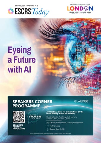
Eyeing a Future with AI SPEAKERS CORNER PROGRAMME Join leading voices for conversations on the latest thinking across the industry Christine Funke, Paul Singh, Erik Mertens, Antoine Labbé, Andreas Cordes Saturday 12 September - Sunday 13 September
SCAN HERE FOR FULL PROGRAMME
11:00 onwards Glaukos Booth E.233 Glaukos, iStent® and iStent infinite® are registered trademarks of Glaukos Corporation. ©2026. PM-EU-0429.
Legendary Haag-Streit optics
Revolutionary projector illumination
Efficient documentation
Powerful “presets”
Optional 3D image & video capture
Optimized viewing angle
Spacious headrest
Optimized control layout
ELARA 900 The slit lamp. Reinvented. See it on booth D.365 at ESCRS
haag-streit.com/elara
Hear Ye, Hear Ye, ESCRS Has Arrived in London “Small cheer and great welcome makes a merry feast”—so speaketh The Bard in 'The Comedy of Errors.'
I
can offer you, dear colleagues and friends, a warm welcome indeed, a merry feast of knowledge, progress, and innovation. And in this magnificent city that never sleeps, with its countless attractions, there will be plenty of cheer! Following yesterday’s iNovation Day and the events organised in collaboration with ESCRS’s partner societies, we are now delving into a programme that can truly be described as unparalleled. As at past ESCRS Congresses, ophthalmic surgeons from five continents will gather in London to catch up on the latest research at the Main and Clinical Research Symposia, Near-Live Surgery programme, and the ever-popular video sessions, along with wet labs, presented posters, and free paper presentations. You will attend not only prestigious lectures, such as the Ridley Medal Lecture on Saturday, but also a wide range of substantial and entertaining events. For example, since we are meeting near one of the world’s oldest continuous democracies, it is fitting ESCRS holds debates that uphold the Westminster tradition, albeit much shorter ones. In the Surgical Pearls video sessions, for instance, speakers must get to the point in just 100 seconds! Hear, hear! Many recent innovations may evoke fear because of the unknown. This is particularly true of artificial intelligence. There are numerous sessions addressing the impact of AI on our daily clinical practice. There is even an AI Summit that will bring clinicians and experts together to examine the opportunities, pitfalls, and ethical issues involved in using AI.
More than anything, the ESCRS Congress is a place where people come together. I am filled with pride at the thought that nowhere else in ophthalmology will so many people gather this year to exchange their experiences, views, and hopes for the future. In a sense, the ESCRS is coming home this year. After all, it was in London that Harold Ridley implanted the first intraocular lens. We will spend five days of learning, joy, and wonder in London following in Ridley's footsteps and those of other pioneers. And if you have some time left while you are in this world-class city, I recommend you visit The Globe on the South Bank. It is a theatre reconstructed in the style of the 1600s—just as it appeared when Shakespeare’s plays were first performed before a jeering, screaming, and often rowdy audience. You’ll be able to catch a performance of 'Much Ado About Nothing.' Could there be a greater contrast to the Congress? We make little fuss—not much ado—about quite a lot, focusing unflinchingly and calmly on what is happening on our stage: eye surgery that is future-proof and serves our patients. Enjoy our Annual Congress, enjoy London! H Burkhard Dick President, European Society of Cataract and Refractive Surgeons
12th SEPTEMBER | ESCRS Today
1
CATARACT/REFRACTIVE
Reading Between the Lines Why critically reading the SVIOL literature matters for everyone. LAURA GASPARI REPORTS
A
n accurate and critical appraisal of scientific studies on simultaneous vision IOLs (SVIOLs) contributes to the best possible outcome for cataract and refractive surgery patients, stresses Filomena Ribeiro MD, PhD. In recent years, simultaneous vision IOL technology has been evolving rapidly, resulting in an increase in published research and clinical studies. However, the quality of evidence is not always consistent, Professor Ribeiro observed. “Rather than simply looking at the conclusions, we need to understand how the study was designed, how outcomes were measured and reported, and how much confidence we can place in the evidence,” she stressed, as unfortunately some studies may be affected by bias or poor methodology. These are not marginal concerns. A study with a small sample size, a short follow-up period, a flawed or inaccurate methodology, or an inconsistent definition of outcomes can lead to conclusions that, while appearing promising on paper, do not necessarily stand up in daily practice. The ability to read studies critically should not be the exclusive preserve of clinical researchers, Prof Ribeiro said. Cataract and refractive surgeons, fellows, and any others involved in patient care should have the practical tools to judge whether published results are reliable enough to influence everyday clinical practice. The ability to identify good findings and trustworthy evidence leads to better treatment for patients, especially when choosing the best lens for them. There is also a need for a better understanding of standardised classifications to translate research into clinical decision making. The increasing number of studies on SVIOLs
2
ESCRS Today | 12th SEPTEMBER
introduces a large number of new technologies and terminologies that may be confusing. Prof Ribeiro noted there has been a definite improvement in the quality and consistency of IOL research, but there is still room for further progress. One of the Society’s key contributions in this regard is the ESCRS Simultaneous Vision IOL Classification system, an important milestone that provides ophthalmologists with a scientifically based framework of standardised terminology that also supports more consistent reporting across future studies. Scientific research is conducted primarily to provide patients with an ever-increasing range of therapeutic and surgical options. Failing to read it critically, or stopping merely at the abstract and conclusions, risks negative consequences for patient care. Congresses, meetings, and scientific societies’ guidelines, Prof Ribeiro said, are there to help people learn these skills and to know which instruments and tools are in place to support them. Research on SVIOLs will continue to grow. What also needs to grow is the responsibility of clinicians to keep refining their abilities to select the most appropriate lens and give their patients the best possible outcome, Prof Ribeiro concluded. “Ultimately, better critical appraisal leads to better clinical decisions, and better clinical decisions lead to better outcomes for our patients.” Filomena Ribeiro MD, PhD, FEBO is head of ophthalmology at Hospital da Luz, Lisbon, Portugal, and the immediate past president of ESCRS.
DON’T MISS
Today’s Congress Highlights Annual General Meeting 08:00 - 08:30 Capital Suite Room 9/10 Arena Debate Mini-monovision is preferred over multifocality in presbyopia cataract surgery 14:00 - 15:00 M2 (Arena)
Main Symposium Modern Management of Cataract and Lamellar Corneal Surgery 11:00 - 13:00 ICC Auditorium Near-Live Surgery Session 14:30 - 16:45 ICC Auditorium
Monofocal+ should be the default standard of care in routine cataract surgery 16:45 - 17:45 M2 (Arena)
Opening Ceremony / Ridley Medal Lecture The flow of cataract surgery 10:00 - 11:00 ICC Auditorium
Arena Forum Complication Solutions in Cornea 15:30 - 16:10 M2 (Arena)
Surgical Pearls Refractive intraocular 08:30 - 09:10 M2 (Arena)
Clinical Research Symposia New insights in Bullous keratopathy: A frequent but neglected corneal disease 08:00 - 10:00 George V Room 1
Refractive cornea 09:45 - 10:25 M2 (Arena)
AI modelling in anterior segment disease 10:30 - 12:30 George V Room 1 Can we make penetrating keratoplasty for keratoconus obsolete by 2040? 14:30 - 16:30 George V Room 1 What’s new in IOL power calculations 16:45 - 18:45 George V Room 1 Independent Medical Education Symposium Phaco Forum: Strategies for Complex Cases 13:00 - 14:00 George V Room 2
Sustainability Symposium Implementation and system-level change: Doing it right for sustainability in ophthalmology 16:30 - 17:30 Capital Suite Room 6/13 Young Ophthalmologists Programme Starting Phaco: Learning from the Learners 08:30 - 12:30 Victoria Room 5/6 Starting Phaco: Learning from the Learners 14:30 - 16:10 Victoria Room 5/6
GLOBAL REFRACTIVE SUMMIT
Head-to-Head Trials Reveal Laser Vision Correction Surgery Differences Examining the evidence from laser vision correction surgery outcomes. CHERYL GUTTMAN KRADER REPORTS
A
n appraisal of published head-to-head clinical trials comparing laser vision correction (LVC) procedures shows that all are safe and effective, although there are some nuanced differences between them, said Edward E Manche MD. Speaking at a session titled “Evidence-Based Refractive Surgery” during the Global Refractive Summit on Friday, Dr Manche reviewed outcomes of prospective, randomised, controlled studies investigating topography-guided (TPG)-LASIK, wavefront-guided (WFG)-LASIK, wavefrontoptimised (WFO)-LASIK, raytracing (RT)-LASIK, and keratorefractive lenticule extraction (KLEx) for myopia/ myopic astigmatism. “When trying to determine the relative safety and efficacy of different treatment options, it is essential to concentrate on results from level-one research publications because, with their rigorous design, these studies typically yield closely matched groups and minimise confounding from differences that can be present in cohorts drawn from retrospective chart reviews or when making cross-study comparisons of groups from single-arm prospective studies,” he explained. The main theme to emerge from his evidence-based review was that there are advantages to the most customised techniques. The greatest volume of available literature compares WFG-LASIK with WFO- or TPG-LASIK, and the results of those studies seem to show some clear differences favouring WFG-LASIK. “From a topline view, all the procedures have predictable refractive accuracy, including for astigmatic correction, and consistently provide high levels of 20/20 uncorrected visual acuity (UCVA),” Dr Manche said. “However, WFG-LASIK is associated with higher rates of 20/16 or better UCVA and superior outcomes for better low-contrast visual acuity (LCVA) and lines gained in corrected distance visual acuity (CDVA), which are endpoints relevant to quality of vision.” Prospective randomised studies comparing RT-LASIK, the newest iteration of LVC, are limited. However, the available data suggests that this technique, which integrates wavefront aberrometry, corneal tomography, and interferometry into its algorithm to generate a highly customised ablation plan, yields better outcomes than both WFO- and TPG-LASIK. Head-to-head studies of LASIK and KLEx are also relatively scarce. In a contralateral eye study Dr Manche conducted using WFG-LASIK and small incision lenticule extraction (SMILE) to treat myopia in eyes with low levels of astigmatism, the LASIK-treated eyes had better LCVA and greater gains in UCVA. As a caveat, however, he noted SMILE was performed using the first generation VisuMax 500 kHz laser (Zeiss).
4
ESCRS Today | 12th SEPTEMBER
Dr Manche also discussed two studies comparing RT-LASIK to SMILE Pro, performed using the VISUMAX 800 femtosecond laser. Both studies are prospective, randomised, contralateral comparisons of RT-LASIK and SMILE Pro that found superior outcomes with the RT-LASIK procedure, which provided higher levels of UCVA (i.e., 20/12.5), greater gains in CDVA, and better astigmatic correction. “There had been a period of relative quietness in LVC surgery in terms of further refinements in technique and hardware innovations, but the introduction of RT-LASIK reminds us to expect ongoing innovations,” Dr Manche said. “More experience is needed with RT-LASIK, but I consider it very exciting and one of the most innovative developments in refractive surgery in the past decade.” Edward E Manche MD is Professor of Ophthalmology at Stanford University School of Medicine and Director of Cornea and Refractive Surgery at the Stanford Eye Laser Center, Stanford, California, US.
We’re willing to bet most eye care professionals don’t realise just how prevalent Demodex blepharitis is. In fact, ~54% of eye care patients in Europe may have Demodex blepharitis (DB).1* *Data from an evaluation of 804 patients from 6 countries in Europe, including 15 clinicians each with ~50 consecutive patients. DB defined by the presence of collarettes.
WHAT ABOUT YOUR PATIENTS? LEARN HOW DB CAN FLY UNDER THE RADAR AT
Find us at ESCRS in London - Booth E.118 Reference: 1. Nanavaty MA, Findl O, Carones F, et al. Proportion of patients with Demodex blepharitis in ophthalmology clinics in Europe: the Eos study. Eye. 2026:40:165-167.
© 2026 Tarsus Pharmaceuticals, Inc. All rights reserved. TARSUS, LOOK AT THE LIDS, THINGS START LOOKING UP WHEN YOU GET YOUR PATIENTS LOOKING DOWN, and any associated logo(s) are trademarks owned exclusively by Tarsus Pharmaceuticals, Inc.
GL--2600026 03/26
12th SEPTEMBER | ESCRS Today
5
GLOBAL REFRACTIVE SUMMIT
The Pursuit of Precision in Refractive Lens Exchange Despite recent innovations, overcoming the problem of refractive surprise in RLE is still a pressing matter. TIMOTHY NORRIS REPORTS
P
ostoperative results and patient satisfaction in refractive lens exchange (RLE) have greatly improved in the last decade, mainly thanks to innovations in IOL power calculation. Despite the efficiency and precision of these calculations, there is still more work to be done, Giacomo Savini MD observed during a presentation at the Global Refractive Summit. “Just as with refractive cataract surgery, refractive surprise in RLE (defined as a prediction error higher than 0.5 D) is still between 10% and 20% of patients, as noted in literature,” he said. “What differs between these procedures is that RLE patients typically have much higher expectations and can be extremely dissatisfied by the slightest difference in the surgical outcome.” And a distressed patient is a distress for the surgeon, Dr Savini emphasised. According to the literature findings, therefore, one or two patients in every ten RLE procedures can be unhappy with the results. He further noted that sometimes a single disgruntled patient is all it takes to ruin a whole working week. Dissatisfaction rates are even higher among the myopes and hyperopes who most commonly undergo RLE. In eyes shorter than 22 mm or longer than 26 mm, the risk of refractive surprise is about 30%. Dr Savini noted these percentages are an improvement from the 40% risk calculated using older formulas a decade ago. However, the risk remains 50% in myopic eyes longer than 30 mm, so he does not recommend performing RLE in this group due to the risk of retinal detachment. Between 2015 and 2025, a new generation of formulas largely replaced the previous options, resulting in substantial improvements in RLE outcomes. A key milestone
6
ESCRS Today | 12th SEPTEMBER
arrived in 2022 with the ESCRS IOL Calculator, containing seven formulas now considered the gold standard for RLE: Barrett Universal II, Cooke K6, EVO, Hill-RBF, Hoffer QST, Kane, and Pearl-DGS. Dr Savini noted that, since its introduction, the ESCRS IOL Calculator is the most logical and accurate solution for eyes requiring RLE. Despite these advances, Dr Savini observed a sense of a plateau in terms of new formulas and predictive precision, noting that more research is needed to overcome the dreaded 10–20% threshold of refractive surprise. Some of the most promising updates in the field are coming from from Zeiss, which has a next-generation AI-based paraxial raytracing calculator in the pipeline. It is the first calculator developed by a company instead of a physicist, he noted. However, it currently supports only a few IOL models. There is also a need for more accurate toric calculators, as refractive surprises remain common when correcting astigmatism. Communication with the patient is still key to avoid having a tough time, he stressed. No matter how many instruments and formulas are available, there will always be a margin of error. A 300-micron difference in lens positioning for an anatomical variability, for example, can lead to a refractive defect. “We are still very far from being perfect,” Dr Savini said. “It is still necessary to talk to the patients and inform them on the risk of a refractive error.” Giacomo Savini MD is an ophthalmologist in private practice at the Studio Oculistico D’Azeglio, Bologna, Italy. giacomo.savini@ startmail.com
®
A NEW PARADIGM IN REFRACTIVE CATARACT CARE WHERE THE OCULAR SURFACE SHAPES THE OUTCOME Pairing the Intensity ® pentafocal IOL with the JETT ® bioelectric modulation platform, so a premium lens can finally deliver on its promise.
OPTIMIZE THE OCULAR SURFACE SUPPORT RELIABLE BIOMETRY REALIZE PREMIUM IOL PERFORMANCE JETT® prepares the surface · Intensity® redefines the vision
H AN ITA L E N SE S
丨
W W W.H A N ITA L EN S E S .COM
丨
JE T T ME D ICAL
丨
HT T P S : // E N .JE T T. EU/
For healthcare professionals only. Please refer to the current Instructions for Use for indications, contraindications, warnings, precautions and complete safety information. Availability, product configuration and approved indications vary by market. For registration and regulatory status, fur ther product information or distribution and par tnership enquiries, contact marketing@hanitalenses.com or info@jett.eu.
12th SEPTEMBER | ESCRS Today
7
CATARACT/REFRACTIVE
Let’s Make a Deal! Leadership, Business, & Innovation programme provides ophthalmologists with negotiation skills they can take from theory to practice. Can you afford to miss tomorrow’s Leadership, Business, & Innovation session, “How to Negotiate”? EuroTimes asked moderator Vincent Qin MD to preview the session.
What negotiation skills will attendees acquire? The theoretical part introduces the fundamentals of principled negotiation, based on the Getting to Yes framework.
EuroTimes: Why should an ophthalmologist learn to negotiate? Vincent Qin: Because ophthalmologists negotiate every day— even when they don’t call it negotiation. How do they obtain more operating room time? How do they convince management to invest in new technology? How do they negotiate with colleagues over responsibilities or on-call schedules? How do they deal with suppliers, practice managers, hospital executives, or staff representatives? Clinical excellence is essential—but it does not automatically translate into influence over resources, strategy, or organisational decisions.
Participants will learn how to: • separate people from the problem; • focus on interests rather than positions; • ask better questions; • create options for mutual gain; • use objective criteria; and • understand their BATNA, or ‘best alternative to a negotiated agreement.’
This session will give participants a practical framework to negotiate more effectively while protecting both personal interests and professional relationships. The session combines a short, practical theory session with an interactive negotiation role-play workshop, allowing participants to immediately put the principles into practice. Will negotiations with private equity investors be discussed? Yes, but as part of a broader, realistic ophthalmology leadership scenario. The workshop is built around a private eye clinic that has recently been acquired by a private equity group. Participants take on different leadership roles, each with different priorities, interests, and constraints. The objective is not to ‘beat’ the private equity investor. It is to learn how to find a solution when different stakeholders have fundamentally different priorities. The skills are directly transferable to real-life situations involving hospital groups, investors, practice owners, management teams, or other external stakeholders.
8
ESCRS Today | 12th SEPTEMBER
We will also explore common negotiation traps such as anchoring, positional bargaining, premature judgement, and fixed-pie thinking. What actually happens during the workshop? This is not a workshop where people simply listen to another presentation. Attendees will be placed in a small group of approximately four participants. Each participant receives a confidential role—CEO, CFO, Chief of Medical Doctors, or Chief of Nurses—and has time to prepare before the negotiation begins. Each role will have their own priorities, concerns, negotiating levers, and BATNA as well as information not shared with the other participants. Then the negotiation begins. For approximately 45–50 minutes, the groups must reach an agreement covering several interconnected decisions. Attendees will need to: • defend their role’s interests; • discover what the other participants really care about; • ask questions and actively listen; • manage conflicting priorities; • create and exchange proposals;
Leadership, Business, and Innovation Session (LBI) How to Negotiate? Sunday, 13 September 14:00–16:00 BST Capital Suite Room 2–4
• make trade-offs; • use objective criteria; • avoid getting trapped in positional bargaining; and • ultimately try to build a package everyone can support. There is no single predetermined answer. The challenge is to create an agreement that is better than the alternatives available to the participants if they fail to reach a deal. After the role-play, the group comes back together for a 30-minute debrief.
We will analyse what happened: • Which negotiating strategies worked? • Who anchored the discussion? • Did participants focus on positions or interests? • Where were opportunities for mutually beneficial trades missed? • How did BATNAs influence the negotiation? • What made it possible—or impossible—to reach an agreement? Why attend? Because negotiation is not only for CEOs, lawyers, or business executives. It is a core leadership skill for modern ophthalmologists. Whether working in a university hospital, public hospital, private practice, hospital group, or private equity-backed organisation, an ophthalmologist’s ability to negotiate increasingly determines their ability to influence investment, resources, working conditions, technology, staffing, and clinical strategy. Come for the theory. Stay for the role-play. Leave with a negotiation framework that can be used the very next day.
REF R AC T I V E & C ATAR AC T S URGERY
ESCRS Booth D.166
WI N TH E
RACE AGA I N ST RI SK
POWE RED BY OCULUS DATA-DRIVEN D IAGNOSTICS
LUNCH SYMPOSIUM
From Diagnosis to Decision: Advanced Corneal Imaging and AI in Clinical Practice Sat, 12 Sep 2026, 1– 2 pm, Capital Suite 14/16
oculus.de/escrs The availability of products and features may vary by country. OCULUS reserves the right to change product specifications and design.
12th SEPTEMBER | ESCRS Today
9
CATARACT/REFRACTIVE
MEET THE ESCRS
T
he European Society of Cataract and Refractive Surgeons (ESCRS) operates as a not-for-profit organisation and registered charity. Founded in 1991, it is a member-led scientific society committed to supporting the ophthalmic community through world-class education, scientific research, clinical registries, publications, fellowships, and year-round professional development. Headquartered in London, ESCRS is shaped by the profession itself. More than 200 board members, committee members, and faculty volunteer their time, expertise, and leadership to develop educational programmes, guide scientific direction, and help shape the future of the Society. Their commitment ensures ESCRS priorities are driven by the needs of ophthalmologists and their patients.
ESCRS Non-Profit Structure and Charitable Status
ESCRS is structured to ensure that all resources are directed toward delivering impact. As a non-profit entity, the Society does not distribute profits to members or stakeholders. Instead, all surplus income is reinvested into initiatives that directly support ophthalmologists and improve patient care. This approach enables ESCRS to deliver meaningful outcomes across the global ophthalmic community, including: • t he development of high-quality educational programmes, courses, and digital learning platforms;
10
ESCRS Today | 12th SEPTEMBER
•d elivery of the Annual Congress and specialist meetings that facilitate knowledge exchange; • f unding of clinical research, grants, and fellowships to advance innovation; •p ublication of peer-reviewed scientific content and journals; and, • support for trainee and early-career ophthalmologists. Through this reinvestment model, every activity contributes to strengthening clinical practice and enhancing patient outcomes.
Independent and Voluntary Governance
ESCRS is governed by a Board of Trustees and supported by committees composed of leading ophthalmic professionals who provide strategic oversight and guidance. All trustees and committee members serve on a voluntary basis and are not remunerated for their roles. This ensures decisions are made solely in the interests of the Society’s mission and the patients it ultimately serves, free from financial incentive. This independent governance model, supported by volunteers, is central to ESCRS’s integrity, reinforcing a culture of professional commitment, accountability, and service to the wider ophthalmic community.
Charitable Status
ESCRS is registered as a charitable organisation under applicable regulatory frameworks. This status reflects the Society’s commitment to delivering public benefit by improving patient outcomes through the advancement of ophthalmic knowledge, skills, and innovation. As a charity, ESCRS is subject to rigorous governance, financial reporting, and compliance requirements. These ensure transparency, accountability, and responsible stewardship of resources, with all activities aligned to the Society’s charitable objectives. At the heart of ESCRS’s non-profit and charitable status is a commitment to investing in education, supporting research, and promoting best practices in cataract and refractive surgery. Through these activities, ESCRS contributes directly to improving standards of care globally. The Society’s independence and reinvestment model create a continuous cycle of impact-advancing knowledge, strengthening clinical practice, and ultimately improving outcomes for patients around the world. Volunteer committee members are central to the Society’s ability to deliver this impact and value. Contributing their time, expertise, and leadership, these clinicians shape educational content, guide research priorities, and ensure
Push button
At the heart of ESCRS’s nonprofit and charitable status is a commitment to investing in education, supporting research, and promoting best practices in cataract and refractive surgery.
programmes remain relevant, practical, and of the highest scientific standard. Their active involvement not only strengthens the quality and credibility of ESCRS initiatives but also reflects a shared commitment across the profession to advancing patient care. This collective, volunteer-driven effort significantly amplifies the Society’s reach and reinforces its role as a trusted global leader in ophthalmology. For more information, please see the latest annual report at https://www.escrs.org/about-escrs/annual-report-2025.
Data processes
AI report generated
iCare DRSplus
iCare RETCAD and DRSplus are CE-marked medical devices under MDR (EU) 2017/745.
12th SEPTEMBER | ESCRS Today 11
CATARACT/REFRACTIVE
Historic Meeting of Ophthalmic Science and Business A unique ESCRS initiative aims to unite science, clinical practice, and industry to shape the next phase of functional vision classification. SEAN HENAHAN REPORTS
M
anaging presbyopia with simultaneous vision lenses (SVLs) was a major topic of discussion at this year’s iNovation Day sessions. Among the speakers was Joaquín Fernández MD, PhD, Executive Secretary of the ESCRS, who coordinates the Functional Vision Working Group that developed the Evidence-Based Functional Classification of Simultaneous Vision IOLs. The group first published a scoping review of the Evidence-Based Functional Classification of Simultaneous Vision IOLs in the Journal of Cataract & Refractive Surgery. It has since expanded to include all of the main international ophthalmology groups, among them ASCRS, the American Academy of Ophthalmology, the Asia-Pacific Association of Cataract and Refractive Surgeons, and the Latin American Society of Cataract and Refractive Surgery. These groups
12
ESCRS Today | 12th SEPTEMBER
formed a global consensus after debating some fine points of terminology at last year’s ESCRS Annual Congress in Copenhagen. The Functional Vision Working Group proposed a classification system based on the depth of field (DOFi) and the degree of visual acuity enhancement from intermediate to near distances provided by different SVLs. The SVL types are divided into partial-DOFi (narrowed, enhanced, and extended) and full-DOFi (steep, smooth, and continuous). Members also agreed to order the subcategories according to the functional benefits they provide to patients. “We started the group because we thought it was important to have a system [that was] not based on opinion, but based on evidence,” Professor Fernández said. “It is based on what is important to the patient.”
We started the group because we thought it was important to have a system [that was] not based on opinion, but based on evidence.
Missing from the consensus were the other main stakeholders, the leaders of the ophthalmic device industry. The London Congress will change that, as industry and ophthalmology meet to finalise a new consensus statement. Prof Fernández reported that the discussion on the next steps in implementing functional vision will be divided into three parts. First, ESCRS President Burkhard Dick will lead a session with the editors of the leading journals on issues affecting peer-reviewed publications and ophthalmology societies. Next, Prof Fernández will lead a discussion with industry executives aimed at defining evidence-based guidelines and
GalaxyH-H-Eurotimes-O.pdf
1
05/08/2026
certainty grading for the functional classification of SVLs. Immediate Past President Filomena Ribeiro will lead the third section, which will focus on patient-centred functional classification, emphasising patient-reported outcomes (PROMs) and clinical relevance. The next phase aims to integrate contrast sensitivity, photic phenomena, PROMs, and task performance in statement updates. “Patients want to know, ‘May I play golf with these lenses?’, ‘May I read fine print with these lenses?’, and ‘Can I drive at night?’” Prof Fernández said. Rather than fight against the International Organization for Standardization and the American National Standards Institute in the premarket arena, Prof Fernández said the goal is to include evidence-based functional classification of SVLs in the post-marketing phase, providing useful information for patients, doctors, and payers. Several sessions during the Congress will address these issues, including a Functional Vision Classification Symposium on Sunday, 13 September, from 08:30 to 10:00, in the Capital Suite, Room 7/8.
09:50
C
M
Y
CM
MY
CY
CMY
K
12th SEPTEMBER | ESCRS Today 13
CATARACT/REFRACTIVE
New IOLs Launching in London An interview with Sunil Vasanth at Alcon.
A
lcon has announced the launch of the Clareon PanOptix Pro IOL at the 2026 ESCRS Annual Congress. The lens offers several refinements over its predecessor, according to company executives. “The new lens is an evolution of the classic Clareon PanOptix platform,” said Sunil Vasanth, vice president surgical Europe at Alcon, in an interview. “We looked at how we could improve the visual quality. That is a function of improving the light utilisation, which in the new lens goes from 88% to 94%. We have also reduced scatter by almost half. As a result of this, we believe visual outcomes will be better, leading to better patient satisfaction.” Early feedback from patients in the US has been positive, he said. Reductions in glare and halo are being reported clinically. He added that the company sees these gains as a means of improving the overall impact of trifocal lenses. When asked about an ideal candidate for this lens, Vasanth noted that the IOL choice is guided by the patient-surgeon discussion but ultimately depends on individual patient needs and expectations. A typical patient would be presbyopic and looking to become spectacle free as much as possible without compromising night-time vision for driving. The lens enters the market amid ongoing efforts to standardise IOL terminology, following the release of the ESCRS Recommendations for Cataract Surgery. Vasanth said Alcon supports this approach and is working closely with ESCRS to improve collaboration with industry on terminology based on actual visual performance. Alcon also supports ESCRS efforts to enhance sustainability, he said. The company is working on ‘packaging of the future’ by using 40% less material than in the past. It is also
14
ESCRS Today | 12th SEPTEMBER
working on new custom packs, digital instructions for use, and improved shipping strategies. “When you look at sustainability, it is about the entire system,” he said. “We want to optimise the entire supply chain, from design, manufacturing, sterilisation, and waste management. We are now thinking in terms sustainability as a core requirement of IOL design.” Other Alcon products with a launch announcement at the Congress include the modified monofocal IOL, Clareon TruPlus, and the UNITY M digital surgical microscope.
A NEW ERA IN DIGITAL SURGERY
Meet Us at Booth #B.300 INDUSTRY SATELLITE SYMPOSIUM Sunday, 13 September 2026 1:00 – 2:00 PM · Captial Suite 7/8
How Enhanced Visualization Is Transforming Ophthalmic Surgery MODERATORS: Steven Thomson and Rosa Dolz-Marco MD, PhD, FEBO SPEAKERS:
D4008976-001 GL.AE26 © Heidelberg Engineering GmbH
Changing the way we see – How Heidelberg OPERA will enhance my ophthalmic surgery Prof. Damien Gatinel, MD, PhD | France Phakic IOL Surgery – Getting it right first time – SafeVault* supported by iOCT imaging Alain Saad, MD | France How ANTERION® supports my surgical decision making Prof. Mohamed Eid, MD | Egypt
Heidelberg OPERA is currently not commercially available in all markets. Can only be made available once it has regulatory approval in your region. * SafeVault is a third-party software solution. It is not part of the ANTERION® platform and is not commercially available as of June 2026.
12th SEPTEMBER | ESCRS Today 15
HENAHAN PRIZE
Henahan Prize Winner Hasan Chican: 'The Hands That See' HASAN CHICHAN MD, MSC, FEBO, MBCHB, FACS
chief complaint. Not visual acuity. Not the Snellen chart. His grandchildren. My attending surgeon, Dr Saad, did something I did not expect. Before the surgery, he sat on the edge of the patient’s bed, not beside it, on it, and held Abu Kareem’s hand. He asked about the grandchildren. Their names. Which one looks most like him. The old man laughed so hard he started coughing. Then Dr Saad said: “Tomorrow you will see that face again, inshallah.” There was no femtosecond laser. No digital OR. The phaco machine was older than me. We had maybe three IOL powers to choose from. But Abu Kareem’s hand did not shake during surgery because Dr Saad kept talking to him, about football, about the weather, about his grandchildren. When the patch came off next day and Abu Kareem looked
Each year, young ophthalmologists are invited to participate in the John Henahan Writing Prize competition by responding to an essay prompt. This year's prompt read as follows: The digital OR, AI algorithms, and robotics notwithstanding, cataract and refractive surgery involve a human relationship between you and your patient. Please describe how an experience or experiences in your early training reminded you of the importance of the human touch, and how this has inspired you in your clinical practice. The following essay scored the highest in this year's competition.
I
remember the smell. Not of the operating theatre, that came later. But the smell of my grandmother’s kitchen in Baghdad, where she would sit by the window every afternoon, squinting at her prayer beads, counting them not by sight but by touch. She had cataracts in both eyes. Dense, white, like two small moons. She never got surgery. She said she could still see what matters, the shape of her grandchildren coming through the door. I thought about her on my very first day assisting in cataract surgery at Al-Shafaa Teaching Hospital in Babylon in 2013. I was a junior resident, terrified, trying to hold retractors with steady hands while mortar rounds shook the windows two blocks away. The patient was a 70-year-old retired schoolteacher named Abu Kareem. He could not see his grandchildren’s faces anymore, he told us. That was his
16
ESCRS Today | 12th SEPTEMBER
But I carried something from Baghdad that no machine could teach: surgery is not just what happens between the speculum and the drape. It is what happens between two people.
at his youngest granddaughter, he started crying. Then Dr Saad started crying. Then I started crying too, and I was just the resident holding the torch. That moment changed something in me, though I did not fully understand it until years later. When I moved to Germany in 2015, I entered a completely different world. Everything was precise, protocolled, efficient. The machines were extraordinary. Outcomes measured in microns. But something felt missing, and it took me time to realise what it was: it was the bed. Nobody sits on the patient’s bed here. In fact, there is no bed—just a chair that moves on a rail into the OR. The system is so efficient that sometimes I would finish a surgery and realise I did not know if my patient had grandchildren at all. I do not mean this as criticism. I learned an enormous amount from European training, and I am deeply grateful. The precision, the safety culture, the evidence-based approach—these are gifts that my training in Iraq could not give me. But I carried something from Baghdad that no machine could teach: surgery is not just what happens
between the speculum and the drape. It is what happens between two people. Now, in my daily practice, I have made small rituals that are my own version of sitting on the bed. I always ask my patients one question before surgery that has nothing to do with their eyes. Sometimes it is about their holiday. Sometimes what they had for breakfast. One elderly Swiss lady told me she was nervous because she promised her cat she would be home by dinner. So I told her we will make sure of that, and during surgery when she tensed up, I said: “Your cat is probably already sitting by the door.” She laughed under the drape and her eye relaxed. These are small moments. They will never appear in a surgical report. No AI algorithm will code them as a quality metric. But I believe they are the difference between a procedure and a healing. My grandmother never got her cataracts removed, but she taught me that seeing is not only about the eye. And Dr Saad, in a crumbling hospital in Babylon with equipment that would not pass any European inspection, taught me that the most advanced instrument
we have is still our ability to connect with another as human beings. I have been trained by machines, and I have been trained by people. The machines taught me how to operate. The people taught me how to care. And when I stand at the slit lamp today, somewhere between the technology that surrounds me and the patient before me, I try to remember: the hand that holds the phaco probe must also be the hand that can hold a patient’s fear. That is the part they cannot automate. And I hope they never will.
As the author of the winning essay, Hasan Chican will receive a €500 bursary and a specially commissioned trophy, awarded during the 2026 ESCRS Congress in London, UK. His essay will also be published in EuroTimes.
cso booth talks
escrs congress • booth n. e255
sep tem ber 12/13 2026 scan the qr code to regIster
SATURDAY 12TH 9:00 Dan Z. Reinstein One device. Endless possibilities: The Italian Swiss Army Knife for cornea & anterior segment 9:30 Andrea Russo Post-cataract aberrometry: making subjective defocus curves more objective 11:00 Giacomo Savini MS-39: redefining biometry and IOL Calculation 12:30 Farhad Hafezi ECO-CAIRS: Surgeon-prepared CAIRS for easier insertion, enhanced sterility, and potentially greater effect SUNDAY 13TH 9:00 Victor Derhartunian Open-Field aberrometry: beyond myopia control 11:00 Riccardo Vinciguerra KAI: The next generation of keratoconus screening 13:00 Ivan Gabric Pyramidal aberrometry: a new perspective on vision quality www.csoItalIa.It
12th SEPTEMBER | ESCRS Today 17
HENAHAN PRIZE
Previous Winners of the John Henahan Writing Prize Reminisce Each year’s John Henahan Writing Prize submissions provide insight into what’s on the minds of young ophthalmologists. Topics have included AI, DEI (diversity, equity, and inclusion), work-life balance, and sustainability in ophthalmology. Theresa Shiu Ting Mak MBChB, MRCEd, FRCEd, FCOphth Consultant Ophthalmologist, CUHK Medical Centre, Hong Kong Time flies. A lot has happened since I received the inaugural John Henahan Writing Prize in 2008—an honour for which I remain deeply grateful today. On the professional front, my journey has been diverse. After 13 years in the public ophthalmology service as a trainee, trainer, and team head, I spent two years practicing administrative medicine and public health. Ultimately, I realised my heart belonged to clinical practice. Returning to the field only strengthened my passion for my specialty. On a personal level, life has been equally eventful. I became a mother of three and developed a deep passion for fencing. I now train and compete regularly, and I am a registered coach with the Fencing Association of Hong Kong, China, upon passing all the required training and assessments. Ophthalmology has evolved drastically over the last two decades. While cutting-edge machines, advanced diagnostics, and premium intraocular lenses have revolutionised patient care worldwide, this rapid technological shift brings a dilemma. I occasionally see trainees who rely so heavily on advanced platforms that they struggle when forced to pivot to fundamental skills, such as converting a
18
ESCRS Today | 12th SEPTEMBER
failing phacoemulsification into a traditional extracapsular cataract extraction. I constantly remind my trainees never to neglect their foundational training. In ophthalmology, as in life, less is often more. Leigh Spielberg MD Vitreoretinal Surgeon, Sint-Jan Hospital, Bruges, Belgium In 2010, I was awarded the John Henahan Writing Prize in the first year of my ophthalmology residency. This award gave me the opportunity to write for EuroTimes, which I did on nearly a monthly basis for 10 years, writing columns and book reviews, and reporting from Europe’s biggest ophthalmology conferences. My most enjoyable writing was inspired by specific moments in the clinic, both in training and after going into practice for myself. Although I no longer write, I regularly come across experiences that make me stop and think, “Hmm, that would make an interesting topic for a column,” such as an unusual diagnosis or an unexpected surgical result. Since 2010, I’ve gone on to finish both my residency and my vitreoretinal fellowship at the Rotterdam Eye Hospital and have been in practice as a vitreoretinal surgeon in Bruges and Gent since 2016. I spent four years in the university
hospital before moving to a non-university training hospital. It has been a very satisfying journey! As for the state of ophthalmology today, it’s always impressive how everything continues to evolve and improve. If we are to believe the experts, AI will revolutionise the practice, but we’ll have to wait and see what the future brings. Clare Quigley MD Consultant Eye Surgeon in private practice at Progressive Vision and in public practice in the Royal Victoria Eye and Ear Hospital and St James’s Hospital, Dublin, Ireland Back in 2017 when I wrote for the John Henahan essay, I reflected on relationships with industry and maintaining independence. This topic is still relevant, particularly in guiding decision making for our patients having cataract surgery. Since exiting formal training in 2023, I have travelled near and far, working in the UK and Australia before returning for a consultant post in Ireland in 2025. The scope of AI in ophthalmology is particularly exciting right now. I believe our jobs are safe for the foreseeable future, but we would do well to adopt our practice as appropriate to improve care and add efficiency with all tools at our disposal. Soosan Jacob MS, FRCS, DNB Director and Chief, Dr Agarwal’s Refractive and Cornea Foundation, Chennai, India Looking back, my journey since my early young ophthalmologist days has been shaped by a continued commitment to learning, teaching, and exploring innovations in cataract, cornea, and refractive surgery, particularly in areas like keratoconus management and secondary IOLs. Ophthalmology continues to evolve, with advances in technology enabling more precise, customised, and safer treatments for our patients. It has been especially rewarding for me to see how innovation has expanded our ability to address complex corneal and anterior segment conditions. The growing global collaboration and the enthusiasm of young ophthalmologists is also very encouraging to see, and this truly does bode very well for the future of our specialty. For more information on the contest and to read previous essays, see www.escrs.org/ eurotimes/john-henahan-writing-prize.
12th SEPTEMBER | ESCRS Today 19
London Looks in
Memorable moments from the ESCRS 2026 Congress, capturing key connections and collaborations.
20
ESCRS Today | 12th SEPTEMBER
12th SEPTEMBER | ESCRS Today 21
CORNEA DAY
A New Paradigm for Keratoconus in Down Syndrome A different keratoconus needs different strategies. LAURA GASPARI REPORTS
K
eratoconus in Down syndrome (DS) patients requires a different approach, with tailored strategies for diagnosis, treatment, and prevention, according to Jorge Alió MD, PhD. In his studies, Professor Alió demonstrated that people with DS always present with abnormal corneas. “They have steeper, thinner corneas and a larger number of high-order aberrations. So, not only are their corneas anatomically different, but optically as well,” he remarked. This distinction may help explain why scientific literature for decades thought the incidence of keratoconus in people with DS was from 5% to 15%. Prof Alió’s recent studies have instead found more than 75% of DS patients with abnormal corneas have keratoconus, while the remaining 25% have other types of corneal irregularities, abnormal thickness, or aberrations. One of the key differences in how keratoconus presents in DS patients involves corneal deformation. In the non-
22
ESCRS Today | 12th SEPTEMBER
One of the key differences in how keratoconus presents in DS patients involves corneal deformation.
DS population, keratoconus presents a bulging of both the anterior and posterior corneal surfaces. In DS keratoconus patients, by contrast, only the anterior apex deviates from the corneal vertex, while the posterior tends to remain stable. Keratoconus in DS patients is more progressive, owing to epigenetic reasons, the high incidence of atopy, and frequent, compulsive eye rubbing, even in the absence of allergies. Moreover, patients with Down syndrome tend to have altered collagen metabolism affecting both the corneal stroma
and Bowman’s membrane, which may be traumatised by the frequent eye rubbing. These factors create a ‘perfect storm’ for keratoconus to progress and aggravate, risking visual impairment and blindness. Diagnosis and treatments pose many challenges in patients with DS, placing early detection and intervention at risk. Sometimes, these patients do not want to cooperate because of communication issues, fear, or sensory overstimulation. Prof Alió suggested performing objective examinations at a distance from the patient using dilated-pupil retinoscopy, automated refractometers, or portable corneal topographers. The recommended age for the examination in patients with DS is between four and six years. All DS patients with keratoconus or any type of corneal ectasia should be treated with cross-linking, Prof Alió advised, as it helps control the disease before contacts or transplants become necessary. Corneal grafts in DS patients with keratoconus, for example, often result in melting due to an abnormal collagen matrix and metalloprotein cascade activation. However, cross-linking may be challenging because of the need for general anaesthesia, which is particularly risky in paediatric patients with DS, given their systemic comorbidities. Prof Alió suggested a partial, standard cross-linking approach to maintain structural integrity while avoiding too much postoperative pain that could disrupt patient compliance. Nevertheless, prevention is the key word to success with these patients. Early screening and proactive treatment with cross-linking are crucial to saving the visual function of people with Down syndrome. Above all, clear and simple communication with patients and their parents is crucial for compliance, especially when managing allergies with antihistamine medications to prevent compulsive eye rubbing. “This way, you stop the histamine cascade activated by allergy and you avoid eye rubbing,” Prof Alió said. “If you speak with the parents and explain in a kind way to patients that eye rubbing is bad, they will accept it and stop it.” Jorge L. Alió MD, PhD is Professor of Ophthalmology at the Miguel Hernández University in Alicante, Spain, and founder of Vissum Miranza, Alicante, Spain.
12th SEPTEMBER | ESCRS Today 23
CORNEA DAY
Is Epi-On the Way Forward? Customised transepithelial procedures promising, but still far from becoming the new gold standard. TIMOTHY NORRIS REPORTS
E
pi-on corneal cross-linking (CXL) may not yet be the gold standard for the treatment of keratoconus, but it is heading in that direction, according to Cosimo Mazzotta MD, PhD. “Epi-on is still struggling to win trust amongst ophthalmologists around the world, and many of the protocols that are available today are still broadly unfamiliar,” he said. Epi-off protocols remain the gold standard for keratoconus treatment, a position reinforced by the Second Global Consensus on Keratoconus and Ectatic Diseases of 2026, in which Professor Mazzotta participated as Group 3 coordinator with Prof Michael Belin and Dr Emilio Torres. The consensus helped define new best practices and a new understanding of keratoconus as a multifactorial disease, while paving the way to a more multimodal diagnostic approach. At the same time, Prof Mazzotta observed that accelerated epi-off is likely to remain the dominant procedure, although there is still no majority agreement on the optimal protocol. However, Prof Mazzotta believes recent advances in epi-on protocols could change this balance. The first step, he said, is to rebuild confidence in the technique by making its mechanisms and potential benefits better understood. One reason for the lack of popularity of epi-on can be traced back to its early development. Original protocols attempted to reproduce epi-off conditions without adequately compensating for the presence of the epithelium. Low oxygen availability, insufficient stromal riboflavin penetration, and inadequate photochemical energy resulted in disappointing outcomes and, in some cases, retreatment.
24
ESCRS Today | 12th SEPTEMBER
Translational research paved the way to a different approach, Prof Mazzotta explained. The introduction of Iontophoresis Plus, for example, combined enhanced riboflavin delivery with higher fluence and pulsed light. A study Prof Mazzotta and colleagues conducted, published in the Journal of Refractive Surgery, demonstrated a consistent demarcation line on optical coherence tomography (OCT) at a meaningful stromal depth, Kmax flattening close to epi-off, and stable keratoconus over time. The findings suggested that epi-on required a different energy balance from epi-off. Higher fluence, pulsed illumination, and active riboflavin delivery appeared to address some of the limitations of the earlier protocols. These results were a huge step forward, Prof Mazzotta said. Findings demonstrated that the limitations of epi-on were not necessarily intrinsic to the technique but could be addressed by understanding and modifying its underlying photochemical and pharmacokinetic mechanisms. Oxygen then became another important piece of the puzzle. In a prospective clinical study published in the Journal of Cataract and Refractive Surgery, Prof Mazzotta and colleagues evaluated customised transepithelial CXL with supplemental oxygen, reporting clinical and OCT outcomes with an epi-on approach. Oxygen is not simply an accessory to the treatment but a fundamental component of the photochemical reaction, particularly when the epithelium remains intact. This research has gained new relevance following the FDA approval of Epioxa (Glaukos) in the United States in October 2025. Epioxa is approved for epithelium-on corneal collagen cross-linking in adults and paediatric patients aged
13 years and older, in conjunction with the O2n System and Boost Goggles. The treatment combines riboflavin photoenhancers with an oxygen-enriched environment. For Prof Mazzotta, the development represents an important validation of the scientific rationale behind oxygen-enriched epi-on CXL. “I am honoured to have been among the pioneers exploring oxygen supplementation in transepithelial cross-linking,” he said. “Our early clinical work helped demonstrate the importance of controlling oxygen availability during the photochemical reaction.” His more recent work has moved towards high-fluence, pulsed-light epi-on protocols at around 10 joules, using either iontophoresis or new-generation riboflavin formulations. The aim is to narrow the remaining efficacy gap with epi-off while preserving the potential advantages of leaving the epithelium intact. The challenge now is to make these advances better known among ophthalmologists, explain the mechanisms behind them, and establish which patients may benefit most. Prof Mazzotta’s involvement in keratoconus extends beyond CXL research. In 2026, he served as president of the third World Keratoconus Congress in Florence. He was also among the authors of the first European Reference Network for Rare Eye Diseases (ERN-EYE) clinical consensus statement on paediatric keratoconus, providing recommendations for diagnosis, monitoring, and management within the European ERN-EYE. The next frontier may be paediatric treatment. “It’s important to be bold but cautious,” he said. In his clinical practice, Prof Mazzotta already uses epi-on in selected paediatric patients, particularly in the better eye, where a less aggressive approach may be desirable. Prof Mazzotta believes the transition towards epi-on can ultimately succeed, provided it never comes at the expense of early diagnosis, eliminating eye-rubbing, timely treatment, and safe retreatment when necessary. “Working together, we can make epi-on the future of corneal cross-linking,” he said. “The goal is not simply to replace epi-off, but to develop a safer, repeatable, and increasingly effective treatment strategy that can be adapted to the patient and to the stage of disease.” Cosimo Mazzotta MD, PhD, FWCRS is Associate Professor of Ophthalmology at University Kore of Enna, Italy.
OA-2000 Optical Biometer
Swept Source OCT biometer with high-speed acquisition and excellent penetration in dense cataracts. Fully automated bilateral measurement, including Placido ring-based topography – completed in under 60 seconds with no manual input required.
Biometry. Fast. Automatic. Complete. Capture axial length, keratometry, ACD, lens thickness and topography in one scan. Fully automated – no manual alignment needed. Consistent, reproducible results for both eyes in under 60 seconds.
New formula options. Optimised outcomes. Comprehensive IOL calculation with new formula options such as Kane and Barret RX will bring your IOL calculation to a next level. Compare up to eight lenses for confident decisions in routine and complex cataract surgery.
Visit our booth D.346 for live expert talks
and discover real clinical insights. Scan the QR code for topics, speakers, and session times.
12th SEPTEMBER | ESCRS Today 25
CORNEA DAY
A Race Against Time Children with keratoconus require a distinct diagnostic and therapeutic approach. LAURA GASPARI REPORTS
T
ime is of the essence in paediatric keratoconus, which requires a different approach to management and treatment than adult disease, with prompt responses and clear strategies essential to preserving children’s eyesight, as Ken K Nischal MD emphasised during a Cornea Day presentation.. Paediatric keratoconus is typically more aggressive, as patients present later, and the disease progresses more rapidly, with a higher risk of visual impairment. Moreover, children younger than the age of eight have a severe risk of developing amblyopia. The situation is particularly delicate because performing a corneal transplant on a child carries risks, as Dr Nischal pointed out. “If children get hydrops and scarring, that’s the real problem,” he said, noting that early detection allows for timely intervention. Diagnosing keratoconus in children is not straightforward: tomography remains the first-line diagnostic tool,
26
ESCRS Today | 12th SEPTEMBER
but many paediatric patients cannot sit still for it, instead requiring a direct ophthalmoscopic dilated exam to check for the oil droplet sign or an early scissoring reflex on retinoscopy. Key warning signs include high astigmatism, frequent spectacle changes, eye rubbing, allergic eye disease, halos without glaucoma, family history, and genetic conditions such as Down syndrome. By the time signs such as fleshy rings, Munson’s sign, or Vogt’s striae appear, however, the child already has keratoconus. The treatment of choice for paediatric keratoconus is the standard and accelerated epi-off, which is strongly supported by the current clinical evidence as effective and safe. Dr Nischal noted that faster and convenient transepithelial or accelerated protocols such as epi-on have not been shown to match the Dresden protocol’s reliable efficacy in children. For instance, the penetration depth of epi-on is 150 µm into the cornea, while the penetration depth in epi-off is 350 µm; more-
The treatment of choice for paediatric keratoconus is the standard and accelerated epi-off, which is strongly supported by the current clinical evidence as effective and safe.
over, accelerated epi-on programmes deliver considerably more energy (11 joules/cm²) compared to Dresden protocol (5.4 joules/cm²). The impact of this extra energy exposure on children with an undiagnosed collagen disorder remains unknown, Prof Nischal said, pointing out it is a risk that may not apply to adults but cannot be ruled out in paediatric patients. Moreover, accelerated epi-off cross-linking only requires 15–20 minutes instead of a full hour, reducing the need for
general anaesthesia and its associated risks for children and developmentally delayed adults. This raises ethical concerns about the use of alternative, less-proven procedures that may expose these patients to anaesthesia unnecessarily, Prof Nischal observed. Managing pain and behaviour in children and developmentally delayed adults poses challenges that require creative solutions. Dr Nischal suggested behavioural therapy to help curb eye rubbing, alongside treating eye allergies and measures to prevent dry eye. In young children and developmentally delayed adults, temporary lid suturing for three days post procedure to control pain without complications proved effective and safe.1 The best approach is, however, a prompt response because with paediatric keratoconus, the stakes are high. “It is a time-sensitive disease and cross-linking is prevention, not refractive surgery. It is aimed at preserving the child’s lifetime visual potential,” Dr Nischal concluded. 1. Conner E, Gagrani M, Lalgudi VG, et al. Cornea, 2025; 44(1): 7–14. doi:10.1097/ICO.0000000000003562
12th SEPTEMBER | ESCRS Today 27
CORNEA DAY
What’s New in CAIRS? Personalised CAIRS serves as an effective option for iatrogenic and other ectasia patients. SEAN HENAHAN REPORTS
S
oosan Jacob MS developed Corneal Allogeneic Intrastromal Ring Segment (CAIRS) surgery as a biocompatible alternative to synthetic intracorneal ring segments. It has evolved considerably since then, becoming popular around the world. She discussed CAIRS during a Cornea Day session. CAIRS is a form of mid-stromal, mid-peripheral intralamellar keratoplasty where donor stroma and Bowman’s membrane are placed within a host channel to reshape the ectatic cornea and make it more regular. This results in improved uncorrected and best corrected visual acuity and decreased refractive error as well as decreased irregular astigmatism. Dr Jacob said it can also potentially delay or avoid keratoplasty. As the procedure has evolved from uniform segments to customised and custom-shaped CAIRS, the precision with which donor tissue is prepared has become increasingly important. A segment that is too wide, too thin, too thick, or poorly matched to the intended plan can alter the flattening response and reduce the treatment predictability. Dr Jacob introduced CAIRS in 2015, which was followed by custom shaping in 2017. CAIRS customisation refers to changing the arc length of a uniformly thick segment. CAIRS custom-shaping, on the other hand, refers to variably changing the CAIRS along its arc length to better suit the patient’s topography. Surgeons can easily prepare and custom shape CAIRS using various nomograms and the CAIRS customiser, among other techniques. Dr Jacob uses her own proprietary nomogram prepared from nearly 2,000 cases. It is a living
nomogram that is constantly updated as data becomes available and is slated for online release soon. CAIRS are available as fresh cut or with a shelf life from eye banks. They can also be prepared from glycerinepreserved corneas. Optigraft CAIRS (Lions World Vision Institute), KeraNatural (VisionGift), and corneal tissue addition keratoplasty are types of CAIRS that are prepared by eye banks.
CAIRS in Post-LASIK Ectasia
CAIRS has now been extensively used for keratoconus and has shown excellent efficacy. It is also being used for post-LASIK ectasia (PLE), where it has been shown to decrease refractive error, improve uncorrected and corrected distance visual acuity, and decrease vertical coma. The generally inferiorly decentred apex in PLE causes negative coma due to a relative inferior phase advance and superior delay, making these eyes ideally suited for CAIRS implantation. “Care should be taken in PLE to preoperatively measure the depth of the LASIK flap at multiple sites in the zone of CAIRS implantation using [anterior segment optical coherence tomography]. The depth of the channel should be kept about 100 microns below the depth of the flap to avoid intersection with the flap,” Dr Jacob explained. “Similarly, during insertion, it should be ascertained that the segment has gone into the channel plane and not inadvertently into the flap plane. This can be done by always going vertically down to the depth of the entry incision before turning horizontally to enter the femtosecond channel. Gently moving the curved Y-rod (Jacob curved-Y, Epsilon, US) horizontally to either side also helps ascertain correct placement, as the rod movement will be restricted by the sides of the channel. If correctly placed within the flap plane, the rod will be seen to move beyond the dissected channel.” A longer column by Dr Jacob on CAIRS will appear in EuroTimes. Soosan Jacob MS, FRCS, DNB is Director and Chief of Dr Agarwal’s Refractive and Cornea Foundation at Dr Agarwal’s Eye Hospital, Chennai, India. She has a patent for special trephines, devices, and processes used to create CAIRS.
Scan the QR code to watch ‘Customized CAIRS Segment Preparation using Jacob CAIRS Customizer.’
28
ESCRS Today | 12th SEPTEMBER
CORNEA DAY
Passing the Baton at Cornea Day A corneal surgery legend is happy to pass his baton thanks to a day packed with corneal wisdom. ANDREW SWEENEY REPORTS
C
ornea Day at the ESCRS Annual Congress is now in its sixteenth year, reflecting an ongoing partnership between the ESCRS Cornea Committee and EuCornea. Throughout those years, José Luis Güell MD, PhD has been directly involved in its organisation—until now. “This is the first year that neither Professor Rudy MMA Nuijts nor myself are directly involved in organising Cornea Day. Originally, it was only a couple of talks, which were mostly clinical cases. The audience was very happy and enthusiastic, and then it grew over the years,” Professor Güell said. “Today, Cornea Day is a comprehensive cornea conference. This year sets a high standard and covers many areas of corneal surgery and other medical problems.” Prof Güell is passing his leadership baton with full confidence. He is particularly keen to highlight preoperative ocular surface optimisation, a topic that is important not only to cataract and refractive surgeons but also in “any intraocular surgery, such as glaucoma or corneal surgery.” The first session of the day, “Cataract Surgery and Corneal Conditions,” covered this topic as well as intraocular lens (IOL) selection during cataract surgery with corneal complications. Prof Güell said he is looking forward to the debate on traditional versus novel approaches. “Traditionally, it has been considered that simple monofocal lenses are the ones to be used for any corneal problem,” he said. “Now we are becoming aware that in some circumstances, other lenses, such as toric or extended depth of focus IOLs, could be considered.”
The second session, “Ocular Surface,” included a focus on dry eye disease. Prof Güell encourages attendees not to consider the disease as an isolated ocular problem. Instead, it should be viewed as one with significant systemic connections, particularly microbiota. Prof Güell also recommends learning more about the management of neurotrophic keratitis, which he describes as the “final problem of a different number of corneal complexities.” He is particularly interested in the standardisation of corneal neurotisation surgery, “once a strange procedure performed by a few specialists, now more frequently encountered.” An update on the EndoArt corneal implant after its first 80 implantations was presented during session three, “Cornea Surgery,” which also covered how regenerative medicine plays a role in limbal deficiency management. The final session covered artificial intelligence (AI) in “Keratoconus and Cross-linking.” “In the last section, I would highlight the use of AI in detecting, diagnosing, and evaluating the progression of keratoconus. Together with biometry, this is one of the areas where AI is gaining a role in anterior segment problems,” Prof Güell said. “Finally, investigate cross-linking procedures. It’s not new, but there are new, modern ways to utilise the concept.” José Luis Güell MD, PhD is head of the Cornea, Cataract, and Refractive Surgery Department at IMO Grupo Miranza in Barcelona, Spain, and past president of the ESCRS.
12th SEPTEMBER | ESCRS Today 29
CORNEA DAY
Standardise Diagnosis, Personalise Treatment How DEWS III is closing the gap between research and practice. LAURA GASPARI REPORTS
T
he Tear Film & Ocular Surface Society Dry Eye Workshop III (TFOS DEWS III) deepened practitioners’ understanding of dry eye disease, refining the standard diagnosis and providing personalised and evidence-based management, according to James S Wolffsohn BSc, PhD. TFOS DEWS III was released last year, condensing the previous DEWS II framework into three reports: one on definition and diagnosis, one on management, and a digest updating the latest dry eye disease (DED) research in key areas of concern, such as pathophysiology, epidemiology, sex, gender, and hormones. As Professor Wolffsohn pointed out, aside from a refinement of the definition of DED, stressing its nature as a disease rather than a syndrome, DEWS III recognises that not all pathophysiological factors are present in every patient. “It is a multifactorial disease, so it is not going to be solved by a single ‘bullet,’” he said, adding that “different people have different forms of the disease.” This drives practitioners toward a thorough consideration of patients’ anamnesis and diagnosis when considering the right treatment. Dry eye is becoming increasingly widespread and, as with all diseases, requires a standardised approach to diagnosis. DEWS III recommends using only the Ocular Surface Disease Index-6 (OSDI-6) questionnaire to assess patients’ symptoms, as it allows for consistency among practitioners. Evaluation of tear film instability with noninvasive tear break-up time is preferred over fluorescein staining, as fluorescein can disrupt the tear film. In terms of ocular surface assessment, fluorescein staining is preferred for the cornea, while lissamine green is preferred for the conjunctiva and lid margins. A combination of symptoms assessed with the OSDI-6 and any one of these signs is sufficient for a fast and standardised DED diagnosis.
30
ESCRS Today | 12th SEPTEMBER
Rather than the previous evaporative-versus-aqueous classification, which had limited clinical use because most patients fell into the ‘evaporative’ group, DEWS III identifies nine main drivers of the disease. These include deficiencies in the tear film’s lipid, aqueous, or mucin/glycocalyx layers); abnormalities in blink and lid closure/lid margins; and ocular surface factors such as anatomical misalignment, inflammation, and cellular and nerve damage. Patients may have more than one driver, Prof Wolffsohn said, which can shift over time. Identifying and monitoring these drivers allows clinicians to choose personalised, evidence-based treatments, replacing the stepwise approach of DEWS II and giving industry a clearer framework for validating new treatments. According to a recent review, despite advancements in research and guidelines such as DEWS III, it typically takes evidence about 23 years to reach clinical practice, a finding Prof Wolffsohn described as shocking.1 For this reason, he stressed the importance of clinicians adopting a single, standardised approach to DED diagnosis, while allowing flexibility in how drivers are subclassified and treated. Moreover, connecting patients through associations to boost compliance and emotional support is crucial. “It is a chronic disease, and it is so common: using the TFOS DEWS criteria, one in three adults and now one in five children warrant a diagnosis of dry eye disease. It means there are a lot of people that we need to help,” he concluded. James S Wolffsohn BSc, MBA, PhD is a Professor of Optometry at Aston University, Birmingham, UK. CITATIONS 1. Wolffsohn JS, Morgan P. Cont. Lens Anterior Eye. 2026; 49(4): 102697. doi:10.1016/j.clae.2026.102697
CORNEA DAY
Balancing the Ocular Microbiome Maintaining a healthy ocular surface is the gateway for preventing and treating eye disease. TIMOTHY NORRIS REPORTS
T
he microbiome plays a key role in the health of the ocular surface, protecting against pathogenic bacteria and modulating immune response—a delicate balance that is essential for preventing infection and the onset of diseases such as dry eye disease (DED). “When we consider the ocular microbiome, we are referring mostly to the ocular surface low-biomass microbiome, the diverse and dynamic community of microorganisms that are present mainly in the deeper folds of the conjunctiva, as well as in the lower and upper lids, and partially on the cornea,” Heleen Delbeke MD, PhD said in an interview. A healthy ocular surface microbiota, maintained through immune tolerance, provides the eye with protection from invading pathogenic microbes. By contrast, an imbalance to this system, known as dysbiosis, can lead to an increased risk of ocular disease. Dr Delbeke noted that a range of factors can disrupt the normal balance between the protective and harmless microorganisms, allowing more harmful bacteria to proliferate. The loss of surface homeostasis promotes inflammation, increasing susceptibility to other diseases. For instance, patients wearing contact lenses experience a well-known change in their ocular surface microbiome, which comes to more closely resemble that of the skin. This might increase the risk of meibomian gland disease, DED, infectious keratitis, and, reportedly, keratoconus, Dr Delbeke pointed out. Regarding keratoconus, she cautioned against drawing conclusions, citing the multifactorial nature of corneal ectasias and the tendency of keratoconus patients
to rub their eyes more frequently. This raises the possibility that changes in the ocular microbiome may be an effect of the disease rather than the cause. There are also many factors that can trigger dysbiosis of the ocular surface. Endogenous factors such as age, genetics, biological sex, and hormonal status are all likely dysbiosis triggers, while exogenous triggers include geographical location, nutrition, and air pollution. She noted that there are ocular biodome differences between patients living in urban and rural areas, those following a healthy diet versus one high in sugar and fat intake, and even those who frequently use swimming pools. There is even a seasonal difference in ocular microbiome composition between winter and summer. According to Dr Delbeke, there is interest in further investigating the mechanisms behind ocular dysbiosis as a means of preventing and treating many ocular diseases. “There has been a little bit of research done on the potential of prebiotic and probiotic foods and supplements, as well as faecal transplants, to reduce symptoms of dry eye disease,” she said. “I think that once we have a clear and precise idea of what a normal ocular microbiome looks like, what the normal variation is, and what a really diseased state looks like, we would be able to intervene.” Heleen Delbeke MD, PhD is a cornea, cataract, and refractive surgeon at University Hospitals Leuven, Belgium. heleen.delbeke@ uzleuven.be
12th SEPTEMBER | ESCRS Today 31
GLAUCOMA
GAT: End of the Road? The current gold standard for IOP measurement may not be perfect. But replacing it is not simply a matter of finding something easier to use. TIMOTHY NORRIS REPORTS
G
oldmann applanation tonometry (GAT) has been central to glaucoma care for decades. It requires contact, topical anaesthesia, and fluorescein, and its readings are influenced by corneal properties. These limitations have naturally driven the search for alternatives that are easier for patients and simpler to integrate into modern clinical workflows. For Luís Pinto MD, PhD, however, the question is whether those alternatives are good enough to replace a measurement on which so much of glaucoma care has been built. “I would be very happy to replace GAT tomorrow if we had something demonstrably better,” Dr Pinto said. “But IOP is too important a number for us to change the way we measure it simply because another method is easier or feels more modern.” Newer technologies have clear advantages. Rebound tonometry is particularly useful in children and in settings where measurements are delegated to technicians. Noncontact devices fit well into triage, screening, and virtual clinics, while instruments such as the Ocular Response Analyzer (Reichert) can provide additional information on corneal biomechanics. “These are useful developments,” Dr Pinto said. “But making a measurement easier to obtain does not necessarily make it a better measurement.” That distinction matters because different tonometers do not necessarily provide interchangeable values. In glaucoma, a difference of only a few millimetres of mercury can influence whether treatment is changed, intensified, or considered successful. “We spend a great deal of time saying that one or two millimetres of mercury matter,” Dr Pinto said. “If that is true, we cannot at the same time be relaxed about changing to devices that may differ from each other by the same amount.” There is also value in having a common reference across clinics and healthcare systems. A patient followed with GAT can move from Lisbon to Beijing or Melbourne and the next clinician has a measurement immediately familiar to them. Such continuity becomes more difficult when different centres use technologies whose readings cannot simply be treated as equivalent. The same applies to the evidence base. Much of what glaucoma specialists know about IOP, progression, and target pressure comes from clinical trials in which pressure was measured with GAT. “When we say that a patient should be at 12, 15, or 18 mmHg, those numbers are not independent of the way they were measured,” Dr Pinto said. “They come from decades of clinical practice and clinical trials using Goldmann tonometry.”
32
ESCRS Today | 12th SEPTEMBER
When we say that a patient should be at 12, 15, or 18 mmHg, those numbers are not independent of the way they were measured. This does not mean that a future device needs to reproduce GAT. A new technology may prove more reproducible, less dependent on the cornea, easier for patients, or even better at predicting progression. If so, there is no reason not to change. “But then we need to understand what those new numbers mean clinically,” Dr Pinto said. “That is a higher bar than simply showing the device is convenient.” Cost and workflow also matter, but for Dr Pinto, the main issue remains confidence in the measurement itself. “The gold standard does not have to be perfect,” he concluded. “It has to be the best reference we have. When something better comes along, with the evidence to support it, I will be very happy to let Goldmann go. But in September 2026, we may simply not be there yet.” Luis Abegão Pinto MD, PhD spoke at Glaucoma Day. He is Head of the Glaucoma Clinic of the Department of Ophthalmology at the Centro Hospitalar Lisboa Norte, Portugal.
GLAUCOMA
Does GAT Belong in a Museum? TIMOTHY NORRIS REPORTS
S
hould Goldmann Applanation Tonometry (GAT) be ditched? According to Professor Anthony Khawaja, the answer is definitely yes. “If we were to step outside of ophthalmology and ask medical colleagues which one of all of these devices is the best way of measuring pressure in the eye, I don’t think any of them would say this tin can,” Prof Khawaja joked. As a matter of fact, the majority of ophthalmologists still rely heavily on GAT to measure intraocular pressure. Considered for many years (and still today) as the gold standard, GAT used to be considered more accurate than standard air-puff tonometry, especially when dealing with thicker corneas. For Prof Khawaja, however, many are the shortcomings of a device that is showing its age. Due to the need for mydriatic eye drops and disposable prisms, GAT is not environmentally sustainable. Additionally, it is very sensitive to corneal artefacts and not as user friendly as more modern devices. GAT is not only quite unreliable, it provides worse results when used by a technician, he said. Underlining that this is one kind of examination that should be delegated, he noted that not using technicians slows down the workflow and creates avoidable waiting queues. Based on his experience at Moorfields, Prof Khawaja underlined how the long-term costs of a more modern device are sensibly lower than using a Goldmann’s. Cost is very important, he said. In the short term, a device like the Corvis ST or the Optical Response Analyzer is surely more expensive; however, they eventually save money thanks to the lack of disposable parts. Based on treating 100 patients a week, it is possible to save up to £50,000 in 10 years and
£100,000 if you have 300 patients a week and an optical response analyser rather than a GAT. Not counting the use of fluorescein, anaesthetic and risk of infection are all things that make Goldmann’s more expensive, he added. GAT also requires a slit lamp, which a staff member must ensure is calibrated correctly, requiring added training and staff time. The biggest elephant in the room, however, is the cornea. It is known that central corneal thickness is just a geometric parameter, not a dynamic parameter. It is important to have a tonometry that better accounts for this. “And we have them,” Prof Khawaja said. At Moorfields Eye Hospital several years ago, Prof Khawaja and his team changed the default tonometry to the corneal-compensated value from the Ocular Response Analyzer, and they haven’t looked back. So, why is ophthalmology not moving on from GAT? “I think this is a dogma,” Prof Khawaja said. “I think when you first start ophthalmology, you are told this is the reference standard, and you don’t hold a challenge in it.” Prof Khawaja is said to be a massive fan of the late Hans Goldmann himself, though he recognises that the technology invented by the Swiss-Austrian ophthalmologist is 70 years old today. “I think it’s time this rusty old tin can is banished to the Museum of Eye Instruments. That can be a redemption,” Prof Khawaja concluded. Anthony Khawaja, MD, PhD, is Professor of Ophthalmology and Honorary Consultant Ophthalmic Surgeon at the University College London, Institute of Ophthalmology and Moorfields Eye Hospital. anthony.khawaja@ucl.ac.uk
12th SEPTEMBER | ESCRS Today 33
UKRAINE
Cataracts and Glaucoma Outcomes Fall, Refractive Surgery Rises in Ukraine Patients are increasingly unable or unwilling to visit ophthalmology clinics, except for soldiers serving on the front line. ANDREW SWEENEY REPORTS
R
ussia’s invasion of Ukraine has severely disrupted the country’s civilian population. And while international focus is often on the military casualties, how many killed and wounded in battle, its impact on civilians must be considered, too. “According to the World Health Organization, more than 3,000 attacks on civilian healthcare infrastructure have been recorded in Ukraine since 2022,” said Volodymyr Melnyk MD, PhD. “This has affected mostly outpatient clinics, hospitals, and other care systems.” With such disruption to healthcare infrastructure, it is unsurprising that Ukraine has seen a significant decrease in the number of recorded patients, particularly among cataract patients, along with poorer prognoses and patient outcomes. Since 2022, Dr Melnyk has experienced a major increase in the number of moderate cataract maturity levels and in patients with a hard nucleus. “They can see, but they did not go for an operation because they still have some vision [and] think they don’t need it,” Dr Melnyk said. “It can be difficult for patients to reach us in wartime conditions. This has led to a large number of cases involving hard nuclei.” While fewer cataract patients are presenting themselves at Dr Melnyk’s clinic, he has observed a significant rise in the number of candidates for refractive surgery. Many are males serving in the Ukrainian Armed Forces, mainly in
34
ESCRS Today | 12th SEPTEMBER
their 40s and 50s, and affected by conditions such as hypermetropia and astigmatism. Although glasses could resolve these issues, they want clear vision on the front lines, where spectacles can be a hindrance. “There are also a growing number of patients with vision of (or less than) 1/20 in both eyes. There are more complex cases, and an increase in complications after surgery too,” Dr Melnyk said. The war has not spared glaucoma treatment either— one of Dr Melnyk’s specialties. Many patients are unable to recognise the disease’s symptoms and live in isolated towns and villages, some them in areas directly affected by the conflict. By the time they present to Dr Melnyk’s clinic, irreversible damage has taken hold. According to a survey by the Society of Ukrainian Ophthalmic Surgeons, 75% of ophthalmologists reported a “significant increase in the number of patients presenting too late for treatment, and that 63% [of patients] don’t follow their treatment instructions. “From 2020 to 2023, we experienced an increase in the rate of blindness by more than three times,” he said. “In 2019, new cases of blindness reached 4,000 people—now, it’s closer to 10,000 patients per year.” Volodymyr Melnyk MD, PhD is the Head of the Society of Ukrainian Ophthalmic Surgeons.
UKRAINE
One in Five Combat Injuries in Ukraine Affects Sight The scale of ocular injuries caused by the war in Ukraine could be unprecedented in military history. ANDREW SWEENEY REPORTS
T
he full-scale Russian invasion of Ukraine is well into its fourth year; hundreds of thousands have been killed or wounded, and millions have been displaced from their homes. Of those wounded, a significant proportion have suffered ocular trauma, according to one of Ukraine’s leading ophthalmologists, Andrii Ruban MD. “While eye injuries were less than 2% in World War II, the proportion increased to 5–7% in the Arab-Israeli conflicts. They reached up to 13% during the 1991 Gulf War (and the recent conflicts in Iraq and Afghanistan) and now reach up to 20% in the Ukrainian war,” Dr Ruban said. “Although the eyes occupy only about 0.1% of the total body surface, they are involved in roughly 10–15% of all battle injuries. This makes the eye disproportionately vulnerable in modern combat. “A high percentage of eye injuries in modern war are caused by explosives, with a pooled incidence of 80% of ocular injuries originating from explosive munitions. In contrast to civilian trauma, up to 25% of ocular war injuries are bilateral.”
Drones and thermobaric weapons
The evolution of modern combat lies behind the increased danger to the eyes. Previously, most ocular injuries were ballistic in nature—direct injuries characterised by shrapnel fragmentation, caused by conventional arms like pistols and assault rifles, as well as armoured vehicles, aircraft, and artillery. Today, high-explosive and fragmentary munitions are employed far more frequently, including grenades, mortars, rocket-propelled grenades, missiles, and aircraft-guided bombs. The Russians are also known for employing thermobaric weapons, a special type of high explosive not governed by conventional blast-wave physics that can travel around corners with significantly greater force. And then there are drones; the war in Ukraine is characterised by the unprecedented deployment of drones. According to Dr Ruban, the availability and low cost of small unmanned aerial vehicles has transformed modern warfare and given rise to new ways of personalised destruction. “Up to 90% of all casualties in the war in Ukraine are from drones, and more than 10,000 drones can be in the air in a single day,” Dr Ruban said. “Anything and anyone can be targeted, including civilians, in a process of individualised killing, and Russian troops [target] evacuation groups.”
Combat wounds away from the battlefield
The injuries directly caused by these munitions are not the only danger, as secondary injuries are just as problematic. These include crushes and fractures caused by collapsed buildings, explosions, burns, and the dust thrown up by debris.
Concussive injuries may be as dangerous as penetrating trauma. To showcase these dangers, Dr Ruban cited a male civilian patient treated at his clinic in Kyiv who suffered multiple facial injuries from glass fragments. The injury to the patient’s left eye was considerable: a penetrating corneal wound and a traumatic cataract due to a rocket attack on his apartment. “He underwent simultaneous suturing of the scalp, face, and eyelids, [as well as] phacoemulsification with intraocular lens implantation. Six months postoperatively, the best-corrected visual acuity of the left eye was 20/20,” Dr Ruban said. “It is impossible to understand injury without understanding the mechanism of blast injuries, as they differ significantly from peacetime injuries. They almost always involve polytrauma and very often include combinations of mechanisms and evolutions—thus conventional thinking in treating them very often fails.”
Polytrauma and polycarbonate
Complex ocular polytrauma, in which the eye is one component of a broader systemic polytrauma, is frequently seen in Ukraine and often complicates treatment. Among open globe injuries, penetrating injuries occur in 72% of cases, penetrating injuries with intraocular foreign bodies account for 44%, and bilateral lesions are present in 22%. The level of knowledge Ukrainian ophthalmologists like Dr Ruban have accumulated over the last four years is considerable, and while that unfortunately means thousands have lost sight, the lessons learned may provide considerable benefit to patients in the long run. In the meantime, he is keen to emphasise that some of the worst injuries can be avoided if more military personnel, and in some cases civilians, wore proper eye protection. “Concussive injuries may be as dangerous as penetrating trauma. Blast causes complex ocular polytrauma. Blast-related damage evolves over time, and the higher the energy, the longer and more extensive the evolution of the injury,” Dr Ruban said. “Small fragments can be effectively stopped by 2 mm polycarbonate tactical goggles. They are not an absolute protection against injury, however. In cases of high-energy injuries, they will not save the eye, but they will reduce the level of overall damage.” Andrii Ruban MD is a former president of the Ukrainian Vitreoretinal Society and a consultant at the Centre of Clinical Ophthalmology in Kyiv.
12th SEPTEMBER | ESCRS Today 35
Annual ESCRS Clinical Trends Survey Underway: Your Participation is Essential Survey participants will be entered into a raffle to receive a free registration for the 2027 ESCRS Annual Congress in Madrid.
T
he 12th annual ESCRS Clinical Trends Survey has been launched at the 2026 Annual Congress in London. This survey builds on the success of prior surveys, which have more than 19,000 participants to date. The survey asks ESCRS members key questions about the issues they face in practice, with the goal of gathering input from a significant proportion of members for review by ESCRS leadership. Building on the 2025 edition, the 2026 survey adds questions on evolving clinical and technological areas, such as retina. Data from previous surveys shaped much of the content for this year’s ESCRS Clinical Forum Independent Medical Education programmes, and results from the 2026 survey will guide future programming and identify new areas of education. The 2026 ESCRS Clinical Trends Survey includes a wide range of contemporary topics, such as: • Cataract extraction • Presbyopia correction • Astigmatism management • Ocular surface disease • Glaucoma • Corneal- and lens-based refractive surgery • Retina • Medical education
36
ESCRS Today | 12th SEPTEMBER
How to participate The survey can be completed in person at the ESCRS Annual Congress in London at the Survey Lounge, located in the Exhibition Area Hall across from the ESCRS booth. The lounge will be open 11–13 September and will provide seating and complimentary refreshments. If you are unable to complete it during the Congress, we encourage you to take this 15-minute survey at tfgedu.questionpro.com/ESCRS2026. Please respond as soon as possible, as the survey closes in October. By completing the survey, and supplying an email address, respondents will be entered into a raffle to win free registration for the 2027 ESCRS Annual Congress in Madrid. Most importantly, responses provide the data that drives future education programming. Respondents who prefer to remain anonymous can still complete the survey and provide ESCRS with valuable feedback.
Want to know more about patient-centred functional vision? Mark your calendar for the ESCRS Functional Vision Classification Symposium Sunday, 13 September 8:30 - 10:00 | Capital Suite Room 7/8
Don’t Miss! GET ON THE BOSS TRACK! SATURDAY
Phaco Complex Cases: Don’t Miss Today’s IME Forum! Join today’s ESCRS Independent Medical Education session exploring challenging phaco cases, from zonulopathy and dense cataracts to white cataracts. Hear practical tips on optimising phaco settings and surgical techniques, supported by real-world cases.
George V Room 2 Saturday, 12 September 2026 13:00–14:00 BST
Building Our Sustainable Society (BoSS) BoSS Symposium
Psychological Safety and Inclusive Leadership: Building Sustainable, High Performing Ophthalmology Teams Sunday, 13 September 2026 11:00—12:30 | Connaught Room 1
BoSS Connect
Who We Are and How Together We Can Make a Difference: The Diversity in Ophthalmology Networking event Sunday, 13 September 2026 13:00—13:45 | South Gallery, Room 22
Chaired by Drs Sorcha Ní Dhubhghaill and Christina Grupcheva, with expert faculty Drs Vincenzo Maurino, Pascal Rozot, and Cristina Peris.
BoSS Course
Expect real-world case presentations, interactive live polling, expert discussion, and insights from the latest ESCRS Clinical Trends Survey.
Sunday, 13 September 2026 16:45—18:15 | George V, Room 1
From Surgeon to Leader: Developing Essential Leadership Skills in Ophthalmology
Gain practical insights into managing challenging cataract cases and optimising your approach for difficult eyes.
SUNDAY, 13 SEPTEMBER SUNDAY
LBI Podium Presentation Skills It’s show time! An all-star cast including Drs David Lockington, Başak Bostanci, and Artemis Matsou will walk you through everything you need to know to up your presentation game. Topics will include prizewinning presentation techniques, ‘death by PowerPoint’, dynamic light board, body language, and social media.
Sunday, 9:00–11:00, Capital Suite Room 2–4
LBI Wealth Management: Optimising Your Personal Finances They don’t teach this in medical school! Christina Grupcheva, Rebecca Emerson, and colleagues share guidance drawn from their own experiences to help you take control of your finances, invest wisely, and plan for the future.
Sunday, 11:30–13:00, Capital Suite Room 2–4
38
ESCRS Today | 12th SEPTEMBER
Functional Vision Classification Symposium 8:30–10:00 | Capital Suite, Room 7/8 Heritage Lecture Ioannis Pallikaris “LASIK: What Next?” 10:30–11:00 | ICC Auditorium ESCRS Video Awards 6:00–16:30 | ICC Auditorium
12th SEPTEMBER | ESCRS Today 39
NOTES
40
ESCRS Today | 12th SEPTEMBER
Meet. Connect. Refresh. The ESCRS Member Lounge is the place where conversations happen and connections grow. Located in the middle of the Exhibition Hall, the lounge is a vibrant and highly visible hub designed to bring the ESCRS community together to connect, collaborate, and unwind.
Experience the heart of ESCRS at the Member Lounge. 12th SEPTEMBER | ESCRS Today
3
DON’T WAIT FOR PROGRESSION Rethinking glaucoma management at the time of cataract surgery SATURDAY 12 SEPTEMBER 13:00-14:00 | CAPITAL SUITE 7/8
MODERATOR HENNY BECKERS
DON’T WAIT FOR CHANGE: Clinical assessment that enables earlier interventional glaucoma Christine Funke DON’T WAIT FOR REFERRAL: Why cataract surgeons must intervene earlier in glaucoma care Pavel Stodulka
SCAN TO SECURE YOUR PLACE
SEE.
DON’T WAIT FOR MORE EVIDENCE: What we’re learning from 6-month iStent infinite® experience José Mª Martínez de la Casa DON’T WAIT FOR PROGRESSION: Protecting the future of glaucoma care Henny Beckers
DISCOVER NEW POSSIBILITIES
iStent infinite® IMPORTANT SAFETY INFORMATION INDICATION FOR USE. The iStent infinite® System is intended to reduce intraocular pressure safely and effectively in adult patients diagnosed with primary open-angle glaucoma, pseudo-exfoliative glaucoma or pigmentary glaucoma. The device is safe and effective when implanted in combination with or without cataract surgery in those subjects who require intraocular pressure reduction and/or would benefit from glaucoma medication reduction. The device may also be implanted in patients who continue to have elevated intraocular pressure despite prior treatment with glaucoma medications and/or conventional glaucoma surgery. CONTRAINDICATIONS: The iStent infinite® System is contraindicated under the following circumstances or conditions: •In eyes with primary angle closure glaucoma, or secondary angle-closure glaucoma, including neovascular glaucoma, because the device would not be expected to work in such situations.• In patients with retrobulbar tumor, thyroid eye disease, Sturge-Weber Syndrome or any other type of condi-tion that may cause elevated episcleral venous pressure WARNINGS/PRECAUTIONS: • For prescription use only. • Intended users are trained ophthalmologists only. • This device has not been studied in patients with uveitic glaucoma. • Do not use the device if the Tyvek® lid has been opened or the packaging appears damaged. In such cases, the sterility of the device may be compromised. • Due to the sharpness of certain injector components (i.e., the insertion sleeve and trocar), care should be exercised to grasp the injector body. Dispose of device in a sharps container. • iStent infinite is MR-Conditional • Physician training is re-quired prior to use of the iStent infinite® System. • Do not re-use the stent(s) or injector, as this may result in infection and/or intraocular inflammation, as well as occurrence of potential postoperative adverse events • There are no known compatibility issues with the iStent infinite® and other intraoperative devices (e.g., viscoelastics) or glaucoma medications. • Unused product & packaging may be disposed of in accord-ance with facility procedures. Implanted medical devices and contaminated products must be disposed of as medical waste. • The surgeon should monitor the patient postoperatively for proper maintenance of intraocular pressure. If intraocular pressure is not adequately maintained after surgery, the surgeon should consider an appropriate treatment regimen to reduce in traocular pressure. • Patients should be informed that placement of the stents, without concomitant cataract surgery in phakic patients can enhance the formation or progression of cataract. ADVERSE EVENTS: The most common postoperative adverse events reported in the iStent infinite® pivotal trial included IOP increase ≥ 10 mmHg vs. baseline IOP (8.2%), loss of BSCVA ≥ 2 lines (11.5%), ocular surface disease (11.5%), perioperative inflammation (6.6%) and visual field loss ≥ 2.5 dB (6.6%). CAUTION: Please see DFU for a complete list of contraindications, warnings, precautions, and adverse events. Glaukos, iStent® and iStent infinite® are registered trademarks of Glaukos Corporation. ©2026. PM-EU-0428.