and Head of the Department of Gastroenterology and Hepatology, Endocrinology, Rheumatology and Nephrology at Klinikum Klagenfurt am Wörthersee, Austria, with expertise in portal hypertension, hepatocellular carcinoma, and
Chronic Systemic Inflammation and Insulin Resistance: A Synergistic Pathway in the Development of Secondary Osteoporosis
Ferah Armutcu
AI in Healthcare: Current and Future Applications in Diagnostics, Therapeutics, and Clinical Decision Making
Antoni
The Limits of ‘Choice’ in Healthcare Access: A Critical Conceptual Perspective on Ethnic Minority Inequalities
Ferozkhan Jadhakhan
Editor's Pick: Recent Advances in Understanding the Endophenotypes of Osteoarthritis
Mobasheri A et al.
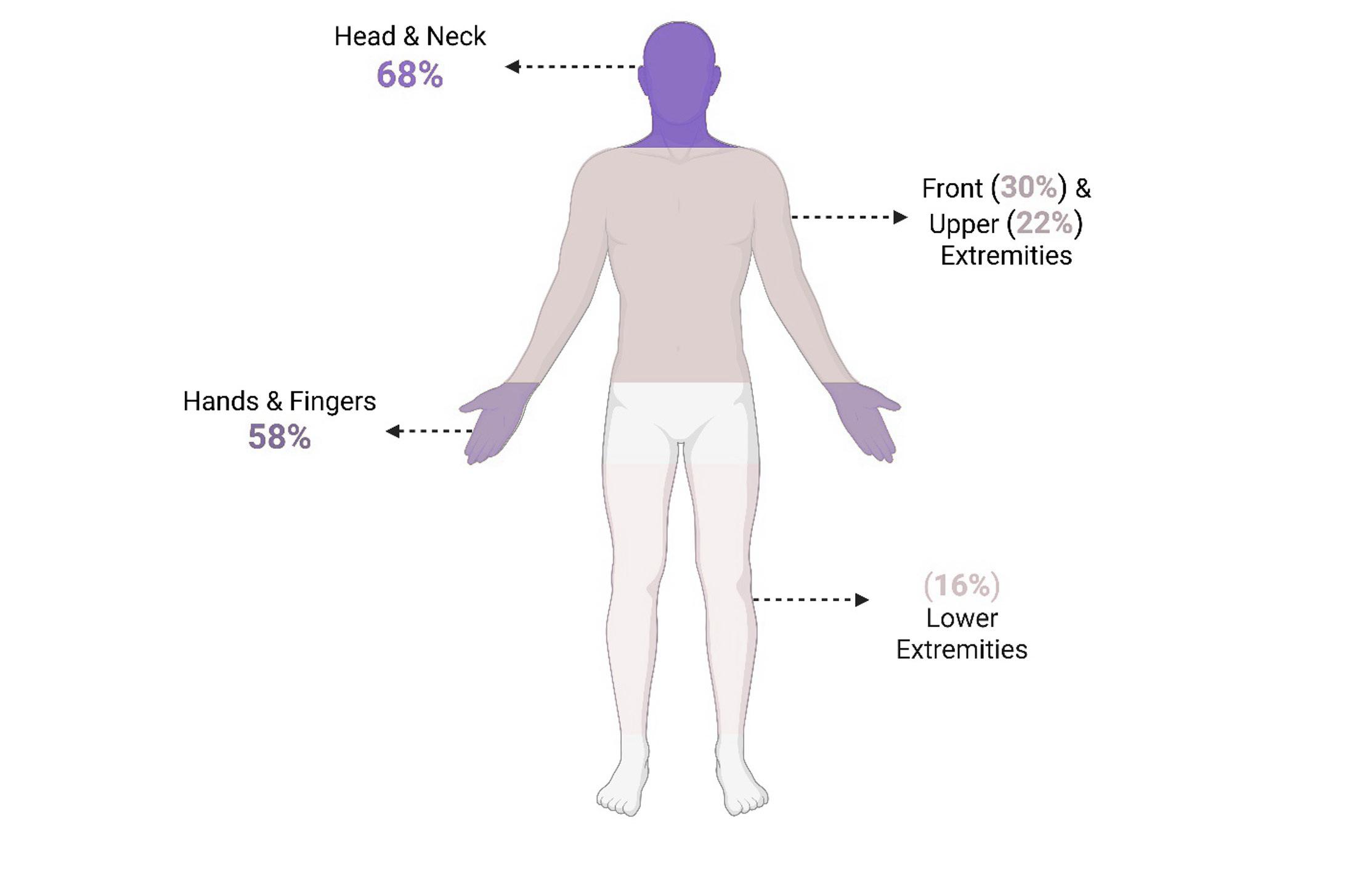
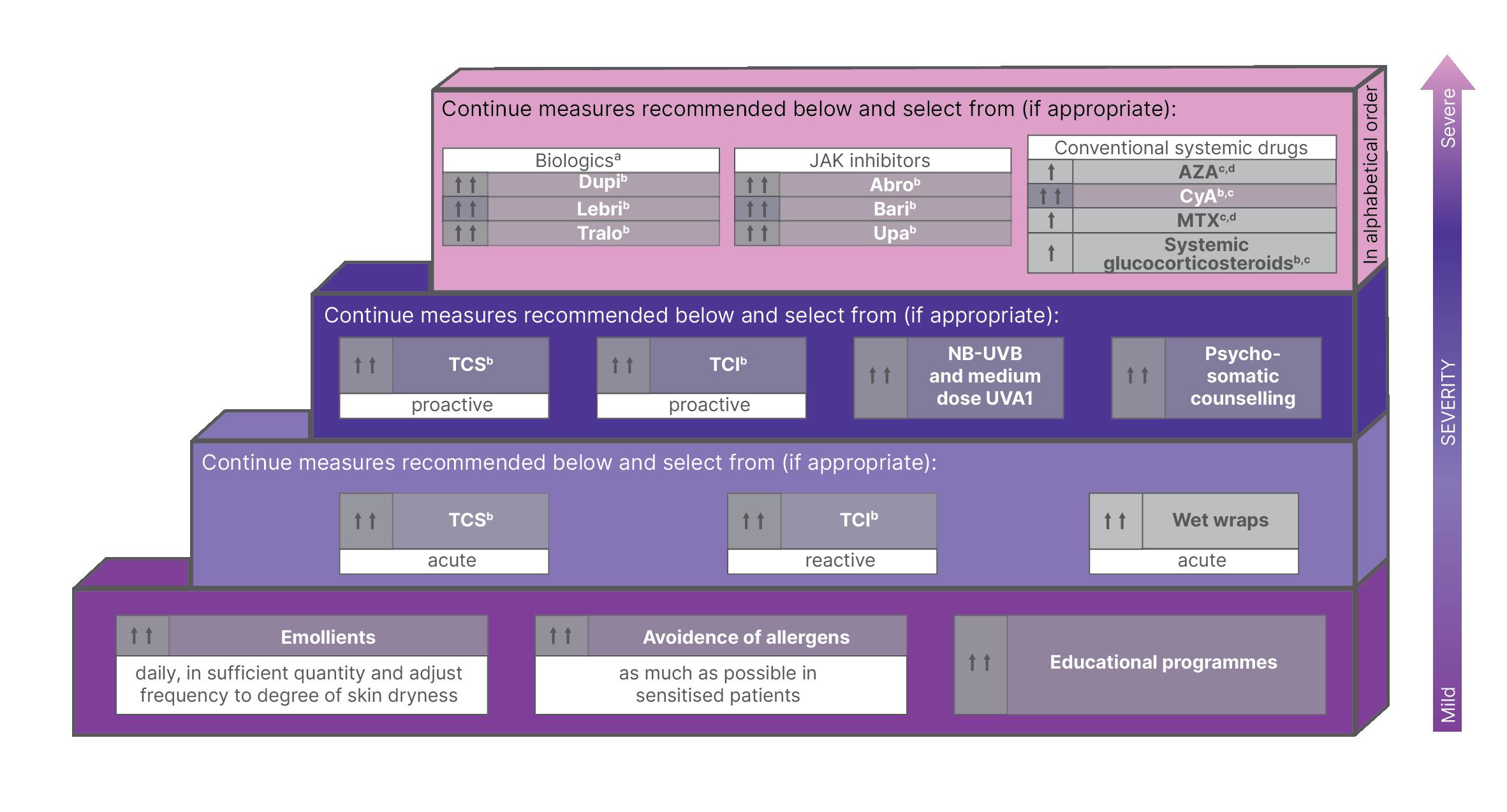
Expert Roundtable: Understanding and Managing High-Burden Areas in Atopic Dermatitis
Association Between Androgenetic Alopecia Severity and Sexual Dysfunction in Male and Female Patients Attending a Dermatology Clinic: An Exploratory Cross-Sectional Study
Özkoca D et al.
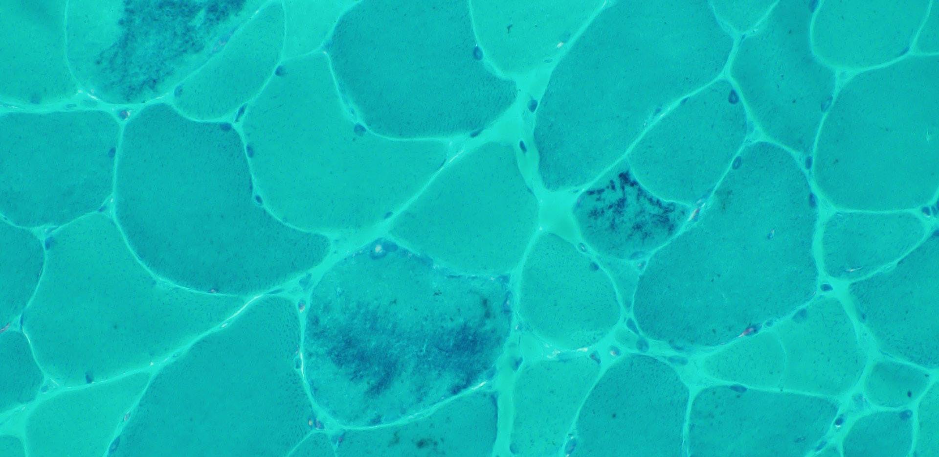
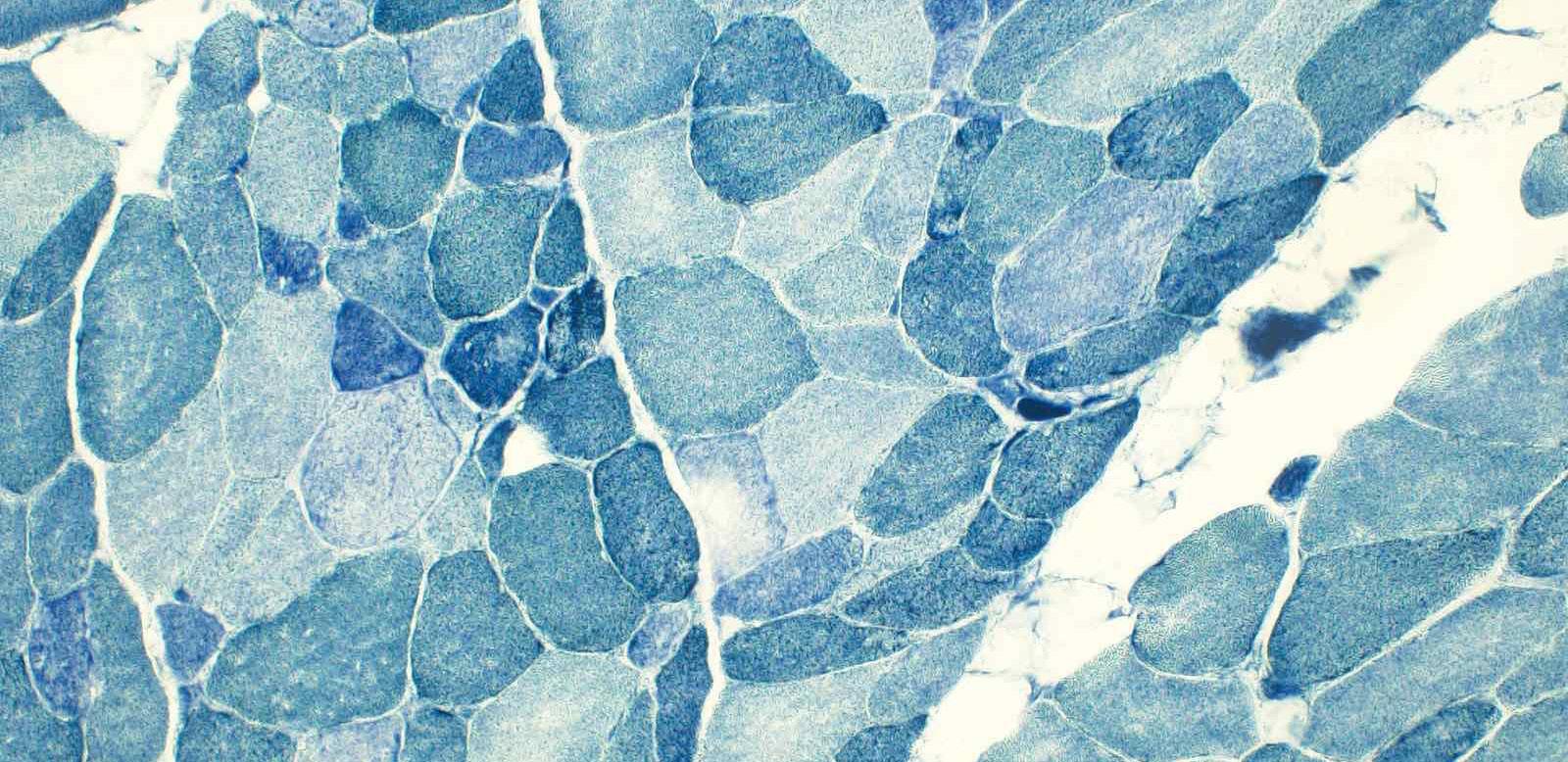
Myofibrillar Myopathy with a Novel LDB3 Variant (c.1804T>C) in a Filipino Mother–Daughter Dyad: A Case Report
Trivilegio MJH et al.
Cervical Screening and HPV Vaccination in Kidney Transplant Recipients
Ward
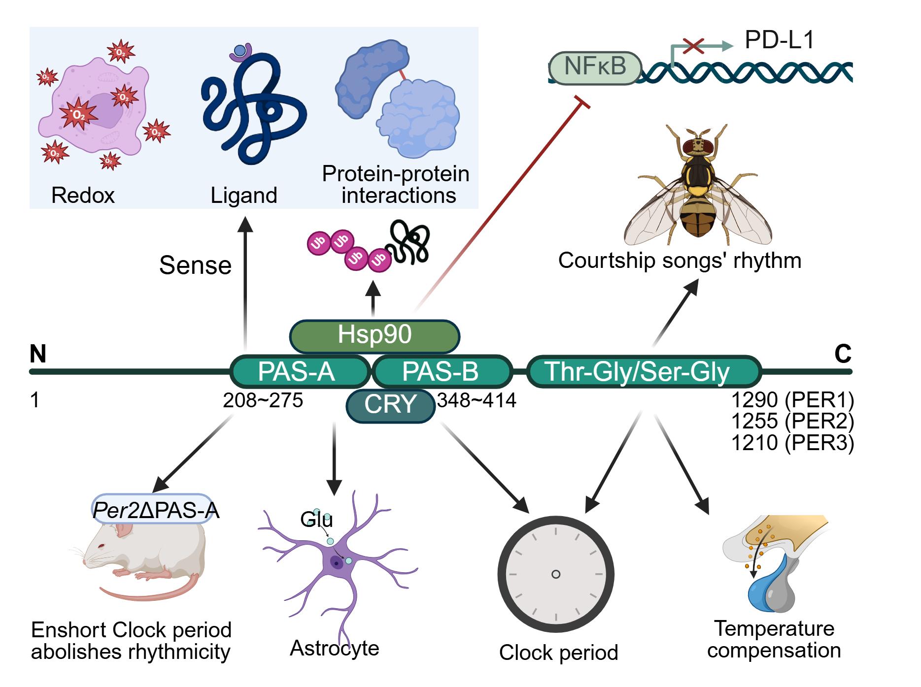
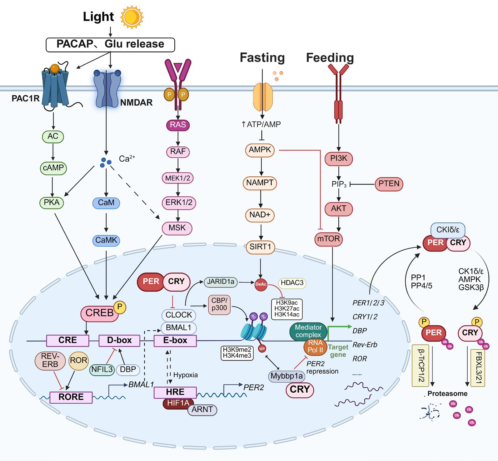
The PER Gene Family: Pivotal Regulators Bridging Circadian Clock Disruption to Multifactorial Pathologies
Li R et
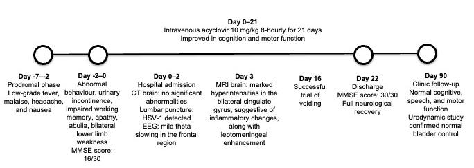
Herpes Simplex Virus Encephalitis Presenting as Frontal Lobe Syndrome: A Case Report
Ngan HT et al.
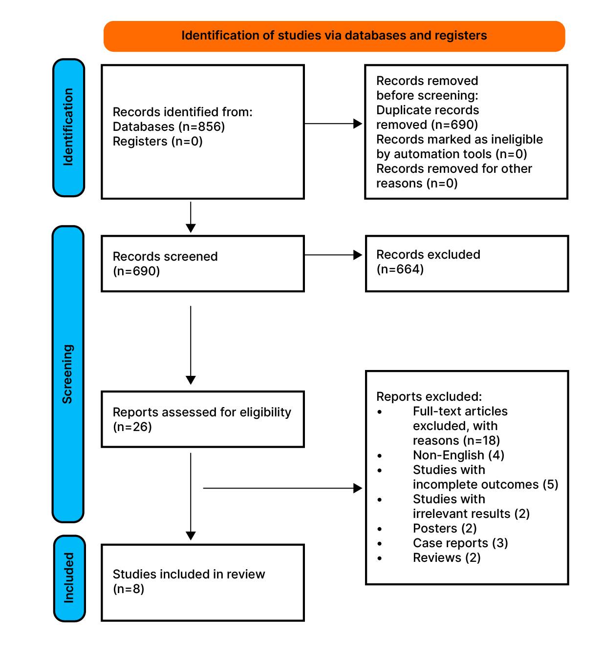
Broncho-Alveolar Lavage for Diagnosis of Pneumocystis jirovecii Pneumonia: A Scoping Review Khan and Osama
"Treatment decisions should be based not only on clinical factors, but also on the morphological features of the excised tissue"
Editorial Board
Editor-in-Chief
Markus Peck-Radosavljevic
Klinikum Klagenfurt am Wörthersee, Austria
Current Chairman and Head of the Department of Gastroenterology and Hepatology, Endocrinology, Rheumatology and Nephrology at Klinikum Klagenfurt am Wörthersee, with expertise in portal hypertension, hepatocellular carcinoma, and HIV-HCV coinfection.
Ahmad Awada
Chirec Hospital Group, Belgium
Prof Sorin T. Barbu
“Iuliu Hațieganu” University of Medicine and Pharmacy, Romania
Prof Abdullah Erdem Canda
Koç University Hospital, Türkiye
Dr Pierfrancesco Agostoni
Ziekenhuis Aan de Stroom (ZAS) Middelheim, Belgium
Dr Fernando Alfonso
Hospital Universitario de La Princesa, Spain
Dr Emanuele Angelucci
IRCCS Ospedale Policlinico San Martino, Italy
Dr George Anifandis
University of Thessaly, Greece
Prof Riccardo Autorino
Glickman Urological Institute, USA
Dr Mátyás Benyó
University of Debrecen, Hungary
Prof Andrew Bush
Prof Ian Chikanza
Harley Street Clinic, UK
Prof Lászlo Vécsei
University of Szeged, Hungary
Imperial College London, UK
Dr Hassan Galadari
United Arab Emirates University, United Arab Emirates
Dr Amir Hamzah Abdul Latiff
Pantai Hospital, Malaysia
Dr Lorenz Räber
Bern University Hospital, Switzerland
Aims and Scope
EMJ, the flagship journal of the EMJ portfolio, is an openaccess, peer-reviewed eJournal, committed to elevating the quality of healthcare globally by publishing high-quality medical content across the 18 clinical areas covered in our portfolio. The journal is published quarterly and showcases the latest developments across these clinical areas.
EMJ publishes peer-reviewed research papers, review articles, and case reports across all therapy areas of the EMJ portfolio. In addition, the journal publishes features and opinion pieces create a discussion around key topics in the field and broaden readers’ professional interests. The journal also features interviews with leading experts in various clinical disciplines.
The journal covers advances within the pharmaceutical arena by publishing sponsored content from congress symposia, which is of high educational value for healthcare professionals. This undergoes rigorous quality control checks by independent experts and the in-house editorial team.
EMJ endeavours to increase knowledge, stimulate discussion, and contribute to the delivery of world-class updates in the clinical realm. We do not publish veterinary science papers or laboratory studies that are not linked to patient outcomes. Further details on coverage can be found here: www.emjreviews.com
Editorial Expertise
EMJ is supported by various levels of expertise:
• Guidance from an Editorial Board consisting of leading authorities from a wide variety of disciplines.
• Invited contributors who are recognised authorities in their respective fields.
• Peer review, which is conducted by expert reviewers who are invited by the Editorial team and appointed based on their knowledge of a specific topic.
• An experienced team of editors and technical editors.
• A team of internal and independent medical writers.
Peer Review
Every review article, case report, feature, and research article published in EMJ undergoes peer review by at least two independent experts.
On submission, all manuscripts are assessed and undergo a technical check by the EMJ Editorial staff to determine their suitability for the journal and appropriateness for peer review. Editorial staff identify appropriate reviewers who are selected based on their specialist knowledge in the relevant area. All peer review is double-blind.
Following review, manuscripts are either accepted without modification, returned to the author(s) to incorporate required changes, or rejected. Editorial staff are responsible for ensuring that necessary amendments to the manuscript have been made, with input from our Editorial Board or the original reviewers where necessary. The Editor of EMJ has final discretion over any proposed amendments. Manuscripts authored by members of the Editorial Board are subjected to the same double-blind process. Short opinion pieces are published following internal review and publication is at the discretion of the Editor. Congress-associated content authored by the EMJ Editorial staff undergoes internal quality control checks. Congress-related content sponsored or funded by our industry partners undergoes quality control checks independently. Industry-supported content that falls into any of
the categories that are eligible for peer review, undergoes the same peer review process.
Submissions
We welcome contributions from professionals, consultants, academics, and industry leaders on relevant and topical subjects. We seek papers with the most current, interesting, and relevant information in each therapeutic area and accept original research, review articles, case reports, and features.
We are always keen to hear from healthcare professionals wishing to discuss potential submissions, please email: editorial.assistant@emjreviews.com
To submit a paper, use our online submission site: https://emj.kriyadocs.com/submissions/submit/emj/emj/login
Submission details can be found through our website: www.emjreviews.com/contributors/authors
Reprints
All articles included in EMJ are available as reprints (minimum order 1,000). Please contact hello@emjreviews.com if you would like to order reprints.
Distribution and Readership
EMJ is distributed through controlled circulation to healthcare professionals in the relevant fields globally.
Indexing and Availability
EMJ is indexed on DOAJ, the Royal Society of Medicine, and Google Scholar®.
EMJ is available through the websites of our leading partners and collaborating societies.
EMJ journals are all available via our website: www.emjreviews.com
Open Access
This is an open-access journal in accordance with the Creative Commons Attribution-Non Commercial 4.0 (CC BY-NC 4.0) license.
Congress Notice
Staff members attend medical congresses as reporters when required.
All information obtained by EMJ and each of the contributions from various sources is as current and accurate as possible. However, due to human or mechanical errors, EMJ and the contributors cannot guarantee the accuracy, adequacy, or completeness of any information, and cannot be held responsible for any errors or omissions. EMJ is completely independent of any event reviews in this issue and the use of the organisations does not constitute endorsement or media partnership in any form whatsoever. The cover photo is of Greece, the location of work for the primary author of Editor's Pick.
Helena Bradbury, Katrina Thornber, Aleksandra Zurowska
Editorial Co-ordinators
Jess Nicholson, Bertie Pearcey, Alena Sofieva
Editorial Assistants
Niamh Holmes, Josh Warren Lister, Roli Omamuli, Nonyelum Okonkwo, Alexander Perkins
Reporter
Anna Caldwell
Creative Director
Tim Uden
Design Manager
Stacey White
Senior Designers
Tamara Kondolomo, Owen Silcox
Designers
Shanjok Gurung, Fabio van Paris
Junior Designers
Fraser Hoey, Helena Spicer, Caleb Wylie
Head of Marketing
Stephanie Corbett
Chief Executive Officer
Justin Levett
Chief Commercial Officer
Dan Healy
Founder and Chairman
Spencer Gore
Welcome
Dear Readers,
It is my pleasure to welcome you to issue 11.2 of EMJ. This edition focuses on chronic diseases, and explores the impact of ongoing conditions on patient wellbeing, treatment pathways, and the provision of healthcare.
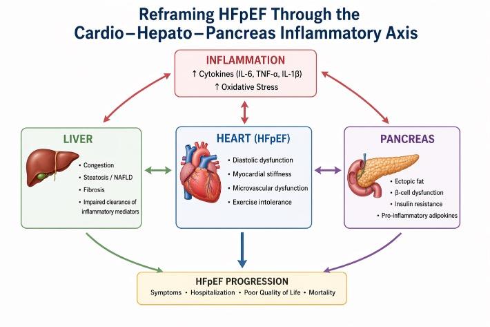
The issue opens with a detailed examination into the endophenotypes of osteoarthritis, biomarker development, and treatment responses, before shifting focus to the diagnosis of pneumocystis pneumonia in patients who are immunocompromised. This theme is expanded across a range of feature articles spotlighting chronic inflammation and insulin resistance, AI in voice disorder therapies, the cardio–hepato–pancreas inflammatory axis, and rheumatoid arthritis.
As ever, EMJ is proud to present two pieces of original research. This issue includes a retrospective cohort study that explores cervical screening and HPV vaccination in kidney transplant recipients, and a cross-sectional study that details the associations between sexual dysfunction and androgenetic alopecia severity. In addition, we present three notable case reports, detailing cases of myofibrillar myopathy in a Filipino mother–daughter dyad, herpes simplex virus Type 1 presenting as frontal lobe syndrome, and disseminated tuberculosis in an immunocompetent adult.
This issue also features interviews with Vikram Patel and Roy Taylor, providing valuable insights into the integration of mental health into broader chronic disease care, and how understanding of disease reversal in Type 2 diabetes could reshape clinical practice.
I would like to extend my sincere thanks to our editorial board, authors, and peer reviewers for their continued dedication and expertise, as well as Katrina Thornber (Editorial Lead, EMJ, London, UK) for her extensive contributions to this issue. I hope you find this issue both insightful and relevant to your clinical practice.
Sean Boyle Editor
Editorial enquiries: editor@emjreviews.com
Sales opportunities: salesadmin@emjreviews.com
Permissions and copyright: accountsreceivable@emjreviews.com
Reprints: info@emjreviews.com
Media enquiries: marketing@emjreviews.com
Stay at the Forefront of Modern Medicine
High-level perspectives. Global experts. Essential updates.
Join us for conversations with the minds shaping healthcare's future. Gain the distilled insights you need to lead in your field and make maximum impact.
Jonathan Sackier: Non Executive Director & CMO, AiM Medical Robotics, Florida, USA
Saranya Ravindran: Paediatric Emergency Medicine Registrar, Imperial College Healthcare NHS Trust
Catherine Glass: Associate NHS GP and Senior Appraiser, NHS England
Foreword
Dear Colleagues,
Welcome to the second EMJ Flagship issue of 2026. This edition focuses on chronic diseases, a theme that extends across every specialty of medicine and continues to shape healthcare systems worldwide.
With an ageing population, the global burden of chronic disease continues to grow. These conditions are no longer viewed solely as isolated diagnoses, but as complex, long-term challenges that influence quality of life, healthcare delivery, and societal wellbeing. From cardiovascular and metabolic disorders to neurological, rheumatological, and psychiatric conditions, chronic diseases demand sustained clinical innovation, multidisciplinary collaboration, and patient-centred care.
In this issue, we explore several important dimensions of chronic disease research and management. Our feature articles examine chronic systemic inflammation and insulin resistance in secondary osteoporosis, the evolving understanding of heart failure with preserved ejection fraction, and rheumatoid arthritis as the final stage of a dynamic disease continuum. Further articles explore the long-term burden and clinical complexity of chronic conditions across diverse therapeutic areas, including neurocognitive, neuromuscular, dermatological, and infectious diseases.
Collectively, they emphasise the diversity and complexity of chronic disease, while reinforcing the importance of continued research, early diagnosis, and integrated care approaches.
With the growing prevalence of chronic conditions worldwide, the challenge facing modern medicine is not only how to treat disease, but how to support long-term health, function, and quality of life for patients over time.
Chronic diseases demand sustained clinical innovation, multidisciplinary collaboration, and patient-centred care
I am grateful to our authors, reviewers, and Editorial Board for their expertise and continuous commitment. I hope this issue encourages thoughtful engagement with both the promise and responsibility that accompany medical advancement.
Prof Markus Peck-Radosavljevic
Professor of Medicine and Chairman, Department of Gastroenterology and Hepatology, Endocrinology, Rheumatology and Nephrology, Klinikum Klagenfurt am Wörthersee, Austria
Managing Endometriosis-Associated Pain: A Two-Step Approach
AT THIS year’s International Society of Gynaecological Endocrinology (ISGE)
Congress 2026, held in Rome, Italy, Vitaly F. Bezhenar, Head of the Department of Obstetrics, Gynaecology, and Neonatology at First Pavlov State Medical University of St. Petersburg, Russia, reviewed the current approach to the management of endometriosis-associated pain. He explored the recommended two-step treatment strategy, compared different postoperative hormonal therapies, and discussed the role of long-term medical management in reducing recurrence and improving quality of life in women with endometriosis.
THE BURDEN AND LONG-TERM MANAGEMENT OF ENDOMETRIOSIS
Recent research indicates that deep infiltrating endometriosis requires a structured approach and long-term postoperative treatment.1 Such treatment helps slow the pathological processes that create an aggressive peritoneal environment, drive cellular changes, and ultimately contribute to recurrence.
Endometriosis has a major impact on women’s quality of life. Around 70% of women with the condition experience chronic pelvic pain of varying severity, and one in two experience infertility.2,3
SURGERY AND RISK OF RECURRENCE
Most patients with endometriosis undergo surgery, although it does not address the underlying cause of the disease.4 Surgery aims to remove as much diseased tissue as possible, relieve pain, improve quality of life, and reduce the risk of recurrence.4
Surgery can lead to complications, particularly when the bowel, bladder, or uterus is involved.4 It may also reduce
ovarian reserve, increasing the risk of iatrogenic premature ovarian insufficiency.4,5 Recurrence rates can reach 50%, even after successful surgery.4,6 Bezhenar noted: “Unjustified repeat surgeries without longterm anti-recurrence therapy may worsen the disease and reduce ovarian reserve.”7,8
POSTOPERATIVE HORMONAL THERAPY
Bezhenar presented data showing that postoperative treatment plays a key role in preventing endometriosis recurrence.9-12 His analysis linked repeat surgery to the absence of hormonal therapy. In the study, fewer than half of patients had not undergone previous surgery, while 14% had one prior operation, 11% had two, and 2% had three or more.9-12
Most recurrences occurred after hormonal therapy was discontinued. Around 80% of patients stopped treatment themselves, while in 22% of cases it was stopped by the gynaecologist.13
TWO-STEP APPROACH
The Russian Society of Obstetricians and Gynaecologists (RSOG) recommends a
combined two-step approach: surgery when indicated, followed by postoperative hormone-modulating therapy for at least 6 months if pregnancy is planned, and longer if it is not.14 Laparoscopic surgery is preferred for deep endometriosis, and treatment should always be individualised.14
Treatment decisions should be based not only on clinical factors, but also on the morphological features of the excised tissue. In Bezhenar’s study, microscopic foci of endometrioid heterotopia were found in 50% of samples taken from macroscopically intact peritoneum.15 Bezhenar concluded: “These findings support the need for postoperative hormonal therapy to prevent disease recurrence.”
CONSERVATIVE MANAGEMENT AND PAIN CONTROL
In a separate study of conservative strategies for endometriosis-associated pain, including non-pharmacological, antiinflammatory, and hormonal treatments, 71% of women reported no symptom improvement.13 A further 23% reported only slight improvement, while significant pain reduction was seen in just 5% of patients.13 Complete pain relief was achieved in only 4%.13
Bezhenar compared these findings with another study of 178 patients who had undergone surgery for endometriosis.11 After surgery, 82 patients (46.1%) received gonadotropin-releasing hormone (GnRH) agonists, 58 (32.6%) received dienogest, and 21 (11.8%) received combined oral contraceptive (COC) pills containing dienogest. A further 17 women (9.5%) were planning pregnancy and therefore received no hormonal therapy. Postoperative treatment lasted 4–6 months.11
The study assessed the proportion of patients reporting significant reductions in endometriosis-associated pain after different treatments. The highest rates were seen with GnRH agonists (40%) and dienogest (38%). Patients receiving these treatments also reported improved quality of life and sexual function.11
PROGESTAGENS AS FIRST-LINE THERAPY
According to the RSOG, progestagens are recommended as first-line therapy.14 They can be given continuously to promote glandular epithelial atrophy and stromal decidualisation, although cyclic use may be considered for patients planning pregnancy.16
Bezhenar and colleagues reported that COCs are not suitable for anti-relapse therapy in endometriosis and should be used only for contraception.17 In their study, patients who received cyclic COCs containing 30 mcg ethinylestradiol (EE) and dienogest had the lowest pregnancy rates and the highest recurrence rates of pelvic endometriosis.
All recurrences after cyclic therapy with 30 mcg EE and dienogest were associated with endometrioid infiltrates identified during repeat laparoscopy that had not been detected at the initial surgery.17
DIENOGEST VERSUS COCs
The authors compared the effects of dienogest 2 mg with those of COCs. Dienogest was associated with a moderate reduction in oestrogen levels while maintaining concentrations within the therapeutic window (30–60 pg/mL).18 It was also shown to reduce several forms of endometriosis-associated pain, including pelvic pain, dysmenorrhoea, dyspareunia, dysuria, and dyschezia.
In contrast, COCs may disrupt the oestrogen–progestin balance when oestrogen doses exceed physiological levels, potentially promoting endometriosis progression.18 Although COCs may reduce dysmenorrhoea, they were not shown to relieve dyspareunia or non-cyclical pelvic pain. Women using COCs for dysmenorrhoea were later diagnosed with severe infiltrative endometriosis.18
QUALITY OF LIFE AND RECURRENCE OUTCOMES
A real-world study showed that dienogest 2 mg was associated with a significant reduction in chronic pelvic pain and improved quality of life in women with endometriosis.19 The ENVISION study included 865 Asian women from 36 clinics across Asia.19
Improvements were reported across all quality-of-life domains during treatment with dienogest 2 mg.19 Quality of life was measured using the Endometriosis Health Profile-30 (EHP-30), which assessed pain, self-control, emotional status, social function, and self-esteem.19 The greatest improvement was seen in pain scores (78.4%), followed by self-control (70.5%), emotional status (61.3%), social function (55.4%), and self-esteem (42.1%).19
A systematic review and meta-analysis found that postoperative treatment with dienogest 2 mg was associated with a significantly lower risk of endometriosis recurrence than no hormonal therapy.20 Recurrence occurred at a rate of 0.89 per 1,000 woman-months in women receiving dienogest, compared with 5.46 per 1,000 woman-months in controls who received no therapy.20
References
1. Gordts S et al. Pathogenesis of deep endometriosis. Fertil Steril. 2017;108(6):872-85.
2. Dunselman GA et al. ESHRE guideline: management of women with endometriosis. Hum Reprod. 2014;29(3):400-12.
3. Ministry of Health of the Russian Federation. Clinical guidelines: endometriosis. 2024. Available at: https://drive.google.com/file/d/14XuaZ zgwocmmXOXVzeVT60QK4WokgbpH/ view. Last accessed: 13 May 2026.
4. Chapron C et al. Rethinking mechanisms, diagnosis and management of endometriosis. Nat Rev Endocrinol. 2019;15(11):666-82.
5. Gusev DV et al. Endometriosisassociated infertility: various aspects of pathogenesis. Gynecology. 2020;22(3):34-8.
6. Ouchi N et al. Recurrence of ovarian endometrioma after laparoscopic
Over a mean follow-up of 28.5 months, only two recurrences per 100 treated women were reported in the dienogest group. By comparison, 29 recurrences per 100 women were observed over a mean follow-up of 35.7 months in women who received no treatment.20
Overall, the probability of recurrence after surgery was significantly lower in patients treated with dienogest than in those who did not receive hormonal therapy (p<0.001).20
CONCLUSION
Bezhenar concluded that endometriosis is a chronic, inflammatory, hormone-dependent condition that requires long-term, comprehensive management, including postoperative therapy. Progestogens remain the first-line treatment, and continuity between inpatient and outpatient care is essential to optimise outcomes and reduce recurrence risk. He emphasised that postoperative therapy should continue for at least 6 months, and that there is no such thing as ‘mild’ endometriosis.
excision: risk factors and effect of postoperative hormonal treatment. J Obstet Gynaecol Res. 2014;40(1):230-6.
7. Harada M et al. High incidence of endometriosis in infertile women with a history of surgically treated benign ovarian tumors. Gynecol Endocrinol. 2011;27(9):717-20.
8. Shah A et al. Catamenial pneumothorax: a rare case of thoracic endometriosis. Abstract A6687. ATS International Conference, 18-23 May, 2018.
9. Adamyan LV et al. Endometriosis: diagnosis, treatment, and rehabilitation. Clinical guidelines. 2013. Available at: https://minzdrav.alania. gov.ru/sites/minzdrav/files/media/ documents/files/2019-07/00002_ Эндометриоз%202013.pdf. Last accessed: 13 May 2026.
10. Bezhenar VF. Hormonal therapy in the treatment of endometriosis. Innovative Pharmacotherapy. 2022;2(6):9-12.
11. Bezhenar VF et al. The rationale for long-term hormonal therapy of endometriosis after surgical treatment. Obstetrics and Gynecology (Russia). 2021;4:134-42.
12. European Society of Human Reproduction and Embryology (ESHRE). Endometriosis guideline. 2022. Available at: https://www.eshre. eu/Guideline/Endometriosis. Last accessed: 13 May 2026.
13. Bezhenar et al. Analysis of the effectiveness of various management approaches in patients with endometriosis and rationale for long-term personalized dydrogesterone therapy within a combined treatment strategy. Prob Reprod. 2024;30(2):102-11.
14. Sukhikh GT et al. Algorithms for the management of the patients with endometriosis: a consensus statement of experts from the Russian Society of Obstetricians and Gynecologists. Akush Ginekol. 2023;5:159-76.
15. Rybakova MG et al. Morphological characteristics of surgical specimens in nerve-sparing and conventional techniques for deep infiltrating endometriosis. J Obstet Women's Dis. 2019;3-4:26-31.
16. Busacca M et al. Guidelines for diagnosis and treatment of endometriosis. Ital J Gynaecol Obstet. 2018;30(2):7-21.
17. Bezhenar VF et al. Comparison of the effectiveness of various immunomodulatory therapy regimens after surgical treatment of external genital endometriosis. Prob Reprod. 2015;4:89-98.
18. Murji A et al. Use of dienogest in endometriosis: a narrative literature review and expert commentary. Curr Med Res Opin. 2020;36(5):895-907.
19. Techatraisak K et al. Effectiveness of dienogest in improving quality of life in Asian women with endometriosis (ENVISIOeN): interim results from a prospective cohort study under real-life clinical practice. BMC Womens Health. 2019;19:68.
20. Zakhari A et al. Dienogest and the risk of endometriosis recurrence following surgery: a systematic review and metaanalysis. J Minim Invasive Gynecol. 2020;27(7):1503-13.
Championing Women’s Voices in Menopause: Broadening the Clinical Lens
This symposium, organised by the Women's Health Academy, which is funded and facilitated by Bayer, took place on 6th March 2026 as part of the International Society for Gynecological Endocrinology (ISGE) Annual Congress held in Rome, Italy
Support: This non-promotional article has been funded and facilitated by Bayer and is intended for healthcare professionals only. This article includes the views, opinions, and quotations as expressed by the symposium speakers.
Chairperson: Andrea R. Genazzani1
Speakers: Rossella E. Nappi,2,3 Claudio N. Soares,4 Michelle Jacobson5,6
1. Department of Clinical and Experimental Medicine, Division of Obstetrics and Gynecology, University of Pisa, Italy
2. Department of Clinical-Surgical, Diagnostic and Paediatric Sciences, University of Pavia, Italy
3. IRCCS San Matteo Foundation, Pavia, Italy
4. Queen’s University School of Medicine, Kingston, Canada
5. Coven Women’s Health, Toronto, Canada
6. Department of Obstetrics and Gynaecology, University of Toronto, Canada
Disclosure:
Genazzani has been a member of the speaker’s bureau for Abbott, AlfaSigma, Bayer, Besins Healthcare, Bionorica, Exeltis, Gedeon Richter, Mithra, Organon, Sérélys, Se-cure, Theramex, and Viatris.
Nappi has received consulting fees from Besins Healthcare, Fidia, and Vichy Laboratories; and honoraria from Abbott, Astellas, Bayer Healthcare, Exeltis, Fidia, Gedeon Richter, Ibsa, Merck & Co, Novo Nordisk, Shionogi Limited, Theramex, and Viatris.
Soares has received research grants from Atai Therapeutics, Clairvoyant, Canadian Institutes of Health Research, Diamond Therapeutics, Eisai, Ontario Brain Institute, Ontario Centre of Innovation, Otsuka, and SEAMO; and has been an advisory board member for AbCellera, Astellas, Bayer, Eisai, Idorsia, Lundbeck, and Otsuka.
Jacobson has received research grants from the Canadian Institutes of Health Research and the United States Department of Defense; and consulting fees from AbbVie, AbCellera, Astellas, Bayer, Biosyent, Duchesnay, Eisai, Hologic, Idorsia, Knight, Kye, Lilly, Lupin, Merck, Pfizer, Sanofi, and Searchlight.
Acknowledgements: Writing assistance was provided by Helen Boreham, HB Medical (UK) Ltd, Wetherby, UK.
Disclaimer: The Women’s Health Academy is funded and facilitated by Bayer. All case studies presented are fictitious; patient symptoms may vary. This material is intended for healthcare professionals only.
Keywords: Counselling, hot flushes, individualised care, menopause, quality of life, sleep disturbances, vasomotor symptoms (VMS), Women’s Health Academy.
Citation: EMJ. 2026;11[2]:14-22.
https://doi.org/10.33590/emj/755OG471
Meeting Summary
PARTNERSHIP
During this symposium at the International Society for Gynecological Endocrinology (ISGE) Annual Congress, leading experts in women’s health and members of the Women’s Health Academy explored the spectrum of menopause symptoms and highlighted the need for an individualised and woman-focused approach to menopause care in clinical practice. The Women’s Health Academy is a multidisciplinary expert group that aims to improve women’s healthcare by advancing education, expanding access to evidence-based resources, and putting women at the centre of care.
Rossella Nappi, Professor at the University of Pavia in Italy, began the session by underscoring the impact of menopause on women’s overall health and quality of life, and the importance of integrated symptom recognition that includes vasomotor symptoms (VMS), sleep, mood, and cognitive function. The expert panel, including Andrea Genazzani, Professor at the University of Pisa, Italy; Claudio Soares, Professor from Queen’s University School of Medicine in Kingston, Canada; and Michelle Jacobson, Assistant Professor from the University of Toronto, Canada, then discussed a selection of fictional patient cases. These cases illustrate the need for consultations centred on women’s goals, expectations, and needs to determine the best path to symptom relief and long-term health outcomes. For clinical practice, the experts recommended adopting a simple three-step approach to menopause counselling: 1) identify key symptoms and concerns; 2) focus on symptom relief; and 3) tailor the approach to the individual patient.
The Women’s Health Academy
This symposium at ISGE was delivered by the Women’s Health Academy, a group comprising over 30 international multidisciplinary experts that is dedicated to improving reproductive health and menopause outcomes for women.1 The Women’s Health Academy is funded and facilitated by Bayer and governed by an independent Executive Committee.
As Nappi explained, the role of the Women’s Health Academy is to champion women’s healthcare, fostering community and knowledge sharing among healthcare professionals and women to shape future directions in care. By prioritising women’s voices, it aims to create an environment in which women feel informed and empowered throughout their lives, from puberty to
menopause and beyond. As part of its mission, the Women’s Health Academy seeks to address the issues that restrict women’s access to healthcare, challenge taboos, and support the next generation of healthcare professionals in delivering clinical best practice, including menopause care.1
Menopause Symptoms
Every woman is different, and menopause is a personal experience, stressed Nappi. Women may present with a variety of symptoms, which are sometimes misdiagnosed as being unrelated to menopause and which women themselves may accept as an inevitable part of the ageing process.1,2
Common symptoms of menopause include VMS, e.g., hot flashes during the day and/ or night sweats at night, and difficulty sleeping. Vaginal symptoms of menopause such as dryness and painful intercourse can occur. Women can also experience menstrual changes as they move towards menopause, including irregular or abnormal bleeding and heavy bleeding, which may result in iron deficiency and anaemia. Menopause can also be associated with musculoskeletal symptoms such as joint pain and soreness.1,3
Beyond the physical manifestations, menopause is linked to a range of associated psychological symptoms, which may include anxiety, poor memory, inability to concentrate, depressed mood, irritability, decreased sexual desire, sleep dysfunction, and impaired cognitive function (commonly known as brain fog).1,3
The Impact of Menopause
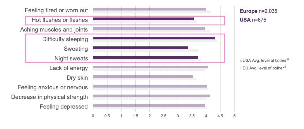
Nappi highlighted how the symptoms of menopause can impose a significant burden on patients.1 An international survey conducted from December 2019–February 2020, which included 2,035 women in Europe, 675 in the USA, and 750 in Japan, all aged 40–65 years, assessed the prevalence of symptoms associated with menopause and their impact on quality of life and daily functioning.4
Women ranked their symptoms on a scale from 0 (not bothersome) to 6 (extremely bothersome), with highly bothersome denoted by a score of 4 or above. VMS and sleep disturbances were identified as among the most common and/or disruptive menopause symptoms in this survey among women from Europe and the USA (Figure 1).1,4 Sleep problems were considered to be a highly bothersome menopause symptom, with a ranking of 4.4
ᵃRanked from 0 (not bothersome) to 6 (extremely bothersome), ≥4 is highly bothersome. Note that the actual base varies per symptom. Only selected symptoms are shown above. Dark purple bars denote VMS and sleep-related symptoms in the USA population.
Adapted from Nappi et al.4
Avg: average.
Figure 1: International survey of menopause symptoms and associated level of bother.1,4
in Europe and 4.3 in the USA. Hot flushes or flashes, the hallmark VMS of menopause, were ranked over 3.5 on the bothersome scale in both Europe and the USA. Women in Europe experienced an average of 4.6 episodes of hot flushes/flashes and/or night sweats per day compared with 3.8 episodes per day in the USA.4
As Nappi outlined, the prevalence of VMS associated with menopause is influenced by many factors. These include psychological symptoms, smoking, diet, and BMI. VMS can also be impacted by external factors such as climate and the cultural context in which the woman lives.1,5-16
In addition to VMS, sleep disturbances are experienced by many women during menopause. Sleep disturbances are often associated with VMS but can be unrelated. Estimates indicate that between 40–60% of women suffer from disturbed sleep during the menopausal transition.17,18 Nappi stressed the importance of addressing sleep disturbances in midlife women due to their negative impact on physical and mental health, and quality of life.19 Sleep
health is also recognised as one of the eight key pillars of cardiovascular (CV) health.20 During menopause, sleep disturbances are particularly associated with an increased number of nighttime awakenings and wakefulness after sleep onset, causing more sleep fragmentation.21,22 The prevalence of these menopause-related sleep issues appears to be broadly similar across different countries and ethnicities.4
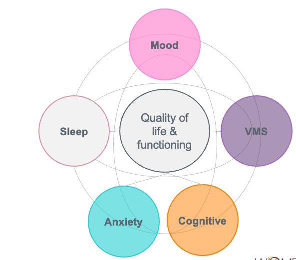
In order to better help patients, healthcare professionals need to understand the interconnected symptom burden during the menopausal transition, Nappi explained (Figure 2).1 VMS, mood alterations, sleep disturbances, anxiety, and cognitive symptoms can frequently overlap.1,23,24
Menopause symptoms also have a direct effect on women’s daily lives, impacting them socially, economically, and functionally.2,25,26 The best way to address this symptomatology is therefore using a biopsychosocial model, Nappi suggested.1
Figure 2: The interconnected symptom burden during the menopausal transition.1,23
VMS: vasomotor symptoms. Anxiety
Menopause Counselling
A major goal of the Women’s Health Academy is to empower women to actively participate in the management of their own menopause care.1,27 This is particularly important as menopause can mark a critical period in a series of risk factors related to cardiometabolic, vascular, musculoskeletal, and mental health, Nappi elaborated.1 Empowering women to participate and make decisions about their own menopause management requires healthcare professionals to provide access to realistic and balanced information, and challenge misconceptions around menopause related to stigma and gender-based ageism.1,27
As part of the empowerment process, Nappi stressed that it is important for healthcare professionals to recognise that many women will require help to address their menopause-related symptoms. Access to a supportive, well-informed clinician willing to listen and provide treatment as needed is therefore key. Decisions around menopause treatment should be made as part of a well-balanced and shared decision-making process. Setting realistic expectations from treatment is also crucial, Nappi pointed out.1
To facilitate women-centric menopause counselling in clinical practice, Nappi proposed a series of key principles for healthcare professionals to consider. Firstly, it is important to recognise the heterogeneous presentation of menopause symptoms. For the individual patient, the priority is to identify the most disruptive symptoms that require immediate attention. Healthcare professionals should provide personalised, evidence-based counselling, and consider whether any symptoms may have a more serious underlying cause. As part of this process, it is also important to offer tailored support strategies and personalised conversations, considering the women’s specific needs and preferences.1
Ultimately, the goal should be to facilitate women-focused menopause counselling through three key steps, Nappi summarised (Figure 3).1 Firstly, identify the most disruptive symptoms and the biggest health concerns. Secondly, focus on symptom relief using the most effective options for
those symptoms and concerns. Thirdly, tailor the approach based on the patient’s preferences and nuances from their own medical history to achieve symptom relief and treatment adherence.1
To illustrate the application of this 3-step menopause counselling model in clinical practice, experts presented and discussed a series of fictional case studies.1 “This approach is quite simple but it is novel, designed to help you as the clinician and your patient,” Jacobson explained.1
These are fictitious cases and proposed outcomes are based on clinician experience only. Individual cases may vary.
Case Study 1: Postmenopausal Woman with Genitourinary Syndrome of Menopause and Sexual Dysfunction
Firstly, Nappi presented the case of the fictional patient Elizabeth, a 68-year-old postmenopausal White woman who had her last menstrual period 16 years ago.1 The patient is retired, has two children, is a non-smoker, and has a BMI of 27 kg/m2 with gynoid weight distribution. Medical history reveals no relevant underlying conditions or gynaecological history, although she has a history of recurrent urinary tract infections and has previously received menopause hormone therapy (MHT) for VMS.1
Nappi outlined the main symptoms experienced by this patient as being genitourinary symptoms, including stress incontinence, sexual dysfunction, and frequent urination at night. Genitourinary symptoms are responsible for daily discomfort (not only during periods of active urinary tract infection), including irritation and pain. Stress incontinence has led to feelings of embarrassment, and she has low mood due to sexual dysfunction. Frequent urination at night also contributes to her fragmented sleep. As Nappi explained, the concern with this patient is that her symptoms may worsen over time without treatment, leading to detrimental effects on her long-term psychological wellbeing and quality of life. The patient herself has expressed a desire to seek therapy and is open to all eligible treatment options.1
1. Identify symptoms/concerns
What are the most disruptive symptoms you are experiencing?
What are your biggest health concerns at the moment?
2. Focus on relief
What are the most effective options for the relief of those symptoms/concerns?
The expert panel discussed the application of the 3-step counselling model to this patient. For Step 1, experts emphasised the importance of deciphering the highest priority symptom from the ‘laundry list’ of menopause-associated symptoms that women often present with. Given the limited consultation time allocated to each patient, Jacobson suggested that these questions could be posed to women in advance if possible, for example, through questionnaires/surveys or in nurse-led consultations. “Preparation in advance can lead to a more fulsome and productive consultation,” she advised. For this particular patient, the panel agreed that genitourinary syndrome of menopause symptoms, and associated low mood and sleep disturbances, appeared to be having the greatest impact on quality of life.1
“Once we know what is bothering the patient, we don’t need to discuss every treatment option for menopause but focus on treatment that is going to improve her symptoms,” Jacobson asserted. In Step 2 for this patient, Nappi indicated that for genitourinary syndrome of menopause symptoms she would recommend pelvic floor exercises, alongside other treatment options as appropriate (Nappi, personal communications).1,28
3. Tailor the approach
What is the option that addresses her needs and preferences (also the nuances from her own medical history) to rapidly achieve relief and treatment adherence?
Step 3 involves tailoring treatment to each individual woman. For example, Nappi suggested that, in her experience, if the patient had comorbid rheumatoid arthritis and lacked dexterity, then oral MHT therapy may be preferrable to topicals.1 “These nuances can guide treatment,” Nappi advised (Nappi, personal communications).1
Case Study 2: Perimenopausal Woman with VMS and Sleep Disturbances Impacting Mood
The second fictional case study presented by Soares was Carol, a 45-year-old perimenopausal Black woman.1 She had made an appointment with her primary care physician after experiencing severe VMS, sleep disturbances, and mood changes over the past year. The patient works full time and has two children, aged 11 years and 13 years. Her last menstrual period was 6 months ago and she has no underlying relevant conditions or gynaecological history. She is a non-smoker with a BMI of 33 kg/m2 and an android weight distribution.1
In terms of symptoms and their impact, the patient is experiencing sleep disturbances 3–4 times per week, and severe VMS 5–6 times per week. The specific number of hot flashes per day is irrelevant here, Soares
Figure 3: Recommended 3-step approach to menopause counselling.1
explained, if the patient is reporting that she is significantly bothered by them. During the transition to menopause, it is important to “respect patients’ wisdom to know what is affecting their quality of life more than we do,” Soares stressed. The hot flashes during the day are causing embarrassment and discomfort at work, and interrupted sleep at night. In turn, interrupted sleep is impacting the patient’s ability to work and complete simple day-to-day tasks and is lowering mood. Together, these symptoms are impacting her long-term psychological wellbeing and quality of life, compromising relationships and work performance.1
Again, the panel reiterated the importance of asking the right questions to ensure that the most disruptive symptoms associated with menopause are not missed. For this case study, the panel observed that the main bothersome symptom appears to be VMS. Hot flashes are in turn having a detrimental effect on the patient’s sleep. Soares confirmed that further tests may be required in some instances to explore other potential causes, but advice on sleep hygiene and treatment options to relieve VMS should still be offered as a key first step. For example, sleep apnoea affects approximately 35% of pre-menopausal women, impacting sleep.29 It is the job of the healthcare professional to explore this and other potential comorbid conditions, Soares reiterated.1
As this patient is receptive to therapeutic options, the panel agreed that treatment should focus on the relief of VMS, which have been identified as the most impactful symptom. In response to an audience question, Soares advised against simply prescribing magnesium for this patient due to insufficient evidence for its role as a sleep aid.30 Rather, he highlighted that the primary goal of treatment should be to minimise VMS, as this was indicated by the patient as the most bothersome symptom.1
Case Study 3: Postmenopausal Woman Recovering from Breast Cancer
Finally, Jacobson presented the fictional case of Helen, a 52-year-old Asian woman recovering from breast cancer and receiving ongoing treatment with tamoxifen.1 She has
been menopausal for 1 year (unrelated to breast cancer treatment), has no known gynaecologic conditions or cardiovascular disease risk factors (gynoid weight distribution and BMI of 18.5 kg/m2), but has a family history of breast cancer. The patient works part time, has one adult child, and is a non-smoker. She has sought help from her primary care physician after struggling with VMS, characterised by daily hot flashes and sleep disturbances.1
Cancer survivorship is a complicating factor in this case, Jacobson stressed, and it is important to know the full details of the patient’s breast cancer history and their risk tolerance in order to recommend appropriate treatment. To this end, a detailed breast cancer history should be taken during the appointment. In particular, it is important to establish the chronology and timing from diagnosis, as women’s attitudes to survivorship may change over time. For many women who have recently undergone treatment for breast cancer, their key priority is to avoid any interventions that will increase the risk of recurrence, Jacobson emphasised. Understanding recurrence risk is therefore vital in order to make suitable menopause treatment recommendations. Jacobson highlighted features of the breast tumour that are important in this regard, such as stage (including size of tumour and presence of negative or positive axillary nodes); oestrogen receptor (ER), progesterone receptor (PR), and human epidermal growth factor receptor 2 (HER2) status; oncotype score (if applicable); and results of any multigene panel tests. She stressed that establishing the type of breast cancer treatment the patient received and the duration of any ongoing treatment is also vital to direct suitable care for menopause symptoms (Jacobson, personal communications).1
Most importantly for Step 1 of this postcancer menopause consultation, we need to know what symptoms the patient is having and which of these are impacting her most, Jacobson reiterated. In this case, the patient is experiencing menopause symptoms, including severe VMS and sleep disturbances (4–5 times per week),
as well as anxiety and fear over breast cancer recurrence. She feels exhausted and irritable after daily VMS and frequent awakenings at night. These menopause symptoms are also contributing to worsening anxiety and fear over recurrence of breast cancer, which may impact the patient’s long-term psychological wellbeing and quality of life.1
Steps 2 and 3 of the menopause counselling model are paramount in this case, Jacobson explained, as the patient wants treatment to relieve her symptoms and is turning to her physician for advice on what is acceptable. The priority for the patient is concern about breast cancer recurrence, so we must take this into consideration, Jacobson advised, and use this to guide the treatment conversation. In this case, systemic MHT is unlikely to be a suitable treatment pathway due to its potential association with an increased risk of breast cancer recurrence, particularly in women with hormone receptor-positive disease.31 The fact that the patient’s symptoms began after starting tamoxifen therapy suggests that VMS may be due to its anti-oestrogen effects rather than pre-existing VMS prior to breast cancer diagnosis, Jacobson explained (Jacobson, personal communications).1,32
Overall, the panel agreed that in a patient like this with specific contraindications or considerations around MHT, it is important to tailor the approach and focus on the most bothersome symptom to gain maximum quality-of-life improvement. By starting with treatment for the most impactful symptoms, in this case VMS, it may be possible to improve many of the symptoms that are “downstream on the patient’s list,” Jacobson advised. Panel members pointed out that patients who have had cancer often think that adverse events associated with breast cancer treatment must be accepted in order reduce cancer recurrence risk. It is
References
1. Genazzani AR et al. Championing women’s voices in menopause: broadening the clinical lens. Symposium. ISGE Annual Congress, 4-6 March, 2026.
2. Aljumah R et al. An online survey of postmenopausal women to determine
therefore important to explain that options are available to treat menopause symptoms in these cohorts of patients.1
Summary
Menopause is a highly individual experience that requires tailored solutions. Treatment approaches should therefore be personalised and guided by a woman’s symptoms, risk profile, health history, and personal preferences, rather than by recommendations applied uniformly to all women. A 3-step approach to menopause counselling should be adopted to identify both the physical and psychological symptoms of menopause, respecting the diversity of the woman’s experiences and needs. Step 1 is to identify the most disruptive symptoms and biggest health concerns, Step 2 is to focus on effective options for symptom relief, and Step 3 is to tailor the approach to the patient’s individual needs and preferences.1
It is important to individualise treatment, as there is no ‘one-size-fits-all’ approach for menopause care, Soares reiterated. The 3-step model is designed to enable clinicians “to efficiently and humanly tailor your menopause consultation and treatment approach to the patient who is in front of you,” he concluded.1
This symposium was highly participative, encouraging attendees to rethink the way they usually approach menopause care and inviting them to listen to their patients and prioritise their concerns. Importantly, it also fostered frank discussions among experts from various disciplines in pursuit of more comprehensive, patient-centred care.1
their attitudes and knowledge of the menopause. Post Reprod Health. 2023;29(2):67-84.
3. Talaulikar V. Menopause transition: physiology and symptoms. Best Pract Res Clin Obstet Gynaecol. 2022;81:3-7.
4. Nappi RE et al. Global crosssectional survey of women with
vasomotor symptoms associated with menopause: prevalence and quality of life burden. Menopause. 2021;28(8):875-82.
5. Thurston RC et al. Vasomotor symptoms and menopause: findings from the Study of Women's Health Across the Nation. Obstet Gynecol Clin North Am. 2011;38(3):489-501.
6. Kronenberg F, Barnard RM. Modulation of menopausal hot flashes by ambient temperature. J Therm Biol. 1992;17(1):43-9.
7. Sievert LL, Flanagan EK. Geographical distribution of hot flash frequencies: considering climatic influences. Am J Phys Anthropol. 2005;128(2):437-43.
8. Avis NE et al. Psychosocial, behavioral, and health factors related to menopause symptomatology. Womens Health. 1997;3(2):103-20.
10. Gold EB et al. Lifestyle and demographic factors in relation to vasomotor symptoms: baseline results from the Study of Women's Health Across the Nation. Am J Epidemiol. 2004;159(12):1189-99.
11. Nagata C et al. Soy product intake and hot flashes in Japanese women: results from a community-based prospective study. Am J Epidemiol. 2001;153(8):790-3.
12. Krebs EE et al. Phytoestrogens for treatment of menopausal symptoms: a systematic review. Obstet Gynecol. 2004;104(4):824-36.
13. Geller SE, Studee L. Botanical and dietary supplements for menopausal symptoms: what works, what doesn’t. J Womens Health (Larchmt). 2005;14(7):634-49.
14. Campagnoli C et al. Climacteric symptoms according to body weight in women of different socio-economic groups. Maturitas. 1981;3(3-4):279-87.
15. Whiteman MK et al. Smoking, body mass, and hot flashes in midlife women. Obstet Gynecol. 2003;101(2):264-72.
16. Chiechi LM et al. Climacteric syndrome and body-weight. Clin Exp Obstet Gynecol. 1997;24(3):163-6.
17. Thurston RC et al. Hot flashes and awakenings among midlife women. Sleep. 2019;42(9):zsz131.
18. Salari N et al. Global prevalence of sleep disorders during menopause: a meta-analysis. Sleep Breath. 2023;27:1883-97.
19. Kravitz HM, Joffe H. Sleep during the perimenopause: a SWAN story. Obstet Gynecol Clin North Am. 2011;38(3):567-86.
20. Lloyd-Jones D et al. Life's essential 8: updating and enhancing the american heart association's construct of cardiovascular health: a presidential advisory from the American Heart Association. Circulation. 2022;146(5):e18-43.
21. Baker FC et al. Sleep problems during the menopausal transition: prevalence, impact, and management challenges. Nat Sci Sleep. 2018;10:73-95.
22. Coborn J et al. Disruption of sleep continuity during the perimenopause: associations with female reproductive hormone profiles. J Clin Endocrinol Metab. 2022;107(10):e4144-53.
23. Khalaf A et al. Exploring symptom clusters across the menopausal stages - systematic review and meta-analysis. Sex Reprod Healthc. 2025;45:101137.
24. Soares CN, Shea AK. The midlife transition, depression, and its clinical management. Obstet Gynecol Clin North Am. 2021;48(1):215-29.
25. Cronin C et al. Using digital health technologies to manage the psychosocial symptoms of menopause in the workplace: a narrative literature
review. Issues Ment Health Nurs. 2021;42(6):541-8.
26. Sarrel P et al. Incremental direct and indirect costs of untreated vasomotor symptoms. Menopause. 2015;22(3):260-6.
27. Hickey M et al. An empowerment model for managing menopause. Lancet. 2024;403(10430):947-57.
28. Mercier J et al. Pelvic floor muscle training as a treatment for genitourinary syndrome of menopause: a single-arm feasibility study. Maturitas. 2019;125:57-62.
29. Wang Y et al. Menopause and obstructive sleep apnea: revealing an independent mediating role of visceral fat beyond body mass index. BMC Endoc Disord. 2025;25(1):21.
30. Mah J, Pitre T. Oral magnesium supplementation for insomnia in older adults: a systematic review & metaanalysis. BMC Complement Med Ther. 2021;21(1):125.
31. Poggio F et al. Safety of systemic hormone replacement therapy in breast cancer survivors: a systematic review and meta-analysis. Breast Cancer Res Treat. 2022;191:269-75.
32. Morga A et al. The burden of illness of treatment-induced vasomotor symptoms in individuals with breast cancer: a systematic literature review. J Clin Med. 2025;14(8):2601.
Burden and Mechanism of Chronic Cough in People Living with Idiopathic Pulmonary Fibrosis
Support: The publication of this article was funded by Trevi Therapeutics.
Interviewees: Tejaswini Kulkarni,1 Matthew Drake,2 Steve Ashton3
1. The University of Alabama at Birmingham, USA
2. Oregon Health and Science University, Portland, USA
3. Person living with IPF, Ohio, USA
Disclosure: Kulkarni has received consultation/advisory board fees from Avalyn Pharma, Brainomix, Boehringer Ingelheim, Bristol Myers Squibb, IMVARIA, Insmed Therapeutics, Mediar Therapeutics, PureTech Health, Rein Therapeutics, Trevi Therapeutics, United Therapeutics, and Vicore Pharma. Drake has received consulting fees from Trevi; advisory board fees from AstraZeneca, GSK, Trevi, Chiesi, and Regeneron; and speaker honorarium from AstraZeneca. Ashton has participated in patient advisory boards for Trevi Therapeutics, Inc and has received support for educational initiatives related to disease burden.
Acknowledgements: Medical writing assistance was provided by Amanda Barrell, freelance medical writer, Brighton, UK.
Disclaimer: The opinions expressed in this article belong solely to the named interviewees.
Chronic cough is common and often debilitating in patients with idiopathic pulmonary fibrosis (IPF), affecting most people living with the condition. It is typically persistent and difficult to control, and can cause significant physical consequences, including pain, sleep disruption, and fatigue. In severe cases, it can cause vomiting and even rib fractures. Beyond the physical burden, chronic cough has a profound psychosocial impact, contributing to embarrassment, social withdrawal, and reduced quality of life. Despite this, it is frequently overlooked in clinical care. Limited effective treatment options mean it is often deprioritised in favour of more measurable aspects of disease, leaving many patients without adequate support or relief.
However, growing scientific understanding is beginning to change this landscape. Chronic cough is increasingly recognised not simply as a consequence of lung disease, but as a neuropathic condition involving dysregulation of peripheral and central nervous system (CNS) pathways. This shift in thinking is helping to identify new therapeutic targets and is driving the development of potentially more effective treatments.
PHARMA
PARTNERSHIP
As a result, the future of care in IPF may look very different, with the possibility of personalised medicine, multidisciplinary care, and a greater emphasis on recognising and managing the full impact of chronic cough on patients’ lives.
This work will discuss the burden and mechanisms of chronic cough, as well as the future of IPF care, with two leading experts in the field and an Ohio grandfather, whose chronic cough forced him to retire from a job he loved.
INTRODUCTION
IPF is a form of interstitial lung disease.1 It is a chronic, progressive lung disease that affects around 3 million people worldwide, and is more prevalent in those over the age of 50 years.1,2 It is characterised by progressive scarring of the lungs and is often associated with cough, dyspnoea, and reduced quality of life.1 Chronic cough, defined as a cough that lasts for more than 8 weeks, is common in IPF.2,3 Affecting up to 85% of people living with the condition, cough can develop prior to a diagnosis of IPF and is often the initial reason people seek help from their doctor.3-5 “Many of our patients report it as a dry, or non-productive, cough that is persistent throughout the day. Some, however, especially those who are former smokers or have concurrent airway disease, report a cough that can be productive of some clear sputum,” said Tejaswini Kulkarni, Associate Professor of Medicine, Director of the Interstitial Lung Disease Program, and Associate Medical Director of the Lung Health Center, The University of Alabama at Birmingham, USA.
Patients often describe cough as the most burdensome aspect of their IPF, and it has a wide-ranging impact.6 It can cause chest pain, lightheadedness, syncope, sleep disturbance, vomiting, and even rib fractures. Patients also report embarrassment, and social and professional isolation.3 “It is incredibly debilitating for people,” said Matthew Drake, Head of the Division of Pulmonary, Allergy, and Critical Care; and the Edwards Professor of Pulmonary Medicine at the Oregon Health and Science University, Portland, USA. “This constant urge
to cough throughout your day will prevent you from doing things like speaking in front of others, because you realise you are going to cough the minute you start to vocalise words.” This is something Steve Ashton knows only too well. The grandfather of four and former police officer felt forced to abandon his second career as an accident reconstructionist due to chronic cough. “I used to teach and give presentations. But when you are at the podium and you just start coughing, it is humiliating,” he explained.
Despite the impact chronic cough can have on physical and mental health, treatment options are limited.3,5,7 There are no approved therapies to specifically target IPF cough; approved IPF medications, such as antifibrotics, have not been evaluated for cough.5 Standard antitussives, including over-the-counter cough drops, neuromodulators, gabapentin, and pregabalin, and non-pharmacological approaches, including speech therapy or physiotherapy, are often ineffective.5,7 In many cases, Kulkarni said, this had led to healthcare professionals simply not talking to patients about cough and its impact.
In recent years, however, advances in research have started to open a window to the mechanisms of chronic cough.2 With this deeper understanding comes the potential for a new treatment landscape that may help to tackle this significant unmet need in IPF care, Kulkarni and Drake believe.
MECHANISM OF CHRONIC COUGH
Chronic cough is the most predominant and, in many cases, most burdensome part of living with IPF, said Drake.2,6
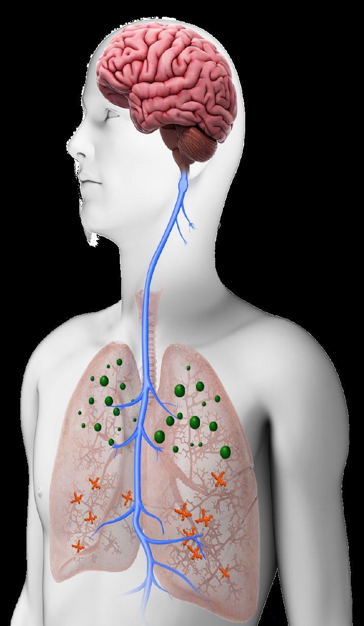
“For some, it may be even more troubling than shortness of breath, because it is so persistent,” he added. In recent years, there has been a “seismic shift” in the scientific understanding of chronic cough. “We have recognised that it is not just a pulmonary problem: it is a consequence of dysfunction between the peripheral nerves in the airways and their connections to the brainstem,” he said. In essence, it is a neuropathic problem that involves peripheral airway triggers, as well as how signals are sensed and responded to centrally, he went on.7-9
A cough happens when sensory nerves in the airways are activated by stimuli and send a signal to the CNS. This, in turn, triggers an alteration in the respiratory pattern to produce a cough. This reflexive pathway is intended to protect the lungs from foreign materials, and, in general, the stronger the stimuli, the stronger the urge to cough.7 “Unlike acute cough, which is protective, chronic cough is maladaptive,” said Drake, adding that chronic cough is caused by increased and amplified signals between the peripheral nervous system (PNS) and the CNS.7-9 “The sensory nerves in the airways, those that travel up to our brain and trigger things like reflexive cough when we choke or aspirate something, are really central to chronic cough.”2,9 When these pathways become dysfunctional, it heightens their response to both noxious and innocuous stimuli and lowers the CNS’s threshold for triggering cough, thereby leading to cough hypersensitivity, he explained.9 “Patients who experience chronic cough describe having an urge to cough, or an actual production of a cough, in relation to innocuous stimuli: things like breathing cold air, laughing, or talking, will trigger a cough. This constant tickle in the throat, that urge to cough, is at the cornerstone of cough hypersensitivity. Mechanistically, we think this is due to changes in the threshold for nerve activation and successive nerve triggering.”9
Expanding on this, Drake explained that, historically, airway sensory nerves have been classified as either nociceptors, those that respond to chemical
signals, or mechanoreceptors, those that respond to physical deformation, pressure, or movement of the airways.10 Advanced research techniques, such as single-cell genomics, however, have provided a deeper understanding of this complex network and revealed a variety of subtypes within those two main classifications.10 “Not only has it shown us that we have this incredibly rich diversity of sensory nerves in the airways, but it has also allowed us to trace those connections to where they integrate with the brainstem, and understand how the brainstem interfaces with higher cortical neurones. In essence, it has given us a tremendous amount of information about how this system functions, both in health and how it begins to change and evolve in disease.”10
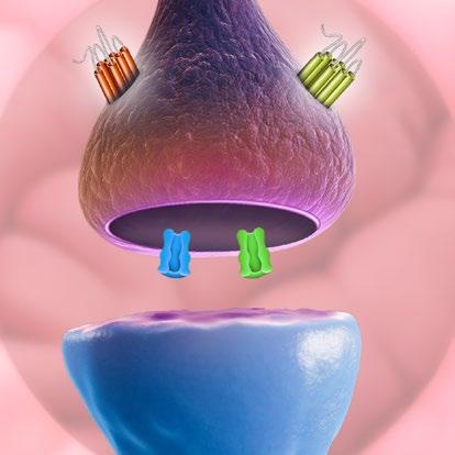
Such work has uncovered a range of receptors and ion channels that contribute to chronic cough.10 At the peripheral level, adenosine triphosphate, for example, which is released by injured or activated airway epithelial cells, acts on the nerve fibres through purinergic receptors, such as purinergic receptor P2X 3 (P2X3), that stimulate the cough reflex.10,11 Transient receptor potential channels, which have been identified in airway sensory neurones, airway epithelial cells, and airway smooth muscle cells mediate chemosensitivity to various endogenous and exogenous triggers.12,13 In addition, some subtypes of voltage-gated sodium channels, which are essential for the action potential in response to all stimuli, are predominantly expressed in coughtriggering airway nerves.14 Drake also explained that opioid receptors, such as kappa and mu, play a pivotal role in both the peripheral and central neural pathways involved in cough. These can be found in high numbers in the CNS and PNS regions that regulate sensitisation.7
In turn, these discoveries have identified new treatment targets, and the therapeutic landscape is evolving rapidly.2,10 Drake highlighted a dual opioid agonist/antagonist approach, which modulates both central
and peripheral neurotransmission involved in cough, via kappa opioid receptor agonism, while limiting side effects, such as respiratory depression and abuse liability, via mu opioid receptor antagonism.7 “Here, the goal is not just stopping the triggering at the source in the airway, but also turning down the volume in the brainstem and cortex to address that key underlying problem of cough hypersensitivity,” he said.
What is currently unclear is why this CNS/ PNS dysfunction develops, said Drake. “We know that the sensory nerves in the airway grow and increase in their overall density, and we know that they become excessively prone to firing and sending those signals to the brainstem. We also know that changes in brainstem and cortical centres occur because of excessive firing, and that leads to this heightened activity level,” he explained.10 Why certain people develop these changes while others do not, however, “is not totally clear.” It may be that some people are born with more nerves, or with differences in the nervous system connections, making them more susceptible, he said.15 In IPF, Drake went on, it is possible that the fibrotic processes in the lungs contribute to excessive cough triggering.16 There is also a suspicion that fibrosis promotes a feedback loop in which the more a person coughs, the more impact it has on the peripheral nerves in the airways, thereby amplifying peripheral responses.17 “The consequence is that the brain is going to modulate the way it processes the signals centrally, to try to accommodate the excessive peripheral cough triggering.” Such a combination, he went on, may have a maladaptive effect, leading to cough hypersensitivity and progressively worsening chronic cough triggering.16,17
CARING FOR A PATIENT WITH CHRONIC COUGH AND IPF
Regardless of the underlying mechanisms, chronic cough has a huge impact on the lives of people living with IPF.3,6
Kulkarni said the majority of the patients she saw in the clinic experience it. “It is not just a cough that might go away. This is something the patient has to live with. It can cause fatigue and disrupt sleep. When it is severe, it can cause chest discomfort or even incontinence in some.”3,6 The negative effects, she went on, often go beyond the physical. “The psychological and social impact is very significant and often overlooked,”3 she explained. People can be embarrassed, avoid social situations, and, in some cases, develop anxiety and depression.3,6 Such feelings were compounded during the COVID-19 pandemic, when coughing in public became a visible sign of infection, she explained. “A lot of patients tell us that cough probably impacts their quality of life more than shortness of breath.”
Highlighting the burden, Kulkarni pointed to a survey of 197 people with IPF and non-IPF interstitial lung disease who experienced chronic cough.18 A total of 63% reported they “often” or “almost constantly” had the urge to cough, while only 4% said coughing always relieved that urge. The most common triggers were respiratory related/ sensation (22%) and exertion/physical activity/body position (22%), while 15% reported unknown triggers, and the cough was unpredictable. Normal everyday events such as talking or laughing (7%) or eating and drinking (7%) were also identified as triggers. Almost 80% reported that, once they started coughing, they continued coughing for more than 30 seconds. In terms of frequency, 35% reported daily coughing bouts, while around 30% reported coughing four or more times a week.18
The survey also demonstrated the significant impact cough had on their lives.18 Almost half (48%) of patients reported that their cough interfered with activities of daily living “sometimes,” “often,” or “daily,” while 46% experienced fatigue or exhaustion as a result of their cough. In 45% of respondents, cough “sometimes,” “often,” or “always” caused them to avoid social situations, while 16% said it
had significantly or extremely affected their ability to maintain physical or intimate relationships. When asked about the emotional impact of chronic cough, 71% reported feeling frustrated, 59% embarrassment, and almost a third (32%) anxiety.18 The data show, said Kulkarni, that chronic cough in IPF is “not minor.” “It has a profound impact on quality of life,” she noted.
Participants’ experience of healthcare with regard to their chronic cough was also explored in the survey.18 The unmet need was clear in the findings. Of the 171 people who answered the question, 56% said they were currently using over-the-counter remedies, and 53% reported using inhalers or nebulisers. Other common treatments included benzonatate (22%), proton pump inhibitors (21%), and glucocorticoids (21%), while 11% reported no treatments and 2% reported speech or cough therapy. Yet 63% of participants said they had experienced no relief, or only partial/ somewhat relief, with current or previous treatments. In addition, only 50% of participants said they felt “heavily involved in decisions about their cough treatment and care.”18
Kulkarni said it was “really important” to routinely assess and proactively address cough, and to involve patients in the decision-making process. “We are challenged by the fact that we do not have many therapeutic options. Because of that, the amount of time spent talking to patients about cough is limited,”3,5,7 she said, adding that healthcare providers tended to focus more on objective data, such as lung function, CT scans, and walk tests, simply because they were measurable. Another challenge is a lack of assessment tools. In the research setting, patient-reported measures such as the Leicester Cough Questionnaire (LCQ) and the Cough Visual Analogue Score (VAS) are often used to quantify severity.19,20 Objective cough monitoring, which utilises wearable technology to measure cough frequency and intensity, is also common in trials.21 There are several factors, however, that limit the use of such
tools in routine clinical practice. Multiitem, structured questionnaire tools, for example, are often not practical from a time standpoint, and monitors are not validated for use in the clinic, Kulkarni explained.22,23
Explaining her own assessment and management method, Kulkarni said she used a stepwise approach. Assessment starts with taking a detailed history, looking at factors such as the duration of cough, the diurnal variations, and triggers. She attempts to identify any common contributors, such as gastrooesophageal reflux, upper airway conditions, or medications.5 She can then suggest treating the underlying cause. “If a patient says their cough is worse after they eat or lay down flat, for example, the cause may be reflux, rather than the IPF itself.” Potential interventions include cough drops or over-the-counter cough medicines, and benzonatate, which reduces the cough reflex. However, efficacy is limited. Low-dose opioids are an option that may help some patients; however, they “come with [their] own challenges,” said Kulkarni. “We have to be mindful of the potential side effects.”5
Despite the challenges and the limited treatment options, there is still much to gain from having a conversation about cough in the clinic. Reflecting on her own experience, Kulkarni explained: “Even if you cannot eliminate the cough completely, improving it just slightly can have a meaningful difference to the patient’s life,” she said. Upcoming and ongoing clinical trials may be an option for some people, and, even where there are no suitable interventions, just having the conversation can help make the person feel validated. “At least they know that I have thought about it and am not ignoring it. Just that can have a psychological impact,” she added.
LIVING WITH CHRONIC COUGH: A PATIENT PERSPECTIVE
Steve Ashton, a motorbike enthusiast and grandfather living in Cincinnati,
Ohio, USA, knows the impact of living with a chronic cough only too well. It had such a huge impact on his ability to speak that he gave up his business, and he routinely chooses to avoid social gatherings. “I have absolutely no control over the cough,” he said. “When it comes, it comes, and I just have to bear with it until it is over. Sometimes it gets to the point where I physically get muscle aches in my back and shoulders, just from the cough itself.”
Like many people living with the condition, he experienced a cough long before his IPF diagnosis, which came in 2021. He said he did not think much of it at first, but it got progressively worse over the course of 5–10 years. “There is a joke in the Cincinnati area that if you do not have a sinus infection, you are definitely not from Ohio,” he said. “All my life, I have had coughs, and they have been treated as sinus infections.” When he was finally diagnosed with IPF, he realised that he had been coughing for some time and that he had the same condition that had led to the death of both his mother and his youngest brother.
While he and Lucie, his wife of 48 years, are very active, regularly enjoying camping, hiking, and cycling, a chronic cough has had a huge impact on his life. After retiring from a 28-year law enforcement career in 2008, he was running an independent accident reconstruction franchise, which frequently involved giving talks and presentations, when he received his diagnosis. However, his cough forced him to retire at the end of 2024. “It was humiliating to stand at the podium in full uniform, trying to talk to 300 or 400 police officers, and just start coughing in the middle of it. I am a very professional person, and it pulled away at my pride,” Ashton explained. “It got to a point where I was so embarrassed, I thought ‘I just cannot do this anymore’. I loved what I was doing, but my personal perception was that people thought less of me.” Describing the feeling, he said: “Picture yourself during the COVID era, when you go into a store, and everybody is wearing masks. The first person who coughs, everyone thinks ‘Oh my
God, we have to get away from you’.” This embarrassment, as well as cough-related fatigue, has also significantly impacted his social life. “I feel like I have isolated myself,” said Ashton. “I do not have the drive to go to see people, because I feel like I am becoming a zombie. I cannot function, and it is humiliating.”
Despite the huge impact chronic cough has had on his life, Ashton said his healthcare team had never addressed the cough with him. “I would mention it to my pulmonologist, but it was like the cough was just a sidebar,” he said. “I started to wonder, ‘Did I really say that, or did I just think I said it?’ It was like it went in one ear and out the other.” Through patient forums, he has found an over-the-counter cough drop which he said, “helps a little,” but “only sometimes.” “I will take those when I’m driving or when it gets to the point where I can’t have a conversation… but there really is nothing else I have found that can control it.” He described the cough drop, as well as drinking mullein tea, as “very, very short stopgaps” that “do not address the issue behind” his chronic cough. “I wish there was something the medical field could do to prepare us for the way life is going to be.”
In terms of triggers, he said there seemed to be “no rhyme or reason for it.” “It just comes and goes on its own,” he went on, adding that each coughing fit could last for anything from 60 seconds to 3 minutes. “It just comes on like a regular cough, but it physically hurts more than a normal cough, and I can hear the wheezing when I cough dramatically.” Asked what he would like to change about his cough, he said he wished it would go away. “If there was some way of managing it, it would give me a piece of my life back. I would love to get back out in public and enjoy life.” Even reducing the cough by 10%, he added, “would be better than nothing.”
FUTURE OF CHRONIC COUGH MANAGEMENT
Ashton’s story may sound very familiar to pulmonologists who work with people living with IPF. The future, however, could
look very different, said Drake. “One of the biggest shifts in the field of chronic cough recently is really conceptual. It says that chronic cough is not just a symptom of other diseases, but a neuropathic disorder: changes in the nervous system that regulate cough lead to a persistent and chronic disease that is incredibly debilitating,” he explained.7-9 “This reframing is important scientifically in terms of how we are thinking about developing new therapeutic strategies, but it is also going to be fundamentally important for the way we address chronic cough clinically.”
As the evidence base deepens, and new agents make their way through pipelines and out into clinical practice, there is a potential for personalised treatments, he went on. “I am hopeful for a day when we have multiple options in this space, and that we will be able to do things like phenotype various forms of cough in a really precise way. Then we would be able to treat patients in a really targeted way, based on biomarkers or other clinical features.” Kulkarni agreed. “As we develop a deeper understanding of the mechanisms of chronic cough, IPF, and its impact on patient outcome, I hope to see a shift towards more targeted, personalised treatments,” she said. “This may be the treatment of comorbidities, or specific treatment [for] cough, or its direct impact.” She hopes a new generation of chronic cough treatments would “truly have an impact on cough while minimising the side effects seen with the currently available options.”5 Drake said the scientific community was currently in the process of “moving down this really transformative pathway.” “It is a very exciting time to be in the field, and I think we are going to be looking at this in a very different way in 5 or 10 years’ time,” he added.
As the research continues to develop, Kulkarni said she would like to see better tools for measuring cough make their way into clinical practice and for IPF care to move towards a multidisciplinary approach that places more emphasis on the patient’s perspective. “I hope we will learn more about speech therapy and involve that in our
multidisciplinary approach, and maybe even getting [the] palliative care team involved earlier to help with the symptoms.” Without this holistic approach, there is a danger of overreliance on objective data such as forced vital capacity, which does not necessarily reflect how the person feels or how the condition is impacting their lives, she believes. “We are just making the numbers look good, without truly having a holistic impact on how the patient feels. Trying to get this multidisciplinary approach, combined with, hopefully, better treatment options in the future, could really help us change that.”
CONCLUSION
Summing up, Kulkarni said that chronic cough in IPF is far more than an inconvenient symptom. It profoundly affects patients’ physical, emotional, and social wellbeing, and frequently overshadows breathlessness in terms of day-to-day burden.2,5,6,19 Ashton’s personal experience of living with chronic cough clearly illustrates how it can erode confidence, limit professional and social participation, and lead to isolation, anxiety, and reduced quality of life.
Despite this, chronic cough has historically been under-recognised and insufficiently addressed in clinical practice.3 Limited treatment options coupled with inconsistent assessment and a tendency to prioritise objective disease markers have contributed to a gap in care.3,19,20 However, recent advances in understanding the underlying mechanisms, particularly the recognition of chronic cough as a neuropathic disorder involving dysregulated peripheral and central neural pathways, mark an important turning point.8-10
Ultimately, understanding the mechanisms and the burden of chronic cough matters, said Drake.
“The fact is that, in the clinic, chronic cough is a very, very prevalent concern. Patients suffer with this for months and years, and we have few to no effective therapeutics for targeting this incredibly debilitating
disease.” It prevents patients from being social, from working, and from living their lives in the way they want, he said.
Now, that could all be about to change. “We are potentially on the cusp of new therapies.
2. Hirons B et al. Cough in chronic lung disease: a state of the art review. J Thorac Dis. 2023;15(10):5823.
3. Mann J. Cough in idiopathic pulmonary fibrosis. Front Rehab Sci. 2021;2:751798.
4. Ryerson CJ et al. Cough predicts prognosis in idiopathic pulmonary fibrosis. Respirology. 2011;16(6): 969-75.
5. Vigeland CL et al. Etiology and treatment of cough in idiopathic pulmonary fibrosis. Respir Med. 2017;123:98-104.
6. Green R et al. The burden of cough in idiopathic pulmonary fibrosis and other interstitial lung diseases: a systematic evidence synthesis. Respir Res. 2024;25(1):325.
7. Birring SS et al. Kappa and mu opioid receptors in chronic cough: current evidence and future treatment. Lung. 2025;203(1):62.
8. Chung KF. Approach to chronic cough: the neuropathic basis for cough hypersensitivity syndrome. J Thorac Dis. 2014;6(Suppl 7):S699.
9. Singh N et al. Peripheral and central mechanisms of cough hypersensitivity. J Thorac Dis. 2020;12(9):5179.
There is a lot of enthusiasm about those that are coming through the pipeline, and that we might finally be able to offer something to the millions of patients who have really been suffering.”
10. Drake MG et al. Airway sensory nerve plasticity in asthma and chronic cough. Front Physiol. 2021;12:720538.
11. Kantar A. P2X3 receptor antagonists in chronic cough: “de gustibus non disputandum est” (there is no arguing about tastes). Chest. 2024;166(5): 911-2.
12. Rouadi PW et al. WAO-ARIA consensus on chronic cough–part 1: role of TRP channels in neurogenic inflammation of cough neuronal pathways. World Allergy Organ J. 2021;14(12):100617.
13. Xia Y et al. Transient receptor potential channels and chronic airway inflammatory diseases: a comprehensive review. Lung. 2018;196(5):505-16.
14. Brozmanova M, Pavelkova N. The prospect for potent sodium voltagegated channel blockers to relieve an excessive cough. Physiol Res. 2020;69(Suppl 1):S7.
15. Shapiro CO et al. Airway sensory nerve density is increased in chronic cough. Am J Respir Crit Care Med. 2021;203(3):348-55.
16. Jones RM et al. Mechanical induction of cough in idiopathic pulmonary fibrosis. Cough. 2011;7(1):2.
17. Song WJ, Chang YS. Cough hypersensitivity as a neuro-immune interaction. Clin Transl Allergy. 2015;5(1):24.
18. Kulkarni T et al. Understanding the burden of chronic cough in patients with interstitial lung disease: insights from a U.S. patient perspective survey. Poster. PFF Summit, 13-15 November, 2025.
19. Trigg A et al. Psychometric validation of the severity of chronic cough diary, Leicester Cough Questionnaire, and a cough severity visual analogue scale in patients with refractory chronic cough. J Patient Rep Outcomes. 2025;9(1):65.
20. Spinou A, Birring SS. An update on measurement and monitoring of cough: what are the important study endpoints? J Thorac Dis. 2014;6(Suppl 7):S728.
21. Galvosas M, Small PM. The value of continuous cough monitoring: a narrative review. J Thorac Dis. 2025;17(11):10571-83.
22. Bottine A et al. A systematic review of the psychometric properties of the Leicester Cough Questionnaires based on the COSMIN guidelines. Respir Med. 2024;231:107739.
23. Chaccour C et al. Validation and accuracy of the Hyfe cough monitoring system: a multicenter clinical study. Sci Rep. 2025;15(1):880.
Interviews
EMJ had the privilege of speaking with two leading experts in chronic disease research and global health: Vikram Patel, Harvard Medical School and Harvard T.H. Chan School of Public Health, Cambridge, Massachusetts, USA; and Roy Taylor, Newcastle University, UK.
Patel discusses the growing global burden of mental disorders as chronic conditions, the integration of mental health into broader chronic disease care, and the challenges of scaling accessible care models across low-resource settings. Taylor reflects on his pioneering work in Type 2 diabetes remission, exploring the mechanisms underlying disease reversal and how changing perspectives on metabolic disease could reshape future clinical practice.
Featuring: Vikram Patel and Roy Taylor
Patel
Paul Farmer Professor and Chair of Global Health and Social Medicine, Harvard Medical School; Professor, Department of Global Health and Population, Harvard T.H. Chan School of Public Health, Cambridge, Massachusetts, USA
Q1Your work has focused on the burden of mental health problems across the life course, their association with social disadvantages, and the use of community resources for their prevention and treatment. What pivotal moments in your early career influenced your shift toward this field?
Some mental health conditions, for example, neurodevelopmental disorders, schizophrenia, and bipolar disorder, are almost always chronic, lifelong conditions
I think that the first such pivotal moment was when I started working in Zimbabwe after training in psychiatry in London, UK, at the Maudsley Hospital, which was then considered to be the mecca for psychiatric residencies, with a highly biomedical orientation on psychiatry which embraced a dominant role of psychiatrists in all aspects of mental health care. At the University of Zimbabwe Medical School, Harare, I began to see the vast differences between where I had been trained and where I was working, and I almost had
to unlearn everything that I had been taught for many years and start from scratch. As an example of these differences, there were just about 10 psychiatrists in Zimbabwe at the time, serving a population of nearly 10 million. The majority of us were foreigners who didn't speak any of the local languages. This was a complete contrast to the Maudsley Hospital, where there were more than 10 psychiatrists just in my corridor of the Institute of Psychiatry. The other big difference was, of course, that the illness narratives of people who were struggling with their mental health were completely different from those in London, primarily the absence of a biomedical framework; the ways people understood their suffering was through a social or spiritual lens, but rarely a biomedical lens. So, the bulk of the clinical work we did was for people with very serious forms of mental health problems, mainly psychoses. You
Vikram
rarely saw the mood, anxiety, trauma-related spectrum, because they were not very visible and did not cause behavioural difficulties that affected other people, and, therefore, you rarely ever saw them in the clinic. This was another contrast with my clinical experience in Britain. Moreover, you never saw any kids, because, in fact, there were no child mental health services at all. Another pivotal experience was related to the AIDS epidemic, which was decimating communities in Zimbabwe at that time. I began to observe how rights-based arguments and actions, for example, addressing issues around equity, the use of community resources, the empowerment of people living with HIV, and most importantly, the activism that people living with HIV were leading to make access to life-saving treatments available, which, at the time, were not available in Africa because these drugs were protected by patent laws. I witnessed the emergence of a rights and equity-focused global health movement around HIV/AIDS, and subsequently
tuberculosis and malaria. I began to recognise that there were intersections with mental health problems, where very similar issues could be encountered, such as stigma, inequities, discrimination, and denial of basic care. These important historical processes inspired me to incorporate these strategies in my future work.
How was care delivery different through a ‘spiritual lens’, as you mentioned?
There was emerging epidemiological data that demonstrated a high prevalence of a whole range of mental health conditions, and yet the people we saw in our clinics were like the proverbial tip of the iceberg. The vast majority of care was being delivered by the traditional medical system, a system that predates biomedicine by centuries and is very deeply integrated with traditional religion. This is true in all parts of the world. In Europe, for example, it was the same until the Enlightenment and the arrival of a biomedical perspective on health.
The original asylums in Europe were built around this emerging theory that people who were disturbed were not bewitched but victims of illnesses of the mind. Thus, I think one has to remember that this has been the history of how mental health problems were understood and cared for in all societies. The advent of biomedicine in Zimbabwe was a relatively recent product of colonial rule, and traditional belief systems remain a dominant way for people to understand and to address their problems. My own research at the time shifted in response to these observations. I worked very closely with traditional healers, for example, I used to sit in their healing areas and observe rituals, document them, ask questions to the healers, and then also to their patients, asking how they understood their suffering and the process of healing and so on. I incorporated those ideas as I developed my own understanding of how culture and context influence both the risk of mental health problems, but also how these are understood, acted upon, and how recovery takes place.
Q2
What parallels and key distinctions do you see between mental health disorders and other non-communicable diseases (NCD) in terms of prevention, management, and policy prioritisation?
A key area of similarity or overlap between mental health conditions and other NCDs is that they often have a chronic and or relapsing course. I'd say the majority of depressive episodes are acute, single episodes, but a significant proportion of people will have recurrent or chronic conditions. Some mental health conditions, for example, neurodevelopmental disorders, schizophrenia, and bipolar disorder, are almost always chronic, lifelong conditions. In that respect, I like to consider these as chronic diseases. Furthermore, there are a lot of comorbidities between these groups of conditions, for example, diabetes and schizophrenia or heart disease and depression. These tend to occur together in the same individual, much more often than by chance, and this is to do with a variety of factors. Most obviously, people
with psychosis take medications that increase their metabolic risk, which in turn contributes to premature mortality, highlighting the need for integrated care. Another important similarity is that the diagnosis in itself doesn't tell you exactly what a person needs for their care. In other words, two people with the same diagnosis may have completely different needs. Diabetes, of course, is a classic example that one cannot apply a one-size-fits-all approach. You can have very mild diabetes, where only lifestyle changes are needed for glycaemic control. At the other end of the spectrum, you can have very severe multiorgan diabetic syndromes which need dialysis, rehabilitation, and complex medication regimes. The same applies to mental illness. I think the whole notion of person-centred care comes from chronic disease care, in which ‘you are not treating the disease, but the person’. Where mental health conditions and NCDs fundamentally differ is that for many NCDs, for example, cancers, cardiometabolic, and autoimmune conditions, we have witnessed dramatic improvements
in our understanding of the pathophysiology of these conditions. While mental health conditions and NCDs do share in common the fact that their aetiology is often the result of an interaction between genetic, environmental, and personal lifestyle factors, for NCDs, many of these factors are much better understood and elucidated. As a consequence of that, we have a number of transformative new therapeutics that have emerged. We have a much better understanding of very specific risk factors, such as smoking, which allows you to design prevention programmes that target those factors. This is much less the case for mental health conditions. I think this takes us to a very important question, that is, the reason for this disparity, which, in my reckoning, is the result of the diagnostic system we use to characterise and study mental health conditions, which relies on clusters of symptoms in patients seen in psychiatric clinics, unlike in NCDs, where we diagnose people according to biomarkers. Many scientists have called for a move away from the syndromic
diagnostic approach, which, as well-intentioned as it was when it was invented 50 years ago, has really led us to a scientific cul-desac. We have not discovered any new disease pathways, prevention targets, or therapeutic targets, so we've got to reimagine how we study mental health problems.
Q3How can healthcare systems better integrate mental health into chronic disease care pathways, particularly in primary care settings?
While there are some unique features about mental health care, there are also some shared principles with other chronic conditions. The most important issue about integrating mental health care within broader NCD care platforms is that this is the best strategy to both destigmatise mental health problems and to provide person-centred care. Instead of the classic specialist biomedical approach, where you have your blood sugar managed by one person, and you get your low mood managed by another person, and you often have to go to totally different places to get these different healthcare tasks done, integration means these health conditions are managed together in the same place, perhaps by different people, but in a coordinated way. So, there's something important to be said about the integration of mental health care in all other platforms of health care and in all countries and contexts. In short, it's a better quality of care when it's integrated, the outcomes are better, and there is abundant evidence to demonstrate that.
Another critical issue is the assumption that mental health care needs mental health professionals. I think perhaps the single most important scientific
advance in the field of psychiatry in the last two decades has been the demonstration that carefully designed brief interventions that target psychological or social mechanisms are effective and, moreover, can be delivered by non-specialist frontline providers, like community health workers, peer support workers, and even lay persons. This evidence converges on two distinct forms of science, one which is related to the design of targeted interventions based on mechanisms, and the other which is based on implementation or delivery science. When these two come together, the result is a demonstration of how making mental health care can be available for everyone with an NCD, no matter what mental health specialist resources are available, because you don't need psychiatrists and psychologists, particularly for the first level of care. Moreover, delivering mental health care in this manner is far more acceptable to patients.
The majority of people with mood, anxiety, and traumarelated problems do not need complex interventions delivered by mental health professionals; person-centredness often implies incorporating models of care like stepped or stratified care to deliver different intensities of interventions and adopting a biopsychosocial model in defining the specific interventions for a particular patient. These models of care recognise that there is a great variation in the severity of conditions and that a range of personal characteristics can moderate the recovery process. Stepped and stratified models of care ensure that you allocate care according to need. The majority of people with depression, for example, are mild–moderate severity, and they can respond
to these brief psychosocial interventions delivered by frontline workers. It's much easier for a diabetes programme, for example, to identify, train, and sustain frontline workers, rather than refer to psychiatrists and psychologists.
Q4
What are the biggest barriers to scaling community-based mental health interventions in low-resource settings today?
I think the most important barrier is related to the political economy of scaling up this approach, because when you are going to allocate money and power to frontline workers, you're taking some money and power away from other providers who have historically dominated the care pathways. This is why this kind of structural reform is much harder to do in wealthy countries, where the interests of mental health speciality services are much more entrenched and numerous. This is why you see some of the most remarkable examples of front-line provider care happening in the least resourced countries in the world, for example, in Africa and Asia, where community health workers are recognised as the major human resource to help improve access to care, not just for mental health problems, but also for maternal and newborn health, care of NCDs, and HIV care. They are widely accepted, and they're fully integrated into health system design. For example, in India, we have more than one million community health workers who are engaged in a range of frontline healthcare tasks, some of whom are now being incorporated within mental healthcare delivery systems. That being said, the Improving Access to Psychological Therapies (IAPT) programme in the UK is actually an example I do consider
to be very similar. For example, a significant number of providers are not clinical psychologists or psychiatrists, but graduates of psychology who undergo very specific training in brief cognitive behaviour therapy. I think in that respect, there is a great degree of similarity with task-sharing models in lowresource contexts. Essentially, what you need is a strong political will to be able to address the pushback you're going to get. But beyond delivering psychological interventions, we also need to acknowledge that mental health problems cannot just be treated like any other medical diagnosis, because these conditions are deeply connected to our social worlds. So, addressing mental health problems without attending to a person's social world is a little bit like attending to, for example, lung cancer without attending to a person’s smoking behaviour. It's missing a hugely important influence, which is associated not only with the emergence of a mental health problem but also profoundly influences its outcome. This is yet another critical advantage that community health workers have over facility-based specialist providers.
Q5Much of your research highlights the relationship between mental health and social disadvantages. What mechanisms most strongly link poverty and mental illness across different settings?
Our brain is constantly being modified and moderated by our social world, and this continues into our last days, but it's especially so in childhood and adolescence, which is the reason for two other key observations: the strong association of adversities in childhood with the risk of mental health problems
in later life and the observation that most mental health problems begin before adulthood. Amongst the key drivers of the association between social disadvantage and mental health problems is the effects of growing up in poverty, both due to direct mechanisms, for example, you're more likely to be exposed to direct damage to your brain which in turn affects your mental health, such as nutritional deficiencies, environmental toxins and infections and because of the challenges faced by low-income parents to be able to provide a nurturing environment for their children. The early years of life are critically important because this is when your brain is most plastic, and nurturing environments catalyse healthy brain development. In environments of neglect or toxic stress, brain development is impaired, and the child’s resilience to deal with difficulties in later life will also be affected. This tells you something about the root causes of mental health problems, that they are developmental in origin. Therefore, if prevention is our goal, we're going to have to look at the social world in particular, in the early years of life.
Another mechanism which operates in adulthood is the daily grind, the precarity, the uncertainty, and the insecurity that are associated with living in poverty. Think about the pandemic. The one defining feature of the pandemic was uncertainty, not knowing what tomorrow would bring, alongside precarity for those already on the margins. It is that uncertainty that acts as a kind of threat to our mental health, and when that uncertainty is unremitting, day after day, week after week, this is an incredibly toxic experience for our brains. The convergence
of early childhood deprivation and later life precarity affects our mental health through different pathways and underpins the strong association between poverty and social disadvantage with poor mental health across the life course.
What are your thoughts on the concept of preventive medicine when it comes to mental health?
Preventive medicine is at the heart of reducing the burden of disease. Let us consider lung cancer, one of the great success stories of preventive medicine, through targeting the individual’s smoking behaviour in multiple ways, from making smoking more expensive to less socially acceptable. That fits very well with the biomedical model. There is a political economy of tobacco, of course, but prevention generally depoliticises the social determinants by focusing on an individual’s behavioural choices. However, prevention of mental health problems is profoundly political choices for the root causes are upstream determinants like poverty, gender, and inequality. Thus, prevention of mental health problems must typically address these determinants. For example, our work in Brazil is showing that when a government decides to tackle absolute poverty by using cash transfers, this leads to direct improvements in health and wellbeing, including dramatic reductions in suicide mortality. Indeed, addressing childhood poverty may be the equivalent of targeting tobacco for lung cancer prevention. As another example, precarity and uncertainty in life are becoming the lived experience for many young people. This is not an accidental occurrence. This is an intentional political choice that governments have made around
the world, for example, privileging the rights of corporations to extract profits over the rights of citizens to secure employment and decent wages. No one across the ideological spectrum would disagree that tobacco is bad, but the role of government in progressive taxation, provision of social services, or universal basic income and health coverage is contested. This is the real challenge, and where mental health is different from NCDs.
Q6 In your critique of global mental health research funding, you described it as ‘too little, too inequitable, too skewed’. What structural changes are needed to correct these imbalances?
First of all, we definitely need more funding, but the funding now needs to follow the evidencebased scientific approaches to characterising mental health and the prevention and care of mental health problems. For example, funding should no longer be hooked onto diagnostic categories which lack any scientific validity;
the National Institutes of Health (NIH) or the Wellcome Trust, two major examples of funders, no longer require mental health research to characterise mental health conditions through the application of the Diagnostic and Statistical Manual of Mental Disorders (DSM)/International Classification of Diseases (ICD) diagnostic systems. What they require is phenotypic characterisation along dimensions of the mental health experience. Secondly, the research needs to incorporate explicitly an equity dimension, quite simply because, as we discussed earlier, these conditions are not equitably distributed. Thirdly, there should be investment in scaling up science. We know what works; we have solutions. However, most of those solutions lie outside the hospital or clinic and outside a narrow biomedical framework. As long as mental health research is dominated by a narrow biomedical perspective, those resources are not going to be allocated to addressing questions about scaling up interventions for prevention and care. This is the
political economy I was referring to earlier, so we have to make mental health research far more diverse in terms of the research actors, the research questions, and where the research is going to take place.
Q7 What does achieving universal health coverage for mental disorders realistically look like in the next decade?
This is very much linked to what we discussed earlier, that is, the integration of mental health care in NCD programmes, because health systems globally are gearing up to address chronic conditions, which have emerged as the leading causes of the burden of disease in most countries of the world. Fifty years ago, most countries were still dealing with infectious diseases, where the one-size-fits-all, diagnosis-driven model dominated, whereas most health systems are now dealing with chronic diseases for which a person-centred approach should be the rule. This offers an amazing opportunity for the full integration of mental health within chronic
disease care, built upon the strong foundation of primary care, the heart of universal health coverage. Hospital specialist care is needed for those who are at the severe end of the spectrum of any health condition, while the majority of care, in particular continuing care, needs to be centred in primary care. The one exception I'll make is for psychosis, which is a rarer condition. People with psychosis often have very complex needs, and I think that mental health specialist care is the right node for coordinating care, but that doesn't mean that all care takes place in a hospital. A lot of the care itself is still delivered in the community, through frontline workers supporting people with psychosis. In contrast, for mood, anxiety, substance use, and trauma-related problems, primary health care should be the node for the coordination of care, so if
you need specialist care, it's the primary care provider who will refer you or seek a consultation.
Q8What advice would you offer to early-career clinicians and researchers who want to contribute meaningfully to global mental health?
Do not be demoralised by the apparent lack of progress in the discovery of aetiology or transformative new therapeutic agents. There has, in fact, been huge progress, but it's not within the narrow psychiatric clinical world. Scientific advances in developmental neuroscience and evolutionary science are helping to explain the early life and ancestral origins of mental health problems. The social and epidemiological sciences have characterised the exposome, the social environments that can either harm
or protect mental health. Clinical and implementation science is demonstrating the effectiveness of brief interventions targeting psychological and social mechanisms which can be delivered by front-line providers with appropriate training and supervision. Knowledge from the lived experience is shedding light on what really matters to people living with mental health conditions. All these bodies of knowledge are pointing to a radically different way of thinking about how we can study mental health problems, identify novel preventive and therapeutic interventions, and deliver person-centred care.
Roy Taylor
Emeritus Professor of Medicine and Metabolism, Newcastle University, UK
For decades, Type 2 diabetes was described as a chronic progressive disease. You argued it can be reversible. When you first proposed the Twin Cycle Hypothesis, how radical did it seem at the time?
Putting together the hypothesis did not seem radical at all, because we had assembled various pieces of new information that were necessary. Then, in a single morning during a brainstorm, it suddenly occurred to me that Type 2 diabetes would fit into a simple pattern.
I spent several months scribbling it down and moving the boxes around, and the Twin Cycle Hypothesis emerged. The critical thing about a hypothesis is that you need to test it to destruction, and, if it will not be destroyed, then it might be right. That is where we are now.
the average and the distribution within populations, and it is very good at that.
When we come down to the level of the individual, however, it falls apart. For instance, you would not go up to a front-row forward of the England rugby team and call them obese, even though they may have a BMI of 34. At the other end of the scale, there are people like me who have had a BMI of 18 throughout life, and putting on weight to a BMI of 21 may cause Type 2 diabetes.
We do, however, have a way of introducing a more relevant personal measurement. This is highly individual and genetically determined. At present, there is no way of detecting it in advance, but the critical measurement is not weight or BMI, it is waist circumference.
The critical measurement is not weight or BMI, it is waist circumference
However, it was revolutionary. It was flying against the tide, because everybody knew, or thought they knew, that Type 2 diabetes was lifelong, inevitably progressive, and would inevitably cause serious complications in later years.
Q2 Your idea of the Personal Fat Threshold challenges the assumption that Type 2 diabetes is mainly a disease of obesity. Why has BMI been such a misleading metric?
BMI has effectively taken over as the standard by which individuals are judged, but what has been forgotten is that it was developed very precisely to measure populations. It describes
Over time, most adults unfortunately put on inches at the waist. If that waist measurement is reduced, typically to somewhere between 36–34 inches, an individual may cross their personal fat threshold, and the diabetes will switch off.
So, that's how the personal fat threshold can be a practically useful measurement.
Q3 What did the ReTUNE study teach us about people who develop Type 2 diabetes despite appearing slim?
This is really important, because, after all our initial studies, we knew that people could reduce their BMI from a high level, even
modestly, and eliminate diabetes. However, we had been faced for a long time with apparently slim people in the clinic who had Type 2 diabetes.
It seemed likely they had the same underlying problem, but that BMI was the wrong metric. In other words, BMI was misleading. So, what we did was take a group of people of normal BMI (between 21–27) with diabetes, and they were given a weight loss diet. They lost weight as expected, and the underlying mechanisms of diabetes returned to normal, exactly as seen in heavier individuals. In the ReTune study, it wasn't just half the people who responded; it was 70%. People who are not very overweight often find it easier to lose a small amount of weight, which is an important message.
And even better, in ReTune, we came out with a really practical answer, because we had the waist measurements of these people. Even after weight loss and remission, when matched with individuals without diabetes and the same BMI, waist
circumference remained higher in those who had previously had diabetes. We could see on the MRI scans that there was still excess fat inside the abdomen, even though people at that time had lost an average of 10% of their body weight. So, individuals developing Type 2 diabetes have a significant excess of fat relative to their personal threshold. Losing around 10–15 kg, or even 8–10 kg in lighter individuals, can bring them below that threshold, restoring metabolic control and avoiding complications. Again, it's such a glittering insight. For people who are slim, just check the waist.
Q4 You showed that remission can occur rapidly after substantial weight loss. Why do some people achieve remission and others do not, even when they lose similar amounts of weight?
There are some other factors. If someone has had Type 2 diabetes for too long, the critical recovery may not occur. The insulin-
producing β cells in the pancreas, when exposed to excess fat for prolonged periods, may be too damaged to recover. How long is ‘too long’ varies. Genetics play a role. Some individuals may become unresponsive after as little as 3 years, while others can have diabetes for over 20 years and still return to normal. I have seen people with 23 years of Type 2 diabetes, treated with tablets, who lost weight, stopped medication on Day 1 of the diet, and maintained normal glucose levels thereafter. We are therefore dealing with individuals, not averages. Epidemiology has limited relevance in the clinic.
Q5 Your early studies used rapid calorie restriction to show that reducing liver and pancreatic fat could put Type 2 diabetes into remission. Do you see glucagon-like peptide-1 medicines as a transformational new chapter in diabetes care, or mainly as a more practical tool for achieving the same biological result?
Well, we're just at the start of this story, so I don't have any definite
views on this, but we can say several things clearly. The first thing is, these amazing medicines achieve what's been wanted for a long time, a direct effect on appetite. When appetite is wound down, everybody loses weight. Now, the importance of that critical observation is that it cuts through much of the confusion that has surrounded obesity research. There's no doubt that when you reduce body weight, you will produce the physiological changes that we have shown. And so, the reason for the improvement in diabetes is exactly the same as what we've shown.
The area is clouded by the manufacturers of these medicines. These drugs are often presented as acting through incretin effects on insulin secretion, but their primary impact is reducing body fat.
What will happen after treatment stops? There haven't been many studies of this so far. Weight often returns, because individuals have not learned how to regulate their intake. That learning cannot be taught didactically; it requires experience. If weight loss is externally imposed via medication, that learning process may be absent. We've got a learning curve here, and that learning curve is absent when people have been taking an injection and are basically having health administered to them artificially. They don't know this essential skill of how much to eat.
So, that's the problem. How can we approach it? It strikes me that there are at least two approaches. One is to try and mimic our food reintroduction programme, so that there is an intensive learning period with careful monitoring of body weight, with management by a doctor and nurse over the first 6 weeks of stopping the
tablet or injection. The second approach is to consider continuing just a low dose of the medicine. Now, we don't know which is better, but there's one thing that's very human: many people with Type 2 diabetes absolutely hate their condition, but many patients wish to avoid lifelong medicalisation, which is an important consideration. This is something that doctors have been slow to realise by and large. This is a problem with using a tablet or a medicine approach. So, I see these drugs as being useful for diabetes potentially, but only when the learning has been appreciated and we've got in place some reasonable long-term measures.
Q6
The number of children and young people with Type 2 diabetes is rising in the UK. Are we seeing a fundamentally different form of Type 2 diabetes in adolescents, or simply earlier exposure to the same drivers?
It is earlier exposure to the same drivers. However, the consequences are more severe. Younger individuals are more susceptible to complications, likely because the vascular endothelium is more vulnerable to hyperglycaemia. Type 2 diabetes at age 20 years can lead to retinopathy within a few years and renal failure within a decade or so. By contrast, onset after age 70 years carries a far lower risk of serious complications. So, Type 2 diabetes in the young is extremely serious and, until politicians and legislators grasp that the food environment has to be legislated for, we'll continue to see this. Prevention is always better than cure, and we know how this can be prevented. It's not through trying to stimulate even more activity. Children leap around quite a lot anyway. What is abnormal is the dysregulated food environment.
The critical thing to mention to your politicians, your legislators, is that excess food is toxic. It's poisonous. While we regulate substances like lead, we have not adequately addressed the toxicity of excess caloric intake.
International food companies have been astonishingly successful in persuading politicians to look the other way. How often has the childhood obesity strategy been abandoned since it was going to be launched in 2017? Even now, with the banning of food adverts that might be seen by young people before 9pm, it turns out to only be the banning of images. The adverts can run if they’re just in print and mention the name of the product. This is the kind of persuasive power that the big food companies have.
We have had a great start with the soft drinks levy on sugar. It's taken 37 tonnes of sugar out of the UK consumption every year, which is spectacular and has had an impact on childhood rates of obesity that is measurable. Politicians have got to be held to account, and we shouldn't be dragged into the folly of assuming that exercise will head this off.
Q7
Looking ahead,what important questions about Type 2 diabetes remain unanswered?
There are many, but, first, what is it about the β cell that causes it to be susceptible to fat? Can we detect these people in advance? Not everyone develops diabetes, even with substantial weight gain. Among White European populations, only about onethird will develop diabetes under such conditions; in some Asian populations, it is closer to half. This reflects genetic differences in personal fat thresholds. So,
what is it that determines that personal fat threshold and the initial susceptibility? The second question is, how can we measure the personal fat threshold? I've tried to measure it to get an index of how we could measure it with blood tests, but attempts to identify inflammatory markers have not yielded clinically useful signals, particularly in relatively lean individuals. So, this is an open question. At present, the most practical method remains observation: individuals can do it for themselves by measuring their waist circumference, substantial weight loss, and monitoring glucose response.
So, those are important things, but I'll circle back round to prevention. Preventing Type 2 diabetes is so important. The economic burden of overweight and obesity in the
UK is estimated at 93 billion GBP annually, according to the Institute of Fiscal Studies. It gives an idea of the extent of what we're turning a blind eye to at the moment.
Q8What practical tools and resources are available to support clinicians?
The NHS England national remission programme is now well underway, although eligibility criteria mean that not all patients qualify. I had lots of inquiries about this, so I put together a toolkit to be used by nurses and doctors in primary care.1
It provides background information, suggested language for patient communication, and structured guidance for implementation. There is also the book ‘Life Without Diabetes’,2
which brings together the evidence and clinical approach, including a dedicated chapter on Type 2 diabetes in young people, and all the proceeds of this book go to Diabetes UK to provide research money not only for what we've already done, but for research in the future.
References
1. Taylor R, Bakhai C. Type 2 diabetes remission: toolkit for general practice. Available at: https://diabetesonthenet. com/wp-content/uploads/4.Remission-toolkit.pdf. Last accessed: 5 May 2025.
2. Taylor R, Life Without Diabetes (2025) 2nd edition, London: Short Books.
acute cough
<3 weeks
It is reported in approximately
Chronic Cough in Patients with Idiopathic Pulmonary Burden, Mechanism,
and
Implications for Treatment
What is Chronic Cough?
Chronic cough, defined as a cough that lasts ≥8 weeks, is highly prevalent in patients with IPF 1,2 % of patients with IPF.3,4
subacute 3–8 weeks
chronic 8 weeks
The mean number of coughs in patients with IPF is 672–905 coughs/day.5,6
Coughs tend to be more frequent and more severe in the daytime than at night.7,8
In a prospective, multicentre study of patients with IPF, greater cough severity (VAS ≥30 mm) at baseline was independently associated with worsening lung function, greater likelihood of disease progression, and reduced transplant-free survival 9
Cough effects on patient QOL
Fatigue4,9
Sleep disturbance7
Urinary incontinence9
Daily functioning impairment4,11
Physical pain7
Cough Hypersensitivity
In cough hypersensitivity, nerve signals become overactive and cause response to normal or subthreshold triggers
Common triggers include:3,12-14
Shortness of breath7,11
Anxiety/ depression7,11
Social isolation/ embarrassment4,7,9,11
Chronic cough impairs quality of life, causes fatigue, negatively impacts sleep, and is socially isolating 4,7,10
References
1. Morice AH et al. Eur Respir J. 2020;55(1):1901136.
2. Ryerson CJ et al. Respirology. 2011;16(6):969-75.
3. Vigeland CL et al. Respir Med. 2017;123:98-104.
4. Green R et al. Respir Res. 2024;25(1):325.
5. Maher TM et al. NEJM Evid. 2023;2(8):EVIDoa2300083.
6. Martinez FJ et al. Am J Respir Crit Care Med. 2022;205(9):1084-92.
7. Mann J et al. Front Rehabil Sci. 2021;2:751798.
Singing, talking, laughing Aerosols, scents, and odours
Changes in body position
Eating, drinking
Sensitisation in Chronic Cough
Central sensitisation15-17
• Mechanisms in the CNS amplify nerve fibre inputs
• Normal afferent nerve fibre activity can produce heightened cough responses
• Reduced cough suppression occurs due to loss of inhibitory nerve activity
Signal transmission15-17
• Amplified nerve signals are sent to the brain
Peripheral
sensitisation
15-17
• Afferent nerve fibres become more excitable due to local tissue damage, mechanical stress, and loss of lung tissue
• Triggering stimuli are more likely to cause afferent nerve activation
8. Key AL et al. Cough. 2010;6:4.
9. Khor YH et al. Am J Respir Crit Care Med. 2024;210(8):1035-44.
10. Hirons B et al. Eur Respir J. 2022;60(suppl 66):7.
11. Graham CS et al. BMC Pulm Med. 2025;25(1):221.
12. Birring SS et al. Lung. 2025;203(1):1-8.
13. Haji A, Ohi Y. Neuroscience. 2010;169(3):1168-77.
14. Singh N et al. J Thorac Dis. 2020;12(9):5179-93.
Pulmonary Fibrosis:
increased cough
The publication of this infographic was supported by Trevi Therapeutics EMJ. 2026;11[2]:42-43. https://doi.org/10.33590/emj/751WS6IA
Role of opioid receptors and cough hypersensitivity
Opioid receptor signalling modulates sensitisation by reducing peripheral and central excitatory neurotransmission and enhancing central inhibitory pathways.15-17
Temperature changes
Exercise
κ and µ opioid receptors in the CNS12,18-21
• κ receptors are present in the nucleus of the solitary tract, where the brain first processes cough signals from afferent nerves
*The receptors in the image sit in higher regions of the brain as well as the brainstem
Excitatory (ascending) Inhibitory (descending)
Peripheral nervous system
Chemoreceptors
Mechanoreceptors
Abbreviations
κ and µ opioid receptors in the lungs12,18-21
• κ and µ opioid receptors are present on vagal afferent nerve fibres
• Opioid receptor signaling in the lung is important for the inhibitory effect on the signals travelling to the CNS
15. Chung KF et al. Nat Rev Dis Primers. 2022;8(1):45.
16. Krajnik M et al. Curr Opin Support Palliat Care. 2014;8(3)191-9.
17. Irwin RS et al. N Engl J Med. 2025;392(12):1203-14.
18. Volkow ND et al. N Engl J Med. 2016;374:1253-63.
Heart failure with preserved ejection fraction (HFpEF) is a complex disease that has been historically difficult to treat and manage. Novel research shifts understanding of HFpEF from a primary heart disease to a systemic syndrome driven by systemic inflammation, congestion, and multi-organ metabolic dysregulation. These three key drivers can all be integrated into the heart–liver–pancreas inflammatory axis, with bidirectional selfamplifying interactions between these organs leading to a vicious cycle with worse clinical outcomes. This paper will unravel the mechanics of this axis and provide important implications from a therapeutic standpoint, shifting the management of HFpEF from cardiac-centric strategies to interventions targeting the underlying systemic inflammation, metabolic dysregulation, and congestion.
Key Points
1. Heart failure with preserved ejection fraction (HFpEF) has been the ‘greatest unmet need’ in cardiology due to its complex pathophysiology, the lack of targeted therapies in the disease, and the futility of cardiac-centric agents.
2. This narrative explores the cardiac–hepatic–pancreatic axis proposed in HFpEF pathogenesis, and offers therapeutic implications illustrated by the benefits of novel therapies such as glucagon-like peptide-1 receptor agonists and sodium-glucose co-transporter 2 inhibitors.
3. Future HFpEF trials should assess metabolic and pancreatic markers to clarify whether this axis represents the true pathophysiology of HFpEF or is just an indirect association.
INTRODUCTION TO THE HEART–LIVER–PANCREAS AXIS
Heart failure with preserved ejection fraction (HFpEF) has historically been a troublesome condition to study and manage because of the variable presentation of the condition and the limited number of disease-modifying therapies to address it. HFpEF has conventionally been thought of as a problem of diastolic dysfunction and stiffness of the heart.1 However, the concept of HFpEF is progressively expanding to encompass a systemic disorder that is mediated by the principles of inflammation, congestion, and metabolism, and not simply a condition of the heart. The integrative theory of a connection between the heart, liver, and pancreas through the principles of a common axis of inflammation and haemodynamics is a significant contemporary contribution to the understanding of the pathophysiology of HFpEF.
DISCUSSION AND OPINION ON THE HEART–LIVER–PANCREAS AXIS AND ITS INFLAMMATORY PATHWAY IN HFpEF
One of the major advantages of this model is the emphasis on venous congestion with low-grade systemic inflammation. Hepatic congestion in HFpEF is more than simply an association with right-sided heart failure; rather, it plays an active pathogenic role in disease progression.2 Elevation of sinusoidal pressures, hepatocellular hypoxia, and secondary fibrosis lead to congestion hepatopathy, which advances
to cardiogenic cirrhosis in end-stage disease.2,3 The inflammatory derangement secondary to hepatic pathophysiology, with increased nitric oxide signalling, cytokine expression, and shifts in the hepatokine profile, generates a vicious cycle that reinforces the pathogenesis of myocardial disease and endothelial damage. Interestingly, this vicious cycle has been borne out in emerging concepts redefining cirrhotic cardiomyopathy not as a new disease, but rather as a clinical manifestation of HFpEF.1-3
Pancreatic exocrine insufficiency (PEI) is prevalent in chronic heart failure but not always evaluated.4 Splanchnic hypoperfusion, venous congestion, autonomic derangements, and inflammation can injure the exocrine functions of the pancreas.4 PEI may result in malabsorption, micronutrient deficiencies, sarcopenia, and cachexia, exacerbating heart failure.4 Additionally, there has been evidence showing that observed weight loss and malnutrition in HFpEF are more complex phenomena that are not solely attributed to heart disease or the ageing process; potential modifications in the gastrointestinal system are also advocated.5 Pancreatic and cardiac inflammatory links are established in acute pancreatitis, in which systemic inflammatory responses (cytokines: IL1β, IL-6, TNF-α) lead to limited diastolic reserve, microvascular ischaemic disorders, arrhythmias, and myocardial depression. Patients with HFpEF with reduced diastolic function and microvascular inflammation are likely more vulnerable to these conditions’ haemodynamic changes.4-6 Observational data linking acute pancreatitis to an increase
in in-hospital mortality among patients with pre-existing heart failure underscore the clinical relevance of this interaction.7-9
The addition of metabolic dysfunctionassociated steatotic liver disease further strengthens the three-organ model. Metabolic dysfunction-associated steatotic liver disease acts as an amplifier of inflammation, oxidative stress, and fibrosis, independently increasing cardiovascular risk and susceptibility to developing HFpEF.10 The secretion of pro-inflammatory adipokines from visceral and epicardial adipose tissue contributes to ventricular stiffness and endothelial dysfunction, whereas hepatic insulin resistance further stresses the endocrine pancreas. From the perspective of the authors, it would appear that HFpEF represents the cardiovascular expression of the metabolic-inflammatory syndrome.10-12
From a therapeutic standpoint, this integrative axis offers several important implications. First, it reinforces the limitations of heart-centric management strategies in HFpEF. Interventions targeting congestion, inflammation, and metabolic dysregulation across organs may yield greater benefit than therapies focused solely on ventricular mechanics. The growing evidence supporting sodium–glucose cotransporter-2 inhibitors and glucagon-like peptide-1 receptor agonists in HFpEF aligns well with this paradigm, given their pleiotropic effects on inflammation, adiposity, and organ crosstalk.13,14
Second, recognition of PEI in heart failure raises the possibility that pancreatic enzyme replacement and nutritional optimisation could represent low-risk, high-yield adjuncts in selected patients as an area deserving prospective evaluation. Importantly, this model also invites a paradigm shift in clinical culture towards interdisciplinary care. Patients with HFpEF frequently traverse cardiology, hepatology, and gastrointestinal services without an integrated framework linking their symptoms and organ dysfunction.14,15 An interdisciplinary care approach incorporating biomarker profiling and monitoring, advanced imaging, and nutritional assessment may allow earlier
identification of patients trapped in this inflammatory tri-organ loop and enable more personalised interventions.
CONCLUSION AND FUTURE DIRECTIONS
Conceptualising HFpEF through the cardiohepato-pancreas inflammatory axis, also known as the heart–liver–pancreatic axis, provides a cohesive and biologically plausible explanation for the complexity of this syndrome. It reframes HFpEF as a systemic disorder driven by congestion, inflammation, and metabolic dysregulation, with reciprocal organ injury perpetuating disease progression. Taken together, while further mechanistic and interventional studies are required, this integrative perspective represents an important step towards more effective, holistic management of HFpEF that acknowledges the heart as part of an interconnected network rather than an isolated, failing pump.
While a direct, well-established link between pancreatitis and HFpEF is limited, the pancreas is relevant to HFpEF largely through shared metabolic pathways, particularly Type 2 diabetes and insulin resistance, which are highly prevalent in HFpEF and contribute to systemic inflammation, endothelial dysfunction, and impaired myocardial energetics underlying exercise intolerance (forward failure).16 Chronic metabolic stress involving pancreatic β-cell dysfunction and ectopic fat deposition can amplify circulating inflammatory mediators that affect both cardiac and hepatic tissues, supporting the broader cardio–hepato–metabolic interaction framework.15,16 Having said that, pancreatitis itself remains an indirect and more speculative component, with its relevance primarily hypothesised through inflammatory overlap rather than robust epidemiologic or mechanistic HFpEF-specific evidence. Figure 1 depicts a proposed cardio–hepato–pancreatic inflammatory axis linking metabolic dysfunction across these organs.
While the hepatic and renal links are well supported by robust clinical and
mechanistic evidence, the pancreatic component is largely inferential, based on shared pathways such as insulin resistance, systemic inflammation, and ectopic fat deposition rather than direct causal data.17 Given the current lack of strong epidemiologic or mechanistic
evidence for pancreatic involvement, future HFpEF trials should include deeply phenotyped populations with metabolic and pancreatic biomarkers to clarify whether this axis reflects true pathophysiology or a secondary association.
1: Reframing HFpEF through the cardio–hepato–pancreas inflammatory axis.
This figure shows HFpEF as a condition driven by a shared inflammatory network between the heart, liver, and pancreas. Inflammation and metabolic stress (like insulin resistance, fatty liver, and pancreatic dysfunction) interact to worsen heart stiffness, reduce exercise capacity, and promote congestion. Over time, these linked organ changes contribute to worsening symptoms, more hospitalisations, and poorer outcomes in HFpEF.
1. Xanthopoulos A et al. Heart failure and liver disease: cardiohepatic interactions. JACC Heart Fail. 2019;7(2):87-97.
2. Hilscher M, Sanchez W. Congestive hepatopathy. Clin Liver Dis (Hoboken). 2016;8(3):68-71.
3. Meyer P et al. Reframing cirrhotic cardiomyopathy: a cardiologist's perspective on an overlooked HFpEF phenotype. Eur Heart J. 2025;DOI:10.1093/eurheartj/ehaf545.
4. Dams OC et al. Heart failure and pancreas exocrine insufficiency: pathophysiological mechanisms and
clinical point of view. J Clin Med. 2022;11(14):4128.
5. Xia T et al. Pancreatic exocrine insufficiency in patients with chronic heart failure and its possible association with appetite loss. PLoS One. 2017;12(11):e0187804.
6. Chen B et al. Association of cardiovascular disease and pancreatitis: what came first, the chicken or the egg? J Clin Med. 2023;12(22):7101.
7. Yegneswaran B et al. Cardiovascular manifestations of acute pancreatitis. J Crit Care. 2011;26(2):225.e11-8.
8. Keller K et al. Cardiovascular comorbidities predict mortality in acute pancreatitis. Int J Cardiol. 2025;435:133409.
9. Garg PK, Singh VP. Organ failure due to systemic injury in acute pancreatitis. Gastroenterology. 2019;156(7):200823.
10. Moon JH et al. Metabolic dysfunctionassociated steatotic liver disease increases the risk of incident cardiovascular disease: a nationwide cohort study. EClinicalMedicine. 2023;65:102292.
Figure
Liver
Heart (HFpEF)
HFpEF progression
Inflammation
Pancreas
11. Targher G et al. MASLD: a systemic metabolic disorder with cardiovascular and malignant complications. Gut. 2024;73(4):691-702.
12. Minetti ET et al. Drivers of cardiovascular disease in metabolic dysfunction-associated steatotic liver disease: the threats of oxidative stress. Front Cardiovasc Med. 2024;11:1469492.
13. Redfield MM, Borlaug BA. Heart failure with preserved ejection fraction: a review. JAMA. 2023;329(10):827-38.
14. Chew NWS et al. Cardiovascular-livermetabolic health: recommendations in screening, diagnosis, and management of metabolic dysfunction-associated steatotic liver disease in cardiovascular disease via modified Delphi approach. Circulation. 2025;151(1):98-119.
15. Ciccarelli M et al. Reciprocal organ interactions during heart failure: a position paper from the ESC Working Group on Myocardial Function. Cardiovasc Res. 2021;117(12):2416-33.
16. Tun HN et al. Heart failure with preserved ejection fraction beyond the heart: exploring the heartliver-pancreas axis. ESC Heart Fail. 2026;13(2):xvag040.
17. Koufakis T et al. Definition, classification, diagnosis, and management of an emerging threat: cardio-renal-metabolic syndrome. Am J Cardiovasc Drugs. 2026;26(1):11-9.
Is it Time to Label ‘At Risk of Rheumatoid Arthritis’ a Disease?
Authors: Laurence Duquenne,1,2 Michele Stanciu,1,2 Andrea Di Matteo,1,2 Kulveer Singh Mankia,1,2 *Paul Emery1,2
1. NIHR Leeds Biomedical Research Centre, Leeds Teaching Hospitals NHS Trust, UK
2. Leeds Institute of Rheumatic and Musculoskeletal Medicine, University of Leeds, UK
*Correspondence to p.emery@leeds.ac.uk
Conflicts of interest:
Duquenne has a patent or copyright pending, issued, or licensed for a Risk Score for Developing Inflammatory Arthritis. Di Matteo has received a grant from Alfasigma USA, with payment made to the institution; and honoraria, royalties, or fees for consultancy, speaker’s bureaus, or expert testimony, etc. from AstraZeneca and Janssen Inc., with payment made to the individual. Mankia has received grants from Gilead, Lilly, Serac Healthcare, Astra Zeneca, Deepcure, and Alfa-Sigma, with payment made to the institution; and honoraria, royalties, or fees for consultancy, speaker’s bureaus, or expert testimony, etc. from Abbvie, ALLin Bio, Alchemab Thertapeutics, AstraZeneca, Engitix, UCB, Lilly, Galapagos, Serac Healthcare, Zura Bio, Deepcure, and Ventus Therapeutic, with payment made to the individual. Emery has received honoraria, royalties, or fees for consultancy, speaker’s bureaus, or expert testimony, etc. from Abbvie, Activa, AstraZeneca, BMS, Boehringer Ingelheim, Galapagos, Gilead, Immunovant, Lilly, and Novartis, with payment made to the individual. Stanciu has declared no conflicts of interest.
Funding statement: The authors received funding in support of this work from NIHR Leeds Biomedical Research Centre, who also supported staff salaries.
Gen AI use: None.
Acknowledgements: Duquenne and Stanciu share first co-authorship for this work.
Peer review: This article was accepted following double-blind peer review.
Received: 27.03.26
Accepted: 15.05.26
Keywords: At-risk of rheumatoid arthritis, pre-rheumatoid arthritis, rheumatoid arthritis (RA).
Rheumatoid arthritis (RA) is increasingly recognised as the final stage of a prolonged disease continuum that may begin years before the onset of clinically apparent synovitis. During this at-risk phase, individuals without clinical synovitis may present with autoantibodies, musculoskeletal symptoms, systemic inflammation, and subclinical synovitis detectable on imaging. Emerging evidence suggests that this phase is associated not only with an increased risk of progression to RA, but also with a substantial clinical, psychological, and socioeconomic burden.
This viewpoint examines evidence on at-risk individuals who frequently report fatigue, pain, impaired function, reduced quality of life, and psychological distress, often comparable to patients with early RA. Imaging studies also demonstrate frequent subclinical synovitis associated with progression risk and functional impairment. Qualitative studies further highlight the emotional burden related to uncertainty and risk awareness. Finally, economic analyses suggest substantial societal costs, largely driven by reduced work productivity. Recent interventional trials have shown that early therapeutic intervention can improve symptoms, imaging findings, and patient-reported outcomes, while reducing progression to inflammatory arthritis in selected populations. These findings support the concept that the atrisk phase represents a biologically and clinically meaningful condition.
Different approaches to defining and classifying the at-risk phase of RA, including stagebased models, are discussed alongside challenges related to terminology. Recognising the at-risk phase of RA as a disease entity could facilitate earlier intervention strategies, improve patient outcomes, and support the development of clearer clinical management pathways.
Key Points
1. Individuals may exhibit consistent predictive risk factors for future rheumatoid arthritis (RA), highlighting a continuum of disease development.
2. This feature article examines the concept of ‘pre-RA disease’, describing how at-risk individuals experience clinically meaningful symptoms and health burden prior to arthritis onset, alongside evidence of favourable responses to early intervention.
3. Patients at risk of RA represent a clinically relevant population with measurable disease burden that should be recognised by clinicians and regulatory authorities, with appropriate consideration given to timely monitoring and treatment strategies.
BACKGROUND
Rheumatoid arthritis (RA) is now recognised as the final stage of a dynamic disease continuum, which may evolve over several years prior to the development of clinical joint swelling, essential for the diagnosis/ classification of RA.1 This process typically starts in the context of genetic and environmental susceptibility, followed by the development of autoantibodies such as anti-cyclic citrullinated peptide antibody (anti-CCP) and/or rheumatoid factor, musculoskeletal symptoms, low-grade systemic inflammation, and subclinical synovitis detectable by imaging. This phase is referred to as ‘at-risk of RA’, and has traditionally also included ‘clinically suspect arthralgia’.1
There is a growing body of evidence that this phase is characterised not only by an increased risk of progression to clinical arthritis but by a significant clinical burden,
which complies with the formal definition of a disease as “a condition of the individual or of one of its parts that impairs normal functioning and is typically manifested by distinguishing signs and symptoms.”2
In line with this, the WHO has created an International Classification of Disease code (ICD-10-CM code R76.81) for abnormal antiCCP alone or with abnormal rheumatoid factor in the absence of RA.
CLINICAL IMPACT
Individuals in the at-risk phase of RA frequently report an evolving spectrum of clinical features and pain characteristics, despite not presenting with clinical joint swelling. Fatigue is a dominant and burdensome symptom in this phase, reported by up to 75% of individuals with seropositive arthralgia, often preceding clinical arthritis by months or years.3
Psychological factors such as stress and
sleep disturbances further contribute to the overall symptom burden.4 Prospective studies have shown that these individuals experience measurable impacts on function, mood, and health-related quality of life using tools such as the Health Assessment Questionnaire-Disability Index (HAQ-DI), often comparable to early RA, established RA, and other diseases like COPD.3,5,6 Importantly, patient-reported pain and general health were similar in those who developed inflammatory arthritis and those who did not.7
THE PRESENCE OF SUBCLINICAL SYNOVITIS
Multiple studies have shown higher rates of subclinical synovitis in at-risk individuals compared with healthy controls, both on MRI and ultrasound, which are associated with progression to RA.8,9 Higher MRI subclinical inflammation (including synovitis, tenosynovitis, and bone marrow oedema) are significantly correlated with higher HAQ-DI scores. Previous work has shown that subclinical inflammation may resolve if autoantibody levels are low and without other risk factors,10 and that absence of ultrasound subclinical synovitis has a high negative predictive value for developing inflammatory arthritis.11 Nonetheless, those who fail to progress also suffer a significant burden during the at-risk phase.
PATIENT PERSPECTIVES
Qualitative studies reveal that at-risk individuals experience distress related to persistent symptoms, uncertainty about disease progression, and emotional strain tied to risk awareness.12,13 Their perspectives are shaped by personal factors such as prior exposure to RA within the family, differing levels of health literacy, and access to information.13,14 Preventive interventions are met with mixed responses. Importantly, individuals were more inclined to accept preventive treatments or predictive testing when experiencing symptoms or when test results could offer precise risk estimates and timelines.13,15
ECONOMIC BURDEN
The economic impact of the at-risk phase of RA is a relevant issue, with increased healthcare use, early disability, and reduced productivity. The cost-effectiveness analysis in individuals with clinically suspect arthralgia and MRI-detected subclinical inflammation estimated total costs from both societal and healthcare perspectives between 43,035 EUR and 45,647 EUR over 2 years, predominantly related to impaired work capacity.16 These findings support early intervention in the at-risk phase of RA and reinforce the argument it is a clinically and economically relevant disease state.
INTERVENTIONAL TRIALS
At present, multiple clinical trials have evaluated medical interventions in individuals at risk of RA.9 The TREAT EARLIER trial showed that methotrexate significantly improved MRI scores, patient reported outcomes including function, and sustained these effects posttreatment, especially in anti-CCP positive individuals.16,17 The APIPPRA trial showed an improvement in all these parameters, including quality of life measures during abatacept treatment period. A significant reduction in RA progression was seen in the treatment group, even after treatment cessation.9,18 The ARIAA trial, also using abatacept, showed a significant improvement in MRI scores (57% versus 31%; p=0.014) and reduced RA progression (35% versus 57%).9
Whilst primary outcomes focused on disease progression such as imaging or clinical changes, intervention studies also demonstrated that symptoms and imaging findings can improve with treatment. Furthermore, when questioned, at-risk individuals reported symptom improvement, inflammation control, and function preservation as valuable targets, increasing the chances of participation.9 The use of composite outcomes including symptom reduction and imaging remission may offer better measures of benefit than a binary ‘progression to RA.’
DISCUSSION
How should the at-risk phase of RA be defined and classified? A key question is whether it should be restricted to those at the highest risk of developing RA, such as autoantibody-positive symptomatic individuals, or whether it should be broader, encompassing all individuals with symptoms and a measurable clinical burden, irrespective of their risk of progression.
One possible approach is to consider stages within the at-risk phase of RA, ranging in ascending risk along the RA disease continuum. Stage 1: asymptomatic autoantibody positivity. Stage 2: symptomatic individuals with or without autoantibodies. Finally, Stage 3: those at the highest risk of progression to RA: those who, in addition to arthralgia, have high level autoantibodies and/or sub-clinical synovitis. This categorisation may help to capture the heterogeneity of this phase while allowing more tailored clinical and research approaches.
References
1. O'Neil LJ et al. Rheumatoid arthritis: the continuum of disease and strategies for prediction, early intervention, and prevention. J Rheumatol. 2024;51(4):337-49.
2. Tikkinen KA et al. What is a disease? perspectives of the public, health professionals and legislators. BMJ Open. 2012;2(6):e001632.
3. Ten Brinck RM et al. Improvement of symptoms in clinically suspect arthralgia and resolution of subclinical joint inflammation: a longitudinal study in patients that did not progress to clinical arthritis. Arthritis Res Ther. 2020;22(1):11.
4. Burgers LE et al. Differences in the symptomatic phase preceding ACPApositive and ACPA-negative RA: a longitudinal study in arthralgia during progression to clinical arthritis. Ann Rheum Dis. 2017;76(10):1751-4.
5. Stack RJ et al. Symptom complexes in patients with seropositive arthralgia and in patients newly diagnosed with rheumatoid arthritis: a qualitative exploration of symptom development. Rheumatology (Oxford). 2014;53(9):1646-53.
A final contentious issue is the terminology. Similarly to ‘pre-diabetes’ being defined by thresholds in haemoglobin A1c and/or blood glucose values, ‘pre-RA’ or ‘pre-RA disease’ could be defined by a certain level of risk. This would constitute a shift from the current denomination of ‘at-risk of RA’, initially used to avoid implying progression to RA. This would have the disadvantage of not including lower risk patients with clinical burden who are unlikely to progress.
Clearly, these suggestions will need to be refined as further evidence emerges regarding disease burden and progression. Nevertheless, they would allow clinicians to work with more homogeneous groups and facilitate studies aimed at improving quality of life for affected individuals. The absence of clear therapeutic guidance in this phase, despite evidence of disease burden and response to treatment, highlights the need for a shift in both classification and clinical management. The fact that the at-risk phase of RA, despite demonstrating a clear disease burden and response to therapy, currently lacks guidance for management suggests that changes are urgently needed.
6. Torlinska B et al. Association of clinical and demographic factors with quality of life, functional status, depression, and fatigue in patients presenting with, and at risk of, rheumatoid arthritis. Rheumatology. 2023;62(Suppl 2):kead104.004.
7. Duquenne L et al. Predicting inflammatory arthritis in at-risk persons: development of scores for risk stratification. Ann Intern Med. 2023;176(8):1027-36.
8. Ohrndorf S et al. Do musculoskeletal ultrasound and magnetic resonance imaging identify synovitis and tenosynovitis at the same joints and tendons? A comparative study in early inflammatory arthritis and clinically suspect arthralgia. Arthritis Res Ther. 2019;21(1):59.
9. Qureshi S et al. Autoantibodies as predictors of progression to rheumatoid arthritis: a systematic review and meta-analysis. RMD Open. 2026;12(1):e006368.
10. Garcia-Montoya L et al. Factors associated with resolution of ultrasound subclinical synovitis in anti-CCP-positive individuals with musculoskeletal symptoms: a UK prospective cohort study. Lancet Rheumatol. 2024;6(2):e72-80.
11. van der Ven M et al. Absence of ultrasound inflammation in patients presenting with arthralgia rules out the development of arthritis. Arthritis Res Ther. 2017;19(1):202.
12. Simons G et al. Systematic review of quantitative preference studies of treatments for rheumatoid arthritis among patients and atrisk populations. Arthritis Res Ther. 2022;24(1):55.
13. Anderson AM et al. Identifying people at risk of rheumatoid arthritis in primary care: a qualitative study. Br J Gen Pract. 2025;75(758):e627-36.
14. Chapman LS et al. 'What is my risk really?': a qualitative exploration of preventive interventions among individuals at risk of rheumatoid arthritis. Rheumatol Adv Pract. a024;8(2):rkae023.
15. Simons G et al. Perceptions of firstdegree relatives of patients with rheumatoid arthritis about lifestyle modifications and pharmacological interventions to reduce the risk of rheumatoid arthritis development: a qualitative interview study. BMC Rheumatol. 2018;2:31.
16. van Mulligen E et al. Is a 1-year course of methotrexate in patients with arthralgia at-risk for rheumatoid arthritis cost-effective? a cost-effectiveness analysis of the
randomised, placebo-controlled TREAT EARLIER trial. Ann Rheum Dis. 2024;84(1):68-76.
17. Krijbolder DI et al. To treat or not to treat? Current attitudes on treatment aimed at modifying the disease burden in clinically suspect arthralgia: a survey among participants of the TREAT EARLIER trial and healthcare professionals. RMD Open. 2023;9(3):e003031.
18. Cope AP al. Long-term outcomes of abatacept in individuals at risk of developing rheumatoid arthritis (ALTO): a randomised, double-blind, placebocontrolled trial. Lancet Rheumatol. 2026;8(3):e171-e80.
Potential Diagnostic and Therapeutic Application of AI in Voice Disorders and Restoration
Authors: *Serena Jiang,1 Oreste Gallo2
1. Department of Otorhinolaryngology, ASST Valtellina e Alto Lario, Sondrio, Italy
2. Department of Otorhinolaryngology, Careggi University Hospital, Florence, Italy
*Correspondence to serena.jiang@asst-val.it
Conflicts of interest: The authors declare there are no conflicts of interest.
Funding: The authors declare they have received no funding for this study.
Gen AI: None.
Peer review: This article was accepted following double-blind peer review.
Received: 14.03.25
Accepted: 20.04.26
Keywords: AI, augmentative and alternative communication (AAC), electrolarynx (EL), laryngectomy, voice rehabilitation.
Total laryngectomy, a sometimes-necessary surgical procedure, results in the loss of glottal sound production, leading to iatrogenic aphonia that can adversely affect a patient's quality of life and social interactions. Traditional post-laryngectomy speech rehabilitation methods, including oesophageal speech, electrolarynx, and tracheo-oesophageal voice prosthesis, often fail to preserve the patient's spoken identity and effective communication. Recent developments in AI technologies, which can decode complex brain activities into language, offer innovative solutions in the rehabilitative process. Integrating augmentative and alternative communication with traditional aids like electrolarynx can create a personalised communication tool. This paper aims to explore the potential of AI-driven speech rehabilitation methods to transform and improve the lives of laryngectomised patients, highlighting the need for continued research to refine these technologies and address ethical and practical considerations for clinical application.
Key Points
1. Technological innovations in AI-driven communication tools have strong clinical relevance, as they can improve patient experience and functional outcomes following laryngectomy.
2. This article provides a clear explanation of existing communication methods, such as electrolarynx devices and tracheoesophageal puncture.
3. Emerging technologies, including augmentative and alternative communication, voice banking, and electromyography-based systems, show promise, but they must be developed with consideration of practical constraints and ethical issues.
INTRODUCTION
The advent of AI tools in the early 21st century was revolutionary in healthcare. Concerning voice, many studies have focused on neurological patients.1 For instance, Moses et al.2 conducted realtime decoding of words and sentences from the cortical activity of an individual with limb paralysis and anarthria due to a brainstem stroke using high-density electrocorticography. Another team demonstrated that visual cortex neural activities indirectly measured by functional MRI could be decoded into phrases or sentences; their novel language decoding AI model, typically used in machine translation, could convert neural activities into human language using a speech synthesiser.
These ‘silent speech’ technologies (functional MRI, electrocorticography, etc.), while promising, have limitations in voicing prediction and practical application outside the lab.
Augmentative and alternative communication (AAC) refers to a group of methods and technologies designed to facilitate communication by utilising artificially generated voices to support or replace speech or writing for individuals with communication difficulties.3
Real-world application of AAC is reported by various newspapers: in July 2024, Jennifer Wexton, who lost her ability to speak clearly due to progressive supranuclear palsy, regained her voice with an AI program that cloned her speaking voice using old recordings; she simply typed her thoughts and played her voice through an iPad. In 2019, James Vlahos created a chatbox powered by AI that could answer questions about his dad’s life using his father’s voice, leading to the app HereAfterAI (HereAfter, El Cerrito, California, USA). In Seoul, South Korea, DeepBrain AI (also headquartered in Palo Alto, California, USA) creates videobased avatars by capturing extensive video and audio to replicate a person’s face, voice, and mannerisms. Given these premises, it is natural to wonder if AI can also help patients with iatrogenic aphonia in communicating with their own voice.
CURRENT COMMUNICATION METHODS
Total laryngectomy (TL) profoundly alters the physiology of respiration and deglutition as well as the production of voice. It consists of the surgical excision of the larynx due to cancer, accidents, burns, or trauma. Laryngeal cancer accounts for 21.6% of all new head and neck cancer cases in Western countries, and is the second most common head and neck malignancy.4 Despite significant advancements in organ-preserving treatments and surgical strategies, preserving laryngeal functions without compromising oncologic outcomes is not always feasible.
A person who has undergone a laryngectomy loses the ability to speak normally while maintaining the ability to breathe through a permanent tracheostomy and eat through the neopharynx. Loss of glottal sound production is the greatest disability for most patients following surgery; a combination of fear of losing their identity, the inability to communicate wishes, and unmet basic needs can impact the therapeutic process.
Nowadays, there are various possibilities for post-laryngectomy speech rehabilitation: erigmophonic voice (oesophageal speech), electrolarynx (EL), and the trachea-oesophageal voice prosthesis, considered the current gold standard of alaryngeal communication in the absence of contraindications. These methods often sacrifice the patient’s spoken identity and hinder communication, reducing patientreported quality of life and affecting family members and caregivers.
ROLE OF EMERGING TECHNOLOGIES
In 2017, Gilbert et al.5 used a permanent magnet articulography system, capturing lip and tongue movements to synthesise speech. During the conversion stage, the sequence of feature vectors extracted from permanent magnet articulography data was processed through a trained recurrent
neural network to predict speech features. The synthesised speech closely resembled the user’s own voice, as the training was based on their personal recordings. However, the training process depended on the availability of parallel data for each patient, which may not always be feasible. This could be due to the patient having already lost their voice or insufficient time between diagnosis and laryngectomy to arrange recordings. In such cases, a ‘donor’ voice from another person, perhaps a relative, could be used as an alternative.
Two years later, Rameau6 conducted a pilot experiment using machine learning to analyse surface electromyography signals from the articulatory muscles of the face and neck, enabling the recognition of silent speech in a patient who has undergone laryngectomy. In addition to developing a recognition algorithm, their goal was to create an application that converts recognised speech into a ‘user-selected computer-generated voice’, allowing for personalised vocal output.6
The use of the aforementioned AAC methods in all patients who have undergone laryngectomy could be considered as an emerging benefit: they could potentially increase patients’ quality of life and decrease the number of patients who are afraid to accept this type of surgery. This may be one of the best options for communication in the days right after TL, or when the alaryngeal speech is dysfunctional.
AAC works through voice banking and text-to-speech devices.4 This process entails recording spoken words and sentences with appropriate intonation and emotional context; these are then stored and organised for future communication. Time plays a crucial role, as TL is often planned shortly after the diagnosis of the malignant disease. To create a high-quality speech corpus, it is necessary to record a substantial number of sentences, ideally in a soundproof studio, although a portable high-quality voice recorder could allow the process to be carried out at home or in the hospital setting; voice recording should be discontinued if the patient’s voice quality begins to deteriorate due to
fatigue. For patients whose voice quality is already affected, previous recordings (e.g., voice messages or videos on social media) can be used. It doesn’t require lengthy training time, and the expenses are limited to the voice recording device and a smartphone or tablet with an application for voice elaboration and synthesis. However, access to such technologies, levels of digital literacy, and the availability of supporting clinical or technical infrastructure may vary significantly across healthcare settings, potentially affecting the feasibility and scalability of voice banking in different populations.
To have an even more rapid and spontaneous way to communicate, the authors propose the integration of this 21st century technology with a late1920s invention: the EL. It represents a viable option for those who experience difficulty, restrictions, or complications with other communication tools. EL voice is monotonous, mechanical, and is perceived as unnatural in its overall sound quality; furthermore, it has limited use in tonal languages, such as Mandarin Chinese.7 This unpleasant auditory-perceptual outcome may have direct implications for social interactions and communication effectiveness. The speech aid is also limited by its manual control, and this condition affects the spontaneity of the speech. Over the years, some improvements have been made. For example, the setting of the fundamental frequency (F0) and range of pitch modulation can now be user-defined; the tones in Mandarin can be produced effectively by the tone-EL, and the quality of rehabilitated language is better than using a conventional EL.7 Researchers have sought to address these limitations using an intra-oral EL or a hands-free EL activated by neck muscle electromyography activity, or by incorporating a mouth-facing camera to control the device’s automatic on/off function.
Integrating the EL voice output with AAC methods creates a personalised speech rehabilitation with the possibility of using the patient’s own voice. It facilitates essential interactions between the patient, medical team, nursing staff, and speech and
language pathologist, while also helping the patient’s caregiver overcome the period of voiceless communication. Although the inability to express emotions nonverbally may be a limitation, the ability to convey wishes, thoughts, and needs through words and sentences in their own voice can serve as a powerful tool for patients against the loss of identity. Therefore, this intervention augments a patient-centred shared decision-making experience.
Evidence on potential negative or unintended psychological impacts, such as emotional responses to hearing a synthesised version of one’s own voice or mismatches between expectations and actual experience, remains limited, but further exploration of these aspects would add depth to the discussion.
Although some view AI in competition with human capabilities as harmless fun, others warn of the potential for misuse in misinformation campaigns and fraud. Authenticity and privacy are significant concerns with AI systems, which often rely on large amounts of personal information. AI algorithms and deep learning models, in particular, are like ‘black boxes’: highly complex and opaque.8 Ensuring transparency and accountability in AI is crucial for building trust and safeguarding ethical behaviour. Additionally, the high computational power required for AI can lead to increased electronic waste and environmental impact.
References
1. Anderer S, Hswen Y. Digital avatars and personalized voices-how AI is helping to restore speech to patients. JAMA. 2024;331(15):1259-61.
2. Moses DA et al. Neuroprosthesis for decoding speech in a paralyzed person with anarthria. N Engl J Med. 2021;385(3):217-27.
3. Repova B et al. Text-to-speech synthesis as an alternative communication means after total
In clinical settings, it is essential to develop and implement AI algorithms ethically and without bias. Clinicians need training to integrate AI tools effectively into their practice while maintaining a patientcentred approach. Addressing extensive data requirements, ethical concerns, and accessibility issues is likewise critical for AIdriven healthcare solutions.
CONCLUSION
AI offers a promising avenue for improving the quality of life and communicative abilities of patients who have undergone a laryngectomy; these innovations could be integrated into existing speech and language therapy pathways by complementing clinician-led interventions with digital tools, and enabling more personalised, continuous support within multidisciplinary care teams. While challenges remain in voicing prediction, data quality, and ethical considerations, the integration of AI in speech rehabilitation represents a step forward in patient-centred care, restoring speech for people who have lost the ability to express themselves with their own voice. Future research should continue to refine these technologies, ensuring that they are accessible, effective, and respectful of patient privacy and autonomy.
laryngectomy. Biomed Pap Med Fac Univ Palacky Olomouc Czech Repub. 2021;165(2):192-7.
4. Lu YA. et al. Seeking medical assistance for dysphonia is associated with an improved survival rate in laryngeal cancer: real-world evidence. Diagnostics. 2021;11:255.
5. Gilbert JM et al. Restoring speech following total removal of the larynx by a learned transformation from sensor data to acoustics. J Acoust Soc Am. 2017;141(3):EL307.
6. Rameau A. Pilot study for a novel and personalized voice restoration device for patients with laryngectomy. Head Neck. 2020;42(5):839-45.
7. Li W et al. Design and preliminary evaluation of electrolarynx with F0 control based on capacitive touch technology. IEEE Trans Neural Syst Rehabil Eng. 2018;26(3):629-36.
8. Wong F et al. Discovery of a structural class of antibiotics with explainable deep learning. Nature. 2024;626(7997):177-85.
Chronic Systemic Inflammation and Insulin Resistance: A Synergistic Pathway in the Development of Secondary Osteoporosis
Author: *Ferah Armutcu1
1. Sanctuary International Visitor Support Scheme, University of Sheffield, UK *Correspondence to drferah@gmail.com
Conflicts of interest: The author has declared no conflicts of interest.
Funding statement: The authors declare they have received no funding for this study.
Gen AI use: In this text, generative AI technologies were used, in part, to check for grammatical errors in some sentences during the creation of the ideas and the text that embodies them.
Peer review: This article was accepted following double-blind peer review.
There is a strong causal link between chronic diabetes, inflammation, and secondary osteoporosis; this link is also often discussed as the diabetic bone disease development process. In this process, where systemic chronic inflammation and insulin resistance (IR) are key contributing factors, diabetes directly disrupts bone remodelling, leading to bone weakness and an increased risk of fractures. Moreover, as IR and low-grade inflammation are also associated with both obesity and metabolic syndrome, diabetes and diabetic bone disease are attracting attention due to their increasing prevalence with age in both sexes, particularly in postmenopausal women. This brief review, highlighting the role of chronic systemic inflammation, IR, and metabolic interactions in the relationship between Type 2 diabetes and secondary osteoporosis, focuses on summarising current developments to provide references for clinical practice in light of recent research data and current literature. Furthermore, increased knowledge regarding diagnostic approaches can contribute to reducing the occurrence of frailty fractures in diabetes through early diagnosis and interventions.
Key Points
1. The synergistic effects of insulin resistance and chronic systemic inflammation in diabetic bone disease necessitate a disease-specific management approach.
2. Given the characteristics of diabetic bone disease, comprehensive risk assessment may help to prevent osteoporotic fractures.
3. Novel therapeutics and interventions may support targeted and personalised fracture prevention strategies.
INTRODUCTION
There is a complex pathological relationship between diabetes and osteoporosis, two of the most common chronic diseases worldwide. The interaction of disease-specific and systemic factors (such as premature ageing, chronic inflammation, and nutritional and metabolic disorders) is among the causal factors involved in skeletal remodelling, which varies from high bone turnover to abnormally low bone turnover. Chronic kidney disease, rheumatoid arthritis, Cushing’s disease, Crohn’s disease, and diabetes are among the leading chronic diseases that may contribute to this variability.1,2 Type 2 diabetes (T2D) in particular, which is characterised by hyperglycaemia, insulin resistance (IR), and chronic low-grade inflammation, is one of the major causes affecting bone health and leading to secondary osteoporosis.3 Remarkably, individuals with T2D face a 20–30% higher overall risk of fractures compared to the general population, with postmenopausal females facing an even more pronounced risk of short-term fractures. Studies have shown a stronger link between T2D and fracture risk, particularly lower extremity fractures, in women and those who have had T2D for a longer period.4
IR has a complex and often detrimental effect on bone mass at different stages of life, transitioning from a potential driver of poor bone development in youth to a contributor to increased fracture risk in older age. Although initially associated with higher bone mineral density (BMD) due to hyperinsulinaemia acting as an anabolic agent, chronic IR ultimately weakens bone quality and increases fracture risk by disrupting normal bone remodelling. It is associated with lower bone mineral content and reduced bone development during critical growth years, particularly in boys. It acts as a negative influence on peak bone mass attainment, increasing the risk of osteoporosis later in life. In the middle age, this relationship is typically ‘two-phased’. Low levels of IR may not adversely affect bone density, but rapidly increasing IR in young adulthood or during
the menopausal transition is associated with faster and accelerating bone loss.5-7
Decreased bone metabolism, chronic inflammation, oxidative stress, and fat accumulation in the bone marrow emerge as key mechanistic factors in inflammation-related IR and diabetic bone disease.8 Chronic systemic inflammation, IR, and secondary osteoporosis are interrelated conditions that form a complex, self-perpetuating vicious cycle and frequently lead to serious musculoskeletal disorders. Notably, the increasing incidence of this triad in both metabolic and chronic inflammatory diseases, such as obesity, metabolic syndrome, and T2D, is a noteworthy phenomenon.9,10
THE CLINICAL IMPLICATIONS OF INFLAMMATION-DRIVEN IR IN BONE HEALTH
The interplay of inflammation and IR serves as a primary driver of diabetic bone disease or secondary osteoporosis, resulting in significant clinical implications, including impaired bone quality despite normal or high BMD, accelerated bone resorption, and high susceptibility to fragility fractures. The chronic, low-grade inflammation associated with IR (common in T2D) disrupts the normal bone remodelling cycle by promoting bone-resorbing osteoclasts while inhibiting bone-forming osteoblasts. The following subheadings and brief descriptions summarise the key pathophysiological examples leading to clinical outcomes and increased fracture risk in diabetic bone disease (Figure 1).1,6,8,11
Paradoxical Fracture Risk
Patients with T2D experience a paradoxical increase in fracture risk, often referred to as the ‘diabetic bone paradox’, where bone fragility occurs despite having normal or elevated BMD. Whilst standard osteoporosis typically involves low bone mass, T2D-related bone disease is characterised by reduced bone quality, altered microarchitecture, and low bone turnover.
Chronic systemic inflammation
Inflammatory cytokines
IL-6, TNF-α
Inflammosome activation
Dysbiosis
Inflammation drives insulin resistance
Insulin resistance drives inflammation
Hyperglycaemia/AGEs
Oxidative stress
NF-κB signalling pathway
Wnt signalling pathway
Diabetic bone structure
Chronic hyperinsulinaemia
insulin resistance
Circulating FFA
Inflammatory cytokines and macrophage infiltration in visceral fat
Osteoblast number
- Impaired osteoblast function
- Osteoblast dysfunction/reduced bone formation
- Increased osteoclast activation -> bone resorption
- Changes in bone marrow and microenvironment
Advanced glycation end products inhibited osteoblast differentiation. IL-6 and TNF-α can promote the proliferation and differentiation of osteoclast precursor cells into mature osteoclasts and accelerate the process of bone resorption.
AGE: advanced glycation end product; FFA: free fatty acids; IR: insulin resistance; NF-κB: nuclear factor kappa-lightchain-enhancer of activated B cells; Wnt: wingless-related integration site.
Impaired Bone Formation (Low Bone Remodelling)
IR prevents osteoblasts from responding to insulin, which is required for normal bone turnover, leading to decrease bone formation rates and impaired fracture healing.
Increased Bone Resorption
(Osteoclast Activation)
In T2D, levels of pro-inflammatory cytokines (particularly TNF-α and IL-6) rise, and this directly stimulates the differentiation and activation of osteoclasts, thereby accelerating bone resorption.
Elevated Sclerostin Levels
Chronic inflammation increases the expression of sclerostin, a glycoprotein that inhibits bone formation by blocking the wingless-related integration site (Wnt) signalling pathway, which is essential for bone formation.
Advanced Glycation End Products Accumulation
Hyperglycaemia and inflammation promote the accumulation of advanced glycation end products in the bone matrix, making collagen brittle and less elastic, which increases fracture risk, especially in the cortical bone.
Figure 1: Pathogenesis of the synergy between chronic inflammation and IR.
Bone Marrow Adiposity
Chronic inflammation and IR cause a shift in the differentiation of bone marrow stem cells, leading to the formation of more adipocytes and fewer osteoblasts. This, in turn, reduces bone density and quality.
Impaired Fracture Healing
Diabetic bone disease leads to an increased incidence of non-union fractures due to impaired osteoblast function and poor vascularisation, as well as prolonged healing times.
CLINICAL ASSESSMENT AND DIAGNOSTIC APPROACHES
In the diagnostic approach to diabetic bone disease, standard bone densitometry and imaging systems are prominent. Patients with chronic inflammation, T2D, and IR should be actively screened for bone health, as they are at high risk of undiagnosed secondary osteoporosis. To diagnose both diabetic bone disease and potential secondary osteoporosis, it is necessary to assess bone quality and integrity using imaging techniques and biomarkers. Dualenergy X-ray absorptiometry (DXA), the gold standard for BMD measurement, is useful for detecting osteoporosis, but it may not fully capture changes in bone quality associated with diabetes.12 Whereas, highresolution CT provides detailed images of bone microarchitecture, including trabecular and cortical bone.13
The Trabecular Bone Score (TBS) is an advanced tissue analysis method that assesses bone microarchitecture using DXA data obtained from the lumbar spine.14 Unlike BMD measurements, which accurately measure bone mass, this method, which assesses bone quality, is preferred for evaluating and predicting the risk of fragility fractures independently of DXA.15 Whilst standard DXA only measures areal BMD, high-resolution peripheral quantitative CT (HR-pQCT) can evaluate both trabecular (inner) and cortical (outer) microarchitectural breakdown, which is often the primary cause of fragility fractures. HR-pQCT is a non-
invasive, low-dose 3D imaging tool that allows clinicians to visualise and measure bone microstructure and volumetric density at peripheral sites like the radius and tibia. In the context of secondary osteoporosis, bone loss triggered by underlying conditions like chronic kidney disease, endocrine disorders, or medication use (e.g., glucocorticoids), HRpQCT provides vital clinical insights that standard testing often misses.16,17
Studies have shown that high-resolution CT can identify trabecular bone loss and structural changes, which are critical in the assessment of diabetic bone disease.8,18 In addition, MRI can image bone marrow and soft tissues, detecting bone marrow oedema and structural abnormalities associated with diabetes. The Traditional Fracture Risk Assessment Tool (FRAX®) often underestimates the actual fracture risk in patients with diabetes, because they often have normal or higher BMD, yet have poor bone quality.19 However, taking into account specifically T2D, FRAXplus® identifies a statistically significantly higher 10-year risk of major osteoporotic fractures and hip fracture compared to risks calculated using the traditional FRAX® model.20 Therefore, this method may provide specific strategies to improve bone health and reduce the risk of fracture not only in postmenopausal women with diabetes, but also in individuals with IR.
Other clinical assessment tools used to evaluate the risk of secondary osteoporotic fractures include biochemical markers. Bone turnover markers not only enable us to assess the activity of osteoblasts and osteoclasts at a wholebody level, but also offer advantages such as being non-invasive, cost-effective, and allowing for multiple measurements over time.21 For example, C-terminal telopeptide (CTX) and procollagen Type I N-terminal propeptide (P1NP) provide insights into bone remodelling dynamics. Recent findings reported that markers of bone formation and bone resorption are lower in patients with T2D compared to those without T2D.8,21 Indeed, the results of a systematic review and
meta-analysis encompassing 66 studies also confirm that T2D is characterised by a condition in which bone metabolism is reduced.22 However, low bone turnover marker levels in patients with T2D do not consistently predict the risk of fracture; therefore, they should not be interpreted as an indicator of low fracture risk in these patients. Meanwhile, a comprehensive meta-analysis conducted by Yang et al.23 in 2024, encompassing 14 carefully selected observational studies, has revealed consistent changes in bone metabolism markers in patients with T2D. A significant reduction of 15.2% in osteocalcin levels and 12.8% in P1NP levels, markers of bone formation, was observed when compared with an age-matched group of non-diabetic controls. In contrast, the 18.4% reduction in CTX, a marker of bone resorption, indicates a generally suppressed bone turnover rather than the high bone turnover profile traditionally associated with osteoporosis.
DISCUSSION
There are complex pathological links and clinical effects between T2D and osteoporosis; in addition to IR and chronic inflammation, bone formation is impaired via hyperglycaemia, advanced glycation end products, oxidative stress, and vascular dysfunction.24 The pathogenesis of diabetic osteoporosis is also related to blood glucose fluctuations. Blood glucose fluctuations can lead to oxidative stress, increased levels of advanced glycation end products and inflammatory mediators, reduced insulin secretion, decreased 1,25-dihydroxycholecalciferol, elevated parathyroid hormone levels, and the occurrence of oxidative stress and autophagy. These factors, in turn, inhibit the function of osteoblasts and osteoclasts, leading to osteoporosis and osteoporotic fractures.25 Even in individuals with normal or high BMD, inflammation-associated IR, characterised by a decline in bone quality, accelerated bone loss, and an increased risk of fractures, is the primary cause of diabetic bone disease. Although it may seem paradoxical, patients with T2D are at significantly higher risk of hip, spinal, and
pelvic fractures, particularly due to the deterioration in bone quality associated with hyperinsulinaemia. In addition to diabetic bone disease, an increase in features associated with metabolic syndrome, such as abdominal obesity, may also indicate an increased risk of fractures linked to IR and chronic inflammation. This is associated with an increased risk of fractures through a complex interaction that impairs bone quality and alters the properties of bone tissue. 7,12
Studies have shown that insulin, which regulates blood glucose levels via the PI3K/protein kinase B (AKT) signalling pathway, promotes cell proliferation and osteogenic differentiation.26 Insulin acts on osteoblasts via membrane receptors, stimulating DNA synthesis, triggering cell proliferation, and promoting the synthesis of osteocalcin and collagen, which are essential precursors for bone formation.27 In addition, insulin facilitates the differentiation of osteoblasts and the maturation of the bone matrix by increasing the expression of the RUNX2 gene, one of the key regulators of bone metabolism.28 In the early stages of T2D, systemic IR and the accompanying compensatory hyperinsulinaemia can lead to an increase in bone mineralisation. However, as the disease progresses to more advanced stages, the function of the pancreas’ β cells declines, and a decrease in BMD is observed alongside reduced insulin secretion.26,29 Existing evidence indicates that IR plays an important clinical role in the development of osteoporosis, and an increasing number of studies have demonstrated a significant negative correlation between IR and BMD. As impaired insulin signalling can suppress bone formation by reducing osteoblast proliferation and collagen synthesis, and consequently lead to a decrease in BMD, tests such as the Homeostatic Model Assessment of Insulin Resistance (HOMAIR) and Triglyceride-Glucose Body Mass Index (TyG-BMI), which serve as markers of IR, are useful in clinical diagnosis.8 For example, the results of a cross-sectional study suggesting that the TyG-BMI may better represent metabolic factors closely associated with bone health compared to the TyG index have shown that higher
TyG-BMI levels are inversely associated with the risk of osteoporosis in patients with T2D.30 In another study, it was reported that a high TyG-BMI is a strong and accessible predictor of osteoporosis, particularly in young men, smokers, and individuals with preserved kidney function.18 Therefore, its integration into clinical practice could enable targeted interventions in highrisk populations. For example, although the pathogenesis of postmenopausal osteoporosis is traditionally addressed from an endocrine perspective, it is largely exacerbated by the effect of chronic, low-grade inflammation. However, it is recognised that secondary osteoporosis, which is associated with symptoms of sarcopenia and frailty in older women, triggers a shift from the single-organ frailty paradigm to the systemic frailty paradigm.31
A decline in metabolic flexibility is a key factor triggering this process; as the ability to switch between lipid and glucose oxidation diminishes with ageing and menopause, IR and increased susceptibility to oxidative stress sustain inflammatory signalling.32 Furthermore, postmenopausal ectopic fat accumulation and changes in the endocrine function of adipose tissue also contribute to IR and systemic inflammation. This, in turn, may influence fat accumulation in the bone marrow and increase osteoimmune activation.8,33 On the other hand, according to Dupuis et al.,34 the association between IR and sclerostin (positive) and osteocalcin (negative), but not with fat mass parameters, suggests that IR plays a predominant role over fat mass in the regulation of osteokines. Therefore, clinically, IR and T2D in postmenopausal women are linked to elevated fragility‐fracture risk (often disproportionate to BMD), arguing for combined metabolic and skeletal risk stratification in postmenopausal osteoporosis management.1,14 On the other hand, dysbiosis, which refers to an imbalance in the gut microbiome, is one of the factors contributing to chronic, low-grade systemic inflammation. This condition accelerates bone resorption while suppressing bone formation, thus contributing to the development of osteoporosis.35 As an imbalance in the gut microbiota significantly contributes to
secondary osteoporosis in T2D by triggering chronic, low-grade systemic inflammation and disrupting nutrient metabolism, restoring microbial balance through diet or targeted treatments has become a new focus in the management of diabetic bone disease.36
Insulin and insulin-like growth factor-1 (IGF-1) deficiencies drive diabetic osteoporosis primarily by creating an ‘anabolic deficit’ in bone remodelling. Their absence impairs osteoblast differentiation and collagen synthesis while dysregulating key signalling pathways (e.g., PI3K/AKT and Wnt/β-catenin) that are essential for building and maintaining bone mass.37 Furthermore, in diabetes, glucolipotoxicity, the combined metabolic stress resulting from high glucose and high lipid levels, is another factor contributing to the development of diabetic osteoporosis via the chronic activation of NLRP3 inflammasome. This excessive activation severely disrupts bone remodelling by halting bone formation and accelerating bone resorption. This leads to skeletal fragility and an increased risk of fractures.38 The potential contribution of IR and IGF-1 deficiency to the development of diabetic osteoporosis suggests that glucagon-like peptide-1 (GLP-1) receptor agonists may play a role in treatment or prevention. GLP-1 receptor agonists and dipeptidyl peptidase 4 (DPP-4) inhibitors indirectly improve bone metabolism by stimulating glucosedependent insulin secretion. Through this incretin-driven pathway, GLP-1 triggers the physiological release of insulin, which is known to possess anabolic (bone-forming) and antiresorptive properties.39 As GLP1 and glucose-dependent insulinotropic polypeptide receptors are also expressed in bone marrow stromal cells and osteoblasts, this implies that these hormones can stimulate bone formation entirely independently of their glucoselowering effects. Similarly, DPP-4 inhibitors also indirectly improve bone metabolism by stimulating glucose-dependent insulin secretion. Whilst some clinical studies suggest that GLP-1 receptor agonists and DPP-4 inhibitors may reduce the risk of fractures, a meta-analysis has not found any significant association between the
use of DPP-4 inhibitors and the risk of fractures.40,41 Indeed, experimental and clinical studies found that GLP-1 receptor agonists had a significant positive effect on bone quality and strength, possibly by improving the blood supply to the bone necessary for bone health.42,43 In addition, potential therapeutic advances in the management of bone-related diabetes include novel drug therapies aimed at improving bone density and strength; these include drugs that may offer targeted benefits, such as selective receptor activator of NFB ligand (RANKL) inhibitors and sclerostin antibodies.44
CONCLUSION
As a result, chronic systemic inflammation and IR act as a synergistic, bidirectional pathway in the development of secondary osteoporosis, creating a state of ‘metabolic
References
1. Zhang X et al. Interaction between diabetes and osteoporosis: imbalance between inflammation and bone remodeling. Osteoporos Int. 2025;36(12):2401-9.
2. Ebeling PR et al. Secondary osteoporosis. Endocr Rev. 2022;43(2):240-313.
3. Srivastava RK, Sapra L. The rising era of “immunoporosis”: role of immune system in the pathophysiology of osteoporosis. J Inflamm Res. 2022;15:1667-98.
4. Naderpour S et al. Association between type 2 diabetes and sitespecific fracture risk: a systematic review and meta-analysis of cohort studies including over 13 million participants. Diabet Med. 2026;43(5):e70276.
5. Karimi F et al. Insulin resistance and bone health in adolescents. Arch Osteoporos. 2021;16(1):66.
6. Shieh A et al. Longitudinal associations of insulin resistance with change in bone mineral density in midlife women. JCI insight. 2022;7(20):e162085.
7. Ravindran S et al. A review of the relationship between insulin and bone health. Biomedicines. 2025;13(6):1504.
8. Sharma P et al. Understanding the impact of diabetes on bone health: a clinical review. Metabol Open. 2024;24:100330.
bone disease’ that often exceeds the fracture risk of age-associated osteoporosis and postmenopausal osteoporosis. Chronic inflammation (e.g., from metabolic syndrome, diabetes, or autoimmune disease) induces IR, while IR fuels further inflammation, together driving accelerated bone loss via increased bone resorption and suppressed bone formation, then diabetic bone disease. Patients with T2D who have chronic inflammation and IR should be actively screened for bone health, as they are at high risk of undiagnosed secondary osteoporosis. To diagnose both diabetic bone disease and potential secondary osteoporosis, it is necessary to assess bone quality and integrity using imaging techniques and biomarkers. Understanding this co-existent causal relationship is important for developing new treatments for conditions that affect both diabetic bone diseases and secondary osteoporosis.
9. Armutcu F, McCloskey E. Insulin resistance, bone health, and fracture risk. Osteoporos Int. 2024;35(11):1909-17.
10. Collins KH, et al. Obesity, metabolic syndrome, and musculoskeletal disease: common inflammatory pathways suggest a central role for loss of muscle integrity. Front Physiol. 2018;9:112.
11. Moayeri A et al. Fracture risk in patients with type 2 diabetes mellitus and possible risk factors: a systematic review and meta-analysis. Ther Clin Risk Manag. 2017;13:455-68.
12. Krugh M et al. Dual-Energy X-Ray Absorptiometry [Internet] (2024) Treasure Island: StatPearls Publishing. Available at: https://www.ncbi.nlm. nih.gov/books/NBK519042/. Last accessed: 24 May 2026.
13. Carballido-Gamio J. Imaging techniques to study diabetic bone disease. Curr Opin Endocrinol Diabetes Obes. 2022;29(4):350-60.
14. Ulivieri FM et al. Utility of the trabecular bone score (TBS) in secondary osteoporosis. Endocrine. 2014;47(2):435-48.
15. Shevroja E et al. Update on the clinical use of trabecular bone score (TBS) in the management of osteoporosis. Osteoporos Int. 2023;34(9):1501-29.
16. Whittier DE et al. Guidelines for the assessment of bone density and microarchitecture in vivo using high-
17. Gazzotti S et al. High-resolution peripheral quantitative computed tomography: research or clinical practice? Br J Radiol. 2023;96(1150):20221016.
18. Liu H et al. Association of triglyceride glucose body mass index with osteoporosis risks in a large Chinese adult cohort. Front Endocrinol. 2025;16:1680775.
19. Armtcu F et al. Fracture risk assessment in metabolic syndrome in terms of secondary osteoporosis potential. A narrative review. Calcif Tissue Int. 2025;116(1):41.
20. Sima O-C et al. 10-Year fracture risk assessment with novel adjustment (FRAXplus): type 2 diabetic samplefocused analysis. Diagnostics. 2025; 15(15):1899.
21. Schini M et al. Bone turnover markers: basic biology to clinical applications. Endocr Rev. 2023;44(3):417-73.
22. Hygum K et al. Mechanisms in endocrinology: diabetes mellitus, a state of low bone turnover - a systematic review and meta-analysis. Eur J Endocrinol. 2017;176(3):R137-57.
23. Yang J et al. Effect of type 2 diabetes on biochemical markers of bone metabolism: a meta-analysis. Front Physiol. 2024;15:1330171.
24. Jiang L et al. Molecular insights into the interplay between type 2 diabetes mellitus and osteoporosis: implications for endocrine health. Front Endocrinol. 2025;15:1483512.
25. Chen F et al. Correlation between blood glucose fluctuations and osteoporosis in type 2 diabetes mellitus. Int J Endocrinol. 2025;2025:8889420.
26. Ma X, Zhang X. Research progress of diabetic osteoporosis: a comprehensive review. Front Endocrinol. 2025;16:1595228.
27. Dixit M et al. Effects of GH/IGF axis on bone and cartilage. Mol Cell Endocrinol. 2021;519:111052.
28. Baniwal SK et al. Runx2 promotes both osteoblastogenesis and novel osteoclastogenic signals in ST2 mesenchymal progenitor cells. Osteoporos Int. 2012;23(4):1399-413.
29. Yuan S et al. Insulin-like growth factor-1, bone mineral density, and fracture: a mendelian randomization study. J Clin Endocrinol Metab. 2021;106:e1552-8.
30. Zhou L et al. Linear inverse association between triglyceride glucose-body mass index and osteoporosis in patients with type 2 diabetes mellitus: a cross-sectional study. Diabetes Metab Syndr Obes. 2026;19:569521.
31. Rao X, Cai X. Aging-driven interorgan crosstalk in postmenopausal osteoporosis: from immunometabolic drift to multisystem frailty. FASEB J. 2026;40(4):e71541.
32. Ang JC et al. Perspectives on whole body and tissue‐specific metabolic flexibility and implications in cardiometabolic diseases. Cell Rep Med. 2025;6(9):102354.
33. Lee AA, Den Hartigh LJ. Metabolic impact of endogenously produced estrogens by adipose tissue in females and males across the lifespan. Front Endocrinol. 2025;16:1682231.
34. Dupuis H et al. Bone phenotype in familial partial lipodystrophy type 2: insulin resistance and sclerostin. Diabetes Metab. 2026;52(3):101762.
35. Armutcu F, McCloskey. Dysbiosis as a driving force for the relationship between obesity and secondary osteoporosis. Obes Med 2025;57:100637.
36. Huang KC et al. Microbiota diversity and its influence on diabetic osteoporosis development. Biochem Biophys Res Commun. 2025;790:152884.
37. Konar M et al. Mechanistic implications of diabetes leading to bone pathology. J Orthop Rep. 2024;4(4):100485.
38. Shen X et al. TLR4 mediates glucolipotoxicity-induced mitochondrial dysfunction in osteoblasts by enhancing NLRP3-MAVS expression and interaction. Int Immunopharmacol. 2025;153:114438.
39. Huang CF et al. The effects of switching from dipeptidyl peptidase-4 inhibitors to glucagon-like peptide-1 receptor agonists on bone mineral
density in diabetic patients. Diabetes Metab Syndr Obes. 2023;16:31-6.
40. Hidayat K et al. Risk of fracture with dipeptidyl peptidase-4 inhibitors, glucagon-like peptide-1 receptor agonists, or sodium-glucose cotransporter-2 inhibitors in real-world use: systematic review and metaanalysis of observational studies. Osteoporos Int. 2019;30:1923-40.
41. Mabilleau G et al. Novel skeletal effects of glucagon-like peptide-1 (GLP-1) receptor agonists. J Endocrinol. 2018;236(1):R29-42.
42. Kvist AV et al. The association between GLP-1 receptor agonists and fractures: a nationwide Danish nested case-control study. Osteoporos Int. 2026;DOI:10.1007/ s00198-026-07950-6.
43. Wu Z et al. Liraglutide, a glucagon-like peptide-1 receptor agonist, inhibits bone loss in an animal model of osteoporosis with or without diabetes. Front Endocrinol. 2024;15:1378291.
44. Tomasiuk JM et al. Osteoporosis and diabetes - possible links and diagnostic difficulties. Reumatologia. 2023;61(4):294-304.
AI in Healthcare: Current and Future Applications in Diagnostics, Therapeutics, and Clinical Decision Making
Author: *Antoni Chan1,2
1. Royal Berkshire NHS Foundation Trust, Reading, UK
2. University of Reading, UK
*Correspondence to antoni.chan@nhs.net
Conflicts of interest: Chan has received payment or honoraria for lectures, presentations, speakers bureaus, manuscript writing, or educational events from UCB, Novartis, AbbVie, and Medac, with payment made to the individual; and support for attending meetings and/or travel from UCB, Lilly, and Novartis.
Data sharing: No datasets were generated or analysed during the preparation of this work
Funding statement: No external funding was received for the preparation of this manuscript.
Author contributions: Chan contributed to the conception, drafting, critical revision, and final approval of the manuscript.
Gen AI use: No generative AI tools were used in the preparation of this manuscript.
Peer review: This article was accepted following double-blind peer review.
AI has transitioned from experimental promise to real-world clinical application across diagnostics, therapeutics, and healthcare delivery. Advances in imaging analysis, predictive analytics, and multimodal data integration now support earlier diagnosis, personalised treatment selection, and proactive disease monitoring. Despite strong technical performance, real-world impact remains limited by data quality, interoperability, governance, workflow integration, and clinician trust. This article reviews current applications of AI across medical specialties, highlights emerging use cases in treatment response prediction and remote monitoring, and examines barriers to implementation. It argues that future impact will depend less on algorithmic innovation and more on clinical integration, human–AI collaboration, regulation, and system-level redesign to ensure safe, equitable, and scalable adoption.
Key Points
1. AI is increasingly embedded in healthcare, addressing rising data complexity, workforce pressures, and the need for earlier, more personalised diagnosis and treatment.
2. This is a narrative review examining current and emerging applications of AI in diagnostics, treatment response prediction, remote monitoring, and clinical decision-making across multiple specialties.
3. AI can enhance clinical precision and efficiency, but meaningful impact depends on integration into workflows, strong governance, clinician leadership, and a focus on trust, equity, and human–AI collaboration, rather than algorithmic performance alone.
INTRODUCTION
Over the past decade, AI has moved from conceptual promise to real-world clinical application. Advances in deep learning, natural language processing, and multimodal data integration have enabled AI systems to interpret medical imaging, predict treatment response, stratify risk, and automate workflows at scale.1 Algorithms now routinely classify radiographs, flag diabetic retinopathy, segment tumours on MRI, transcribe consultations, and predict hospital deterioration.2 However, while technical performance has accelerated, real-world clinical impact has been slower and more fragmented. Translation into routine care remains uneven, constrained by data quality, governance, clinician trust, interoperability, and equity.3
Healthcare is fundamentally a sociotechnical system, and accuracy alone does not guarantee adoption, safety, or value. Successful AI deployment requires alignment with workflows, governance, regulatory frameworks, clinician trust, patient acceptability, and health-system incentives.4 This article explores how AI is reshaping diagnostics and therapeutics across medical specialties using imaging, predictive analytics, and real-world deployment as core examples. It argues that the next phase of impact will depend less on algorithmic novelty and more on clinical integration, validation, human–AI collaboration, and system-level redesign.
AI IN DIAGNOSTICS
AI is already transforming diagnostic pathways across multiple therapy areas, with growing evidence of improved early detection and risk stratification (Table 1). Medical imaging has been the earliest and most mature application of AI in healthcare. Deep learning systems have demonstrated specialist-level performance in detecting breast cancer on mammography, lung cancer on CT imaging, and multiple pathologies across radiographic modalities. For example, an AI system for breast cancer screening demonstrated performance comparable to that of radiologists while reducing workload by approximately 44% in a retrospective evaluation, highlighting potential efficiency gains.5 Similarly, deep learning-based lung cancer screening models have shown reductions in false positives compared to traditional approaches.6
In digital pathology, AI systems increasingly support tumour classification, biomarker quantification, and prognostication.7 Clinical value often lies not in replacing clinicians but in reducing cognitive load, improving consistency, accelerating turnaround time, and reducing diagnostic variation. AI triage systems can flag abnormal scans for urgent review, reducing time-to-treatment for stroke, trauma, and oncological care. Beyond accuracy, AI is reshaping workflow. Automated prioritisation, structured reporting, and decision support tools may help address workforce shortages and rising imaging demand. As chronic disease prevalence increases, scalable diagnostic capacity is becoming essential.
Table 1: Clinical applications of AI in diagnostics across therapy areas.5-7
Oncology
Cardiology
Ophthalmology
Breast cancer screening, lung nodule detection
Mammography, CT, digital pathology
Automated ECG and imaging interpretation ECG, echocardiography, cardiac MRI
Retinal image analysis Fundus photography, OCT
Rheumatology MRI and ultrasound scoring Imaging, clinical data
Respiratory Lung disease phenotyping CT, spirometry, wearable data
Neurology
Stroke detection and triage CT, MRI, digital biomarkers
Improved detection, triage, and workflow prioritisation
Faster diagnosis, early detection of arrhythmias and structural disease
Screening for diabetic retinopathy and macular disease
Earlier diagnosis and disease monitoring
Risk stratification in chronic lung disease
Reduced time to treatment
Personalised screening and risk prediction
Continuous monitoring and early prevention
Prediction of systemic cardiovascular risk
Predictive imaging and personalised disease trajectories
Early identification of exacerbation risk
Predictive neurodegeneration models
AI is increasingly used in diagnostic imaging to support early detection, improve consistency, and optimise clinical workflows. Deep learning and multimodal approaches are expected to enhance predictive and personalised diagnostics across specialties.
OCT: optical coherence tomography.
Ultrasound represents another particularly promising frontier. Operator dependency, variable image quality, and training constraints have historically limited scalability. AI-guided acquisition systems now assist probe positioning, optimise image quality, and automate anatomical recognition, supporting non-expert users in acquiring clinically meaningful images. In inflammatory arthritis and musculoskeletal medicine, AI-assisted ultrasound interpretation has demonstrated potential to standardise scoring, reduce inter-observer variability, and support earlier diagnosis. Similar approaches are expanding in emergency medicine, cardiology, obstetrics, and respiratory care. The next evolution moves beyond single-modality AI towards multimodal fusion, combining imaging, laboratory data, genomics, wearable signals, electronic health records, and patient-reported outcomes. Such systems can contextualise imaging findings within
longitudinal clinical trajectories, improving predictive accuracy and reducing false positives. Multimodal AI may also support earlier identification of disease in at-risk populations. By analysing subtle patterns across multiple data streams, these models could detect preclinical disease and enable preventive intervention.
PREDICTING TREATMENT RESPONSE
Across many specialties, treatment selection remains challenging and is driven largely by population averages from clinical trials rather than individual biology or behaviour. Machine learning models can integrate high-dimensional clinical data, imaging features, and molecular signatures to stratify patients by likelihood of benefit or toxicity.8 In oncology, radiomic signatures increasingly guide immunotherapy and
targeted therapy selection. In chronic inflammatory diseases, predictive models are emerging that estimate response to biologic and targeted synthetic therapies using baseline disease activity,
comorbidities, imaging features, and longitudinal digital biomarkers. These developments are driving a shift towards precision and proactive care across specialties (Table 2).
Flare prediction Remote monitoring and digital biomarkers
Drug discovery Generative and deep learning models
Clinical trials
Decision support
Synthetic control arms and adaptive trials
Clinical decision support systems
Chronic disease management
Target identification and drug design
Improved efficiency and recruitment
Guideline adherence and shared decision-making
Proactive and continuous care
Accelerated discovery pipelines
Faster evidence generation
Improved consistency and safety
Integration into learning health systems
Personalised therapeutics
Real-world and hybrid trials
Real-time adaptive clinical pathways
AI is enabling precision medicine through treatment prediction, risk stratification, and remote monitoring. These approaches may improve outcomes, support personalised care pathways, and accelerate research and drug development.
An important next step is the integration of predictive models into routine clinical decision-making. Rather than producing static probability scores, future systems may provide adaptive recommendations that evolve as new patient data become available. For example, treatment response models could update predictions based on early changes in symptoms, laboratory results, or digital biomarkers, allowing more rapid optimisation of therapy. This dynamic approach reflects how clinicians already think in practice, but may improve precision and reduce delays in achieving disease control. Importantly, these tools may also support shared decision-making. Patients increasingly expect personalised explanations of treatment benefits and risks. AI systems could help visualise outcomes in ways that align with individual
priorities, such as symptom control, longterm safety, work participation, and quality of life. This may strengthen communication and trust between clinicians and patients.
FLARE PREDICTION AND REMOTE MONITORING
Chronic disease trajectories are dynamic, and flares, defined as an increase in disease activity, are often unpredictable. AI-enabled remote monitoring platforms integrate wearable data, patient-reported outcomes, and physiological signals to predict deterioration or flare before clinical escalation is required. In cardiology and respiratory medicine, predictive models anticipate decompensation days in advance. For example, in heart failure, continuous
Table 2: AI in therapeutics and precision medicine.2,8,9
wearable monitoring combined with predictive analytics has been associated with earlier detection of decompensation and reduced hospitalisation risk in prospective studies.10 Similar approaches are emerging in rheumatology, diabetes, and neurological conditions, enabling anticipatory care rather than reactive care. Remote monitoring supports new models such as virtual clinics, patient-initiated follow-up, and proactive outreach. These approaches may improve patient experience while reducing unnecessary healthcare utilisation.
These strategies may also support personalised follow-up pathways. Patients at low risk of deterioration could be monitored remotely, while those at higher risk receive earlier clinical review. Over time, this model may enable a shift towards continuous care rather than episodic care. Furthermore, digital monitoring may empower patients to take a more active role in managing their conditions. More advanced systems simulate patient trajectories under different therapeutic strategies. These digital twin approaches allow scenario testing and optimisation of treatment strategies, but require careful validation, transparency, and governance.
REAL-WORLD DEPLOYMENT
AI performance is constrained by fragmented, incomplete, and biased healthcare data. Interoperability across hospital systems, community care, imaging platforms, and patient-generated data remains a major barrier to scalability. Clinicians adopt tools that improve efficiency and demonstrably improve outcomes. Black-box systems that disrupt workflows or generate excessive alerts rapidly lose credibility. Clear accountability frameworks are essential when AI recommendations influence clinical decisions. Regulatory frameworks are evolving to address adaptive algorithms, post-market surveillance, and algorithm drift management. Prospective validation and health-economic assessment remain essential. Bias in training data can propagate health inequities if not
proactively mitigated. Digital exclusion also risks widening disparities unless addressed through inclusive design and policy. Addressing these barriers is essential to ensure safe, equitable, and scalable deployment of AI in healthcare (Table 3).
Despite promising research performance, implementation remains one of the greatest challenges. Many models fail to demonstrate improved outcomes in routine care. This reflects the importance of organisational culture, infrastructure, and workflow integration. Data fragmentation remains a major barrier. Although many AI models demonstrate strong retrospective performance, real-world deployment frequently exposes limitations. Failures may arise from dataset shift, where models trained on historical data perform poorly in evolving clinical populations. Calibration drift can lead to inaccurate risk estimates over time. Alert fatigue may reduce clinician responsiveness to predictive systems embedded within electronic records. Unmeasured confounding and incomplete data can undermine predictive validity outside research environments. Implementation barriers, including limited clinician buy-in, workflow disruption, regulatory uncertainty, and unclear reimbursement pathways, further constrain adoption. Understanding these multifactorial failure modes is essential if AI is to transition from promising prototype to reliable clinical infrastructure.
Healthcare systems operate across multiple platforms with limited interoperability. Investment in digital infrastructure and governance is essential. Equally important is pathway redesign. Predictive models improve outcomes only when linked to proactive intervention, care coordination, and multidisciplinary teams. AI is therefore a catalyst for service transformation rather than a standalone solution. Another critical factor is organisational readiness. Successful adoption requires leadership, training, and alignment with clinical priorities. Continuous monitoring of algorithm performance after deployment is essential as models may degrade over time. Health economic evaluation will be central to demonstrating that value, efficiency, and
Challenge Clinical implications
Data fragmentation
Bias and inequity
Poor validation
Workflow disruption
Lack of transparency
Governance and accountability
Digital exclusion
Algorithm drift
Reduced reliability and generalisability
Risk of unequal outcomes
Safety concerns
Low clinician adoption
Reduced trust
Unclear responsibility
Unequal access
Reduced long-term performance
System-level impact
Limits scalability
Widening health disparities
Regulatory barriers
Reduced return on investment
Ethical and legal concerns
Implementation delays
Population inequity
Safety risks
Potential solutions
Interoperable digital infrastructure
Diverse datasets and inclusive design
Prospective clinical trials
Co-design and usability
Explainable and interpretable AI
Regulatory frameworks and oversight
Patient engagement and education
Continuous monitoring and recalibration
Real-world implementation requires alignment of technology, clinical workflows, governance, and patient engagement. Key barriers to clinical adoption include bias, poor validation, workflow integration, and data fragmentation. Addressing these challenges through governance, regulation, and stakeholder engagement is essential for safe and equitable implementation.
sustainability will drive adoption. While AI tools may promise efficiency gains, costeffectiveness must account not only for initial development but also integration, validation, cybersecurity, monitoring, and ongoing recalibration of adaptive models. Procurement pathways in public health systems can be slow, particularly where evidence of outcome improvement remains limited. Furthermore, reimbursement models often lag behind technological innovation, creating uncertainty regarding return on investment. Sustainable deployment, therefore, requires alignment between clinical benefit, financial incentives, and long-term maintenance strategies.
HUMAN–AI COLLABORATION
AI is unlikely to replace clinicians, but will redefine professional roles. Routine pattern recognition and administrative tasks will increasingly be automated, allowing clinicians to focus on complex judgement,
shared decision-making, and leadership. Training curricula must evolve to include data literacy, AI governance, and digital leadership competencies. Clinicians will increasingly need to understand not only how to use AI tools but also how to evaluate their performance, recognise bias, and ensure safe, equitable implementation. This represents a shift from passive users of technology towards active stewards of digital innovation. The future workforce will also become more interdisciplinary. Collaboration between clinicians, data scientists, engineers, behavioural scientists, and policymakers will be essential to codesign solutions that are clinically relevant and socially acceptable. Patient and public involvement will be equally important to ensure transparency, trust, and alignment with patient values. These partnerships may foster new models of care that are more adaptive, personalised, and responsive.
Although adoption varies across specialties, shared infrastructure increasingly underpins
Table 3: Barriers and enablers for real-world implementation of clinical AI.3,4,9
diverse use cases, including imaging platforms, predictive analytics engines, and language models.9 This convergence enables faster translational impact across therapy areas. Human oversight remains central. AI identifies patterns but cannot fully interpret context, values, or preferences. Shared decision-making, ethical judgement, and compassionate care will therefore remain fundamental. The goal is not automation alone but augmentation, where intelligent systems support clinicians in delivering more precise, proactive, and patient-centred care.
FUTURE DIRECTIONS
Over the past 2 decades, structural changes in healthcare have created the conditions necessary for AI deployment. The widespread adoption of electronic health records has digitised clinical workflows at scale. At the same time, wearable technologies, advanced imaging, genomic sequencing, and remote monitoring have generated unprecedented volumes of digital health data. National biobanks containing hundreds of thousands of participants have enabled high-dimensional population-level modelling. Meanwhile, diagnostic and therapeutic pathways have become increasingly complex, often exceeding the cognitive bandwidth of individual clinicians. AI has therefore emerged not merely as a technological innovation, but as a response to increasing data density and clinical complexity.
The next decade will likely be characterised by integrated multimodal intelligence, continuous learning systems embedded in care pathways, regulatory maturity, stronger clinician leadership, and an explicit focus on equity and trust. Rather than isolated tools, AI will increasingly operate as part of connected digital ecosystems, linking primary care, specialist services, community care, and patient-generated data. This convergence may enable real-time risk stratification, dynamic
treatment optimisation, and earlier intervention across a wide range of conditions.
Continuous learning systems represent a particularly important shift. By integrating real-world data into routine care, these platforms could refine clinical pathways and improve outcomes over time. In this model, healthcare becomes adaptive, with feedback loops supporting ongoing improvement rather than static guideline-driven practice. Such approaches may be especially valuable in chronic disease, multimorbidity, and ageing populations, where complexity and variability challenge traditional models of care. Large language models are already beginning to support clinical documentation, triage, and knowledge synthesis at scale. Over time, these tools may enhance communication between clinicians and patients, support shared decision-making, and facilitate personalised education. They may also accelerate research through improved literature synthesis, protocol development, and trial design.
At a broader level, AI may support prevention and population health by identifying individuals at high risk and enabling earlier intervention. This could include targeted screening, lifestyle interventions, and community-based care. Beyond incremental integration, AI may drive more fundamental shifts in healthcare delivery. One potential transformation is the decentralisation of diagnosis, with AI-enabled point-of-care tools moving assessment from hospitals into community settings. Continuous risk modelling may replace episodic appointments with dynamic, data-driven care pathways. In therapeutics, adaptive treatment algorithms could evolve in near real time as population-level outcomes are updated, challenging traditional guideline cycles. Conversely, it is also plausible that AI remains largely assistive rather than transformative, constrained by regulatory, ethical, and reimbursement realities. Whether AI radically reshapes care or becomes a sophisticated decision-support layer will depend
less on technical capability and more on governance, trust, and system redesign.
As health systems shift towards value-based and preventive care, such capabilities may become increasingly important. However, equity must remain central. Without careful design, AI risks reinforcing existing disparities through biased datasets, digital exclusion, or unequal access to innovation. Conversely, when implemented thoughtfully, these technologies offer an opportunity to improve access, reduce unwarranted variation, and support more consistent care across regions and populations. Public engagement, transparency, and co-design will therefore be essential to maintain trust and ensure that AI-driven transformation benefits all.
References
1. Topol EJ. High-performance medicine: the convergence of human and artificial intelligence. Nat Med. 2019;25(1):44-56.
2. Rajpurkar P et al. AI in health and medicine. Nat Med. 2022;28(1):31-8.
3. Kelly CJ et al. Key challenges for delivering clinical impact with artificial intelligence. BMC Med. 2019;17(1):195.
4. Wiens J et al. Do no harm: a roadmap for responsible machine learning for health care. Nat Med. 2019;25(9):1337-40.
CONCLUSION
AI is reshaping diagnostics and therapeutics, but real impact depends on integration, governance, trust, and equity rather than algorithmic performance alone. When deployed thoughtfully, AI augments human expertise, accelerates learning, and enables personalised, proactive care at scale. The future of AI in medicine will be defined by partnership between clinicians, patients, and intelligent systems. If guided by strong clinical leadership and public trust, this transformation offers an opportunity to improve outcomes, experience, and sustainability across health systems.
5. McKinney SM et al. International evaluation of an AI system for breast cancer screening. Nature. 2020;577(7788):89-94.
6. Ardila D et al. End-to-end lung cancer screening with three-dimensional deep learning on low-dose chest computed tomography. Nat Med. 2019;25(6):954-61.
7. Bera K et al. Artificial intelligence in digital pathology - new tools for diagnosis and precision oncology. Nat Rev Clin Oncol. 2019;16(11):703-15.
8. Kourou K et al. Machine learning applications in cancer prognosis and prediction. Comput Struct Biotechnol J. 2015;13:8-17.
9. Rouzrokh P et al. A current review of generative AI in medicine: core concepts, applications, and current limitations. Curr Rev Musculoskelet Med. 2025;18(7):246-66.
The Limits of ‘Choice’ in Healthcare Access: A Critical Conceptual Perspective on Ethnic Minority Inequalities
Author: *Ferozkhan Jadhakhan1
1. Department of Life and Sport Sciences, School of Life and Health Sciences, Birmingham City University, UK *Correspondence to ferozkhan.jadhakhan@bcu.ac.uk
Ethical approval: Ethical approval was not required for this study as it is a conceptual analysis based on published literature and does not involve human participants, identifiable personal data, or primary data collection.
Conflicts of interest: The author declares there are no conflicts of interest.
Funding statement: The author declared they received no funding for this study.
Gen AI use: AI language tools (ChatGPT) were used to support sentence-level clarity and editorial refinement. After using this tool, the author reviewed and edited the content as needed and takes full responsibility for the content of the final publication.
Acknowledgements: The author would like to thank colleagues who provided informal feedback on earlier versions of this manuscript. Any remaining errors or omissions are the author’s own.
Peer review: This article was accepted following double-blind peer review.
Healthcare access is frequently framed in policy and practice as a matter of patient choice. This framing assumes that individuals are able to make informed and voluntary decisions about whether, when, and how to seek care. While widely used to interpret patterns of healthcare use, this assumption sits uneasily alongside persistent ethnic inequalities in access and health outcomes, particularly across European healthcare systems characterised by universal coverage but persistent inequities.
Aim:
This paper critically examines the concept of ‘choice’ as it is applied to healthcare access for ethnic minority patients, and advances a theoretically grounded alternative based on structural competence.
Approach:
The paper adopts a critical conceptual approach, drawing on scholarship from public health, medical sociology, and health policy. Rather than offering a systematic review, it uses established theoretical frameworks and selected empirical studies to examine how healthcare access is shaped by social position, institutional practices, and lived experience. It develops a conceptual framework that repositions healthcare access as structurally conditioned rather than individually determined.
Key arguments:
Behaviours frequently interpreted as patient choice, such as delayed presentation, nonattendance, or disengagement from services, are often better understood as responses to constrained circumstances. Language barriers, economic insecurity, administrative complexity, and experiences of discrimination significantly limit the options available to many ethnic minority patients. Building on this critique, the paper elaborates the concept of structural competence as a framework for understanding and addressing these constraints at the level of healthcare systems. Framing access as a matter of choice risks moralising disadvantage and shifting responsibility away from healthcare institutions.
Implications:
Moving beyond individualised notions of choice has important implications for policy, practice, and research. This paper proposes a structurally competent approach to healthcare access, outlining how institutions can operationalise this perspective through service design, accountability, and equity-oriented practice. Addressing ethnic inequalities in healthcare access requires greater attention to institutional responsibility, structural competence, and equity-oriented service design as foundations for meaningful patient autonomy, especially within European systems where formal universality coexists with stratified access. This paper makes a novel conceptual contribution by challenging dominant choice-based explanations of healthcare access and advancing structural competence as an alternative analytical framework for understanding ethnic minority inequalities within European universal healthcare systems.
Key Points
1. This paper argues that healthcare access for ethnic minority patients is too often framed as a matter of individual ‘choice’, despite being shaped by structural inequalities. Language barriers, economic insecurity, discrimination, and institutional complexity constrain meaningful autonomy, making many healthcare decisions responses to unequal conditions rather than freely exercised preferences.
2. Drawing on public health, medical sociology, and structural racism scholarship, the paper critiques behaviourally oriented models of healthcare access that individualise responsibility for unequal utilisation. It advances structural competence as an alternative framework that explains healthcare access as produced through institutional practices, social position, and broader relations of power.
3. The paper proposes a structurally competent approach to reducing ethnic inequalities in healthcare access, emphasising institutional accountability, equity-oriented service design, and reduced administrative burden. It argues that meaningful patient autonomy requires healthcare systems to address the structural conditions that shape access, particularly within European systems characterised by formal universalism but persistent inequity.
INTRODUCTION
Access to healthcare is widely regarded as a cornerstone of equitable health systems. In policy and practice, access is assessed through indicators such as service availability, utilisation rates, and patient engagement. These measures commonly rest on the assumption that individuals make active choices about seeking care and are broadly able to navigate healthcare systems on equal terms.1,2 Missed appointments, delayed presentation, or low uptake of preventive services are interpreted as outcomes of patient decision-making rather than as reflections of systemic barriers.3 For ethnic minority populations, this framing is particularly problematic. A substantial body of research has consistently documented ethnic inequalities in healthcare access and health outcomes across high-income countries.4,5 This pattern is also evident across European contexts, where healthcare systems are organised around principles of universalism and equity, yet continue to exhibit marked ethnic disparities in access, utilisation, and outcomes.6-8 In this paper, the term ‘ethnic minority’ is used as a socially and politically situated category that reflects historically produced patterns of marginalisation, racialisation, and unequal access to social and healthcare resources, rather than simply demographic minority status. Despite this evidence, explanations for unequal access revert to individuallevel accounts, such as cultural beliefs, limited health literacy, or reluctance to engage with services.9 These explanations implicitly position ethnic minority patients as autonomous actors making poor or irrational choices, while diverting attention away from the social, economic, and institutional conditions that shape those decisions. Healthcare access cannot be separated from individuals’ positions within social hierarchies structured by income, education, employment, housing, and legal status.5,10 Ethnic minority groups are disproportionately exposed to socioeconomic disadvantage due to racialised labour markets, residential segregation, and the cumulative effects of discrimination over the life course.11,12 Limited financial resources restrict the ability to absorb indirect costs
of care, including unpaid time off work, transport expenses, childcare, and access to digital appointment systems. Under such conditions, what appears as a choice to delay or forego care often reflects constraint rather than preference. Across Europe, healthcare systems are assumed to deliver equitable access by virtue of universal coverage. However, comparative research demonstrates that ethnic minority and migrant populations continue to experience barriers to care, including unmet need, delayed access, and poorer quality of care. These patterns challenge the assumption that formal entitlement translates into equitable access in practice.13,14
Experiences of marginalisation further shape how ethnic minority patients engage with healthcare systems. Racism and discrimination, both within healthcare settings and across wider social institutions, are associated with reduced trust in providers, poorer communication, and worse health outcomes.4,15 Anticipation of stigma or dismissal can influence decisions to delay or avoid care, behaviours that may be better understood as rational responses to prior harm rather than as disengagement or poor decision-making.16 This paper argues that the concept of ‘choice’ is insufficient and potentially misleading when applied to healthcare access for ethnic minority patients. While previous research4,5 has documented ethnic inequalities in healthcare access, less attention has been paid to how the language of ‘choice’ itself obscures the structural production of those inequalities. This paper’s central contribution is to reconceptualise healthcare access as structurally conditioned rather than behaviourally chosen, advancing structural competence as a framework for rethinking access within formally universal healthcare systems. Framing access as a matter of choice implies that individuals have meaningful alternatives and the freedom to act upon their preferences. This paper’s novel contribution is to reconceptualise healthcare access by directly challenging the assumption of meaningful ‘choice’ in explaining ethnic minority inequalities, and advancing structural competence as a more analytically robust framework for
understanding how access is structurally produced rather than individually chosen. For many ethnic minority patients, however, decisions are made within contexts shaped by economic insecurity, discrimination, institutional complexity, and historical mistrust. In response, this paper advances a conceptual reframing of healthcare access grounded in structural competence, positioning access not as an outcome of individual decision-making but as a product of institutional and structural conditions. By doing so, it develops a more explicit theoretical account of how healthcare systems themselves produce or constrain the conditions for choice. The paper’s contribution lies not simply in critiquing the discourse of patient choice, but in repositioning healthcare access itself as a structurally organised phenomenon.
ANALYTICAL APPROACH AND SCOPE
Conceptual Framework
This paper is a critical conceptual analysis rather than a systematic, scoping, or narrative review. Its aim to interrogate the conceptual assumptions underpinning the use of ‘choice’ as an explanatory framework in health policy and research. The analysis draws selectively on interdisciplinary literature from public health, medical sociology, health policy, and critical race scholarship. Sources were chosen to reflect influential theoretical frameworks such as social determinants of health, fundamental cause theory, and structural racism, as well as robust empirical studies that illustrate how healthcare access is shaped by social position, institutional practices, and lived experience. Empirical research is used illustratively to support conceptual arguments. Structural conditions are treated not as static background variables but as active forces that shape what is framed as patient ‘choice’. This paper therefore contributes a theory-building exercise, synthesising existing critiques into a more explicit conceptual framework centred on structural competence as an alternative to choice-based models of healthcare access. In doing so, the paper moves
beyond existing critiques of individualised access models by explicitly linking debates on patient choice to the structural competence literature. Rather than treating structural competence solely as a clinical orientation, the paper develops it as a broader framework for understanding how healthcare systems organise, constrain, and distribute access.
Conceptualising Choice in Healthcare
Choice occupies a central place in contemporary health policy, particularly in systems shaped by neoliberal governance. Within European health systems, the concept of patient choice has been actively promoted through reforms introducing provider competition, patient mobility, and quasi-market mechanisms. While intended to enhance efficiency and responsiveness, these policies have raised concerns about their distributional effects, particularly for socially disadvantaged and ethnically marginalised groups, as access to and benefits from ‘choice’ are unevenly distributed across populations and shaped by broader structural inequalities in European health systems.17-19 It is commonly presented as both a marker of quality and a mechanism for enhancing accountability, efficiency, and patient empowerment,2 driving system improvement by selecting providers, treatments, and modes of access. Meaningful choice requires available options, comprehensible information, and the practical capacity to act on preferences.20
In practice, these conditions are unevenly distributed. Social determinants such as income, education, employment, and housing strongly influence individuals’ capacity to navigate healthcare systems and exercise choice.5 Health literacy is also an endemic issue across populations and can further constrain healthcare-related decision-making by limiting individuals’ ability to understand, interpret, and act on health information, thereby affecting the exercise of choice beyond ethnic minority groups alone. For ethnic minority patients, these determinants frequently intersect with racism, migration status, and linguistic exclusion, further narrowing the scope
of possible choices. Despite this, patterns of healthcare use are often interpreted through an individualised lens. Non-attendance may be framed as disengagement, and lower uptake of preventive services attributed to cultural preferences. Such interpretations risk pathologising ethnic minority patients while obscuring the institutional conditions that produce exclusion.21
Structural Constraints on Choice for Ethnic Minority Patients
Language and communication
While many ethnic minority patients are fluent in the dominant language, languagerelated barriers remain significant for specific groups, including recent migrants, refugees, and some older adults. Limited proficiency can affect every stage of the healthcare journey, from booking appointments to understanding diagnoses and treatment plans.22 Interpreter services are widely recognised as essential but remain inconsistently available, and reliance on informal interpreters such as family members is common.2 Language discordance undermines informed decision-making and creates power imbalances that constrain meaningful choice. Studies consistently link language barriers to poorer communication, reduced adherence, and worse health outcomes.23,24 When communication support is inadequate, apparent ‘choices’ may therefore reflect constrained responses to institutional failures in communication. Framing these outcomes as individual choices obscures institutional responsibility for providing accessible communication. In European contexts, increasing linguistic diversity linked to migration has intensified these challenges. Although many countries have formal provisions for interpretation services, access remains uneven and often dependent on local resources and institutional priorities.7,25
Economic and Time Constraints
Economic insecurity plays a central role in shaping healthcare access. Ethnic minority patients are disproportionately represented in low-paid and insecure employment, often with limited flexibility to attend appointments during standard working hours.26 Even
where care is free at the point of use, indirect costs such as transport, childcare, and lost income create substantial barriers. Long waiting times, fragmented care pathways, and repeated appointments require sustained investment that may be unmanageable for individuals balancing multiple jobs, caring responsibilities, or unstable housing. Delayed or forgone care under these conditions is better understood as a response to structural constraint than individual preference. Evidence from across Europe indicates that unmet healthcare need remains socially patterned, with lowerincome and migrant populations more likely to forgo care due to financial, administrative, and time-related barriers, even within systems that provide universal coverage.14
Discrimination and Mistrust
Experiences of racism and discrimination within healthcare settings further shape access for ethnic minority patients. Such experiences contribute to mistrust, dissatisfaction, and disengagement and reflect broader patterns of institutional racism rather than isolated interpersonal encounters.4 Patients who anticipate stereotyping or dismissal may delay seeking care or disengage from treatment. These behaviours are often labelled as non-compliance, yet research shows they frequently represent protective responses to prior harm.27 Experiences of discrimination are associated with reduced trust in medical advice and lower uptake of preventive interventions, including vaccination.28 Research across multiple European countries has documented experiences of discrimination in healthcare among ethnic minority populations, reflecting broader patterns of structural racism and contributing to inequalities in trust, access, and outcomes.6,29,30
The Moralisation of Access and Responsibility
Emphasising choice in healthcare access carries implicit moral judgements. Patients who do not engage with services as expected are often framed as irresponsible or undeserving, particularly in discussions of missed appointments
and preventive care.31 Ethnic minority patients are disproportionately positioned within these narratives, reinforcing racialised assumptions about compliance and motivation.32 Such framing shifts responsibility away from institutions and onto individuals, rendering structural barriers invisible and legitimising interventions focused on behaviour change rather than institutional reform. This moralisation risks deepening mistrust and reinforcing existing inequalities.
DISCUSSION
Rather than reiterating existing critiques of individualised models, this paper extends them by integrating these insights within a structural competence framework, offering a more explicit account of how healthcare systems can be reoriented to address ethnic inequalities in access. This paper has argued that framing healthcare access as a matter of patient choice provides an incomplete and, in some cases, misleading account of ethnic inequalities in access and use. While the language of choice is deeply embedded in health policy and service design, it rests on assumptions that are not equally realised across social groups. In particular, it assumes that patients have similar levels of time, financial security, institutional familiarity, and freedom from discrimination conditions that are patterned by ethnicity and socioeconomic position.5,33 Much existing research on healthcare access draws on behavioural models, most notably Andersen’s Behavioural Model of Health Services Use.1 These models have been influential in identifying disparities in utilisation and highlighting differences between population groups. They remain widely used in policyoriented research because they offer a clear structure for analysing access in terms of individual characteristics, enabling resources, and perceived need. However, a growing body of scholarship has questioned whether these models are well suited to explaining inequalities rooted in structural disadvantage.34
One limitation of behavioural models is that they tend to treat structural conditions such
as poverty, racism, or insecure employment as background factors rather than as active forces shaping decision-making. Ethnicity is often included as a demographic variable, but the social processes that produce ethnic disadvantage are left largely unexamined. As a result, behaviours such as non-attendance, delayed presentation, or low uptake of preventive services can appear as individual choices rather than as responses to constrained circumstances. This paper supports earlier critiques which argue that such frameworks risk locating responsibility for unequal access at the level of the individual rather than the system.34 In contrast, social determinants of health frameworks and fundamental cause theory provide a more convincing account of ethnic inequalities in healthcare access. Link and Phelan10 argue that access to flexible resources such as money, knowledge, power, and social connections shapes health outcomes across contexts and over time. From this perspective, healthcare access is not simply a matter of motivation or preference but is fundamentally tied to social position. Marmot5 similarly demonstrates how disadvantage accumulates across the life course, influencing exposure to risk and access to care long before individuals make contact with health services. The concept of choice appears far less neutral or empowering than policy narratives suggest. For individuals with stable employment, flexible schedules, financial buffers, and confidence in navigating institutions, choice may indeed enhance autonomy. For many ethnic minority patients, however, choice operates under very different conditions. Economic precarity, language barriers, insecure immigration status, and prior experiences of discrimination narrow the range of realistic options long before any formal decision is made.11,12 The European context provides a particularly important lens for interpreting these findings. Despite longstanding commitments to universal healthcare, many European systems continue to exhibit persistent ethnic inequalities in access and outcomes. This apparent paradox, universal coverage alongside unequal access, highlights the limitations of frameworks that rely
on individual choice as an explanatory mechanism and underscores the importance of structural analysis.8,13
This paper suggests that, under such conditions, choice can function as a stratifying mechanism. Within European healthcare systems, policies that expand patient choice may inadvertently widen inequalities if they fail to account for differences in resources, health literacy, and institutional familiarity. Individuals with greater socioeconomic and cultural capital are better positioned to navigate complex systems and benefit from expanded choice, while those facing structural disadvantage may experience choice as an additional burden.17,18
Policies that emphasise choice often increase the cognitive, administrative, and emotional work required to access care. Booking appointments, comparing providers, attending multiple referrals, and advocating for oneself all demand time, confidence, and institutional knowledge.31 For patients already facing multiple pressures, these demands can become barriers in themselves. Research on patient choice has shown that individuals with fewer resources are less able to benefit from expanded choice and may experience it as confusing or burdensome rather than empowering.35,36 Experiences of racism and discrimination further shape how ethnic minority patients engage with healthcare systems. A substantial body of evidence shows that perceived and experienced discrimination in healthcare is associated with lower trust, poorer communication, reduced satisfaction, and worse health outcomes.4,15,21 These experiences do not simply affect individual encounters but influence future decisions about whether, when, and how to seek care. Avoidance, delay, or disengagement are often interpreted within policy discourse as poor choices or lack of compliance. However, critical scholarship has highlighted that such behaviours can represent rational responses to anticipated harm, dismissal, or stereotyping.16,27 When patients expect not to be listened to or taken seriously, disengagement may function as a form of self-protection rather than irresponsibility.
Framing these responses as choices made in a neutral context obscures the relational and historical dimensions of healthcare access. The emphasis on choice also carries important moral implications. Patients who do not use services as expected are frequently positioned as irresponsible, disengaged, or undeserving, particularly in discussions of missed appointments and preventive care.31 Ethnic minority patients are disproportionately affected by these narratives, which often draw on racialised assumptions about culture, motivation, or compliance.32 Such framing shifts attention away from institutional practices and places the burden of access on individuals, reinforcing stigma and mistrust.
Structural competence offers a more analytically robust alternative to choicebased approaches because it shifts attention from individual behaviour to the institutional and structural organisation of healthcare access. Unlike cultural competence frameworks, which often focus on improving clinician awareness of difference at the interpersonal level, structural competence examines how policies, administrative systems, funding arrangements, labour markets, immigration regimes, and institutional practices shape clinical encounters and patterns of access.27 In this framework, inequalities in healthcare access are not viewed primarily as failures of patient engagement but as predictable outcomes of structurally uneven conditions. This distinction is important because it changes both the explanatory focus and the locus of responsibility. Choice-based models tend to interpret healthcare utilisation through the lens of individual decisionmaking, whereas structural competence situates those decisions within broader relations of power, constraint, and institutional organisation. In doing so, it provides a framework for analysing how healthcare systems themselves actively produce unequal conditions of access, even within systems formally committed to universalism and equity.
Operationalising Structural Competence in Healthcare Access
Structural competence offers more than a critique of existing frameworks; it provides a basis for rethinking how healthcare systems are designed and evaluated through the lens of healthcare access inequality, and the structural conditions that shape administrative burden in accessing care. To move beyond abstraction, this section outlines how structural competence can be operationalised across three interrelated domains: clinical practice, organisational design, and research. First, at the level of clinical practice, structural competence requires clinicians to recognise how social and institutional conditions shape patient behaviour. Non-attendance, delayed presentation, or disengagement should not be interpreted solely as individual choices but as potential indicators of structural barriers and the unequal administrative burden embedded within healthcare systems. This shift reframes clinical encounters, encouraging practitioners to ask not only why a patient did not attend, but what conditions made attendance difficult or costly. Second, at the organisational level, structural competence involves redesigning services to reduce the burden placed on patients to navigate complex systems. This may include simplifying appointment systems, improving the availability and quality of interpreter services, offering flexible scheduling, and reducing administrative barriers and administrative burden that contributes to structural inequality in healthcare access. In doing so, responsibility for access is repositioned as an institutional obligation rather than an individual task. Third, at the level of research, a structurally competent approach requires moving beyond the treatment of ethnicity as a fixed variable and instead examining the processes through which inequality is produced. This includes greater use of qualitative methods, intersectional analysis, and engagement with critical race theory to understand how healthcare systems reproduce disadvantage. Importantly, operationalising structural competence requires moving beyond diversity rhetoric or culturally sensitive communication alone. A structurally competent approach would
require healthcare organisations to examine how institutional rules, appointment systems, referral pathways, digital access requirements, and workforce practices may systematically disadvantage particular groups. This shifts equity work from the margins of professional training into the core organisation of healthcare systems themselves. Taken together, these dimensions illustrate that structural competence is not simply an abstract ideal but a practical framework for reorienting healthcare systems toward equity and reducing healthcare access inequality. By specifying concrete domains of action, this framework extends existing critiques of choice-based models and provides a clearer basis for institutional and policy intervention. In this sense, it provides a concrete alternative to choice-based models by specifying how institutions can create the conditions necessary for meaningful, rather than nominal, patient autonomy. In European healthcare systems, operationalising structural competence requires aligning equity goals with existing commitments to universalism. This includes embedding equity indicators into system performance frameworks, strengthening accountability for disparities, and ensuring that policies centred on patient choice do not exacerbate existing inequalities.13
This approach aligns closely with the arguments advanced in this paper, as it recognises that access problems are often produced by systems rather than by patients themselves. Importantly, moving beyond choice does not mean rejecting patient autonomy. Instead, it involves recognising that autonomy is shaped by social and institutional conditions. As Kyle et al.35 argue, meaningful choice requires not only options but also the ability to act on them without undue constraint. In contexts characterised by economic insecurity, discrimination, and complex administrative systems, the language of choice risks overstating the degree of freedom patients actually possess.37 The implications of this analysis are clear. For researchers, there is a need for greater caution in using choice-based explanations for ethnic differences in access and utilisation. Treating ethnicity as an independent variable without examining
the processes that produce inequality risks reifying difference rather than explaining it.11 Greater engagement with critical race scholarship, political economy, and qualitative research on lived experience would strengthen analyses of access. For policy and practice, interventions focused solely on changing patient behaviour are unlikely to reduce inequalities. Instead, efforts should focus on reducing institutional barriers, simplifying access pathways, improving communication support, and embedding accountability for equity within healthcare organisations.28,38 Designing systems that work for those facing the greatest constraints is a prerequisite for meaningful choice, not an alternative to it. Overall, this paper reinforces the argument that healthcare access cannot be adequately understood through the lens of individual choice alone. When detached from structural context, choice obscures power relations, moralises disadvantage, and shifts responsibility away from institutions. A structurally informed reframing of access is therefore essential for addressing ethnic inequalities in healthcare in a way that is both analytically robust and ethically defensible.
CONCLUSION
Healthcare access continues to be framed as a matter of patient choice, yet for many ethnic minority patients, choices are shaped and constrained by socioeconomic insecurity, discrimination, and institutional
References
1. Andersen RM. Revisiting the behavioral model and access to medical care: does it matter? J Health Soc Behav. 1995;36(1):1-10.
2. Fotaki M. Towards developing new partnerships in public services: users as consumers, citizens and/or coproducers in health and social care in England and Sweden. Public Adm. 2011;89(3):933-55.
3. Sung B et al. Identifying barriers to outpatient appointment attendance in patient groups at risk of inequity: a mixed methods study in a London NHS trust. BMC Health Serv Res. 2024;24(1):554.
4. Williams DR, Mohammed SA. Discrimination and racial disparities in health: evidence and needed research. J Behav Med. 2009;32(1):20-47.
complexity. Treating access as an individual decision obscures these realities and risks reproducing inequities within healthcare systems. This paper has argued for a shift away from individualised and behaviourally oriented explanations of healthcare access toward a structurally competent framework in which responsibility for equitable access is understood as an institutional and systemic obligation rather than an individual burden. In the European context, where healthcare systems are often assumed to be equitable by design, recognising the limits of ‘choice’ is particularly important. Without attention to structural inequalities, the language of choice risks obscuring the mechanisms through which inequities are reproduced, even within universal systems.8,14 Rethinking choice is not a rejection of patient autonomy but a necessary step toward creating the conditions in which autonomy can meaningfully exist. By operationalising structural competence as a guiding framework, this paper contributes a theoretically grounded and practically relevant approach to addressing ethnic inequalities in healthcare access.
5. Marmot M. The health gap: the challenge of an unequal world. Lancet. 2015;386(10011):2442-4.
6. Bailey ZD et al. How structural racism works - racist policies as a root cause of U.S. racial health inequities. N Engl J Med. 2021;384(8):768-73.
7. Nazroo J, Bécares L. Evidence for ethnic inequalities in mortality related to COVID-19 infections: findings from an ecological analysis of England and Wales. BMJ Open. 2020;10(12):e041750.
8. Mackenbach JP. Health inequalities: persistence and change in European welfare states (2019) 1st edition, Oxford: Oxford University Press.
9. Giebel C et al. A systematic review on inequalities in accessing and using community-based social care in dementia. Int Psychogeriatr. 2024;36(7):540-63.
10. Braveman P, Gottlieb L. The social determinants of health: it’s time to consider the causes of the causes. Public Health Rep. 2014;129(Suppl 2):19-31.
11. Nazroo J. Race/ethnic inequalities in health: moving beyond confusion to focus on fundamental causes. Oxf Open Econ. 2024;3(suppl 1):i563-76.
12. Williams DR et al. Racism and health: evidence and needed research. Annu Rev Public Health. 2019;40:105-25.
13. Crawford A, Serhal E. Digital health equity and COVID-19: the innovation curve cannot reinforce the social
gradient of health. J Med Internet Res. 2020;22(6):e19361.
14. Organisation for Economic Cooperation and Development (OECD). Health at a glance: Europe 2020 –state of health in the EU cycle. 2020. Available at: https://www.oecd.org/ content/dam/oecd/en/publications/ reports/2020/11/health-at-a-glanceeurope-2020_674ebee7/82129230-en. pdf. Last accessed: 26 May 2026.
15. Paradies Y et al. Racism as a determinant of health: a systematic review and meta-analysis. PLoS ONE. 2015;10(9):e0138511.
16. Viruell-Fuentes EA et al. More than culture: structural racism, intersectionality theory, and immigrant health. Soc Sci Med. 2012;75(12):2099-106.
17. Doetsch JN et al. A scoping review on the impact of austerity on healthcare access in the European Union: rethinking austerity for the most vulnerable. Int J Equity Health. 2023;22(1):3.
18. Greener I. Towards a history of choice in UK health policy. Sociol Health Illn. 2009;31(3):309-24
19. Dixon A, Le Grand J. Is greater patient choice consistent with equity? The case of the English NHS. J Health Serv Res Policy. 2006;11(3):162-6.
20. Entwistle VA et al. Supporting patient autonomy: the importance of clinicianpatient relationships. J Gen Intern Med. 2010;25(7):741-5.
21. Institute of Medicine (US) Committee on Understanding and Eliminating Racial and Ethnic Disparities in Health Care; Smedley BD et al (eds.), Unequal Treatment: Confronting Racial and Ethnic Disparities in Health Care (2003) 1st edition, Washington DC: National Academies Press.
22. Al Shamsi H et al. Implications of language barriers for healthcare: a systematic review. Oman Med J. 2020;35(2):e122.
23. Karliner LS et al. Do professional interpreters improve clinical care for patients with limited English proficiency? A systematic review of the literature. Health Serv Res. 2007;42(2):727-54.
24. Woods AP et al. Limited English proficiency and clinical outcomes after hospital-based care in English-speaking countries: a systematic review. J Gen Intern Med. 2022;37(8):2050-61.
25. WHO Regional Office for Europe. Report on the health of refugees and migrants in the WHO European Region: no public health without refugee and migrant health. 2018. Available at: https://www.who.int/publications/i/ item/report-on-the-health-ofrefugees-and-migrants-in-the-whoeuropean-region-no-public-healthwithout-refugee-and-migrant-health. Last accessed: 26 May 2026.
26. Siegrist J. Health inequalities: the role of work and employment. Eur J Public Health. 2020;30(4):620.
27. Metzl JM, Hansen H. Structural competency: theorizing a new medical engagement with stigma and inequality. Soc Sci Med. 2014;103;126-33.
28. Razai MS et al. Covid-19 vaccine hesitancy among ethnic minority groups. BMJ. 2021;372:n513.
29. European Union Agency for Fundamental Rights. Second European Union minorities and discrimination survey: main results. 2017. Available at: https://fra.europa.eu/en/ publication/2017/second-europeanunion-minorities-and-discriminationsurvey-main-results. Last accessed: 26 May 2026.
30. Nazroo JY et al. Where next for understanding race/ethnic inequalities in severe mental illness? Structural, interpersonal and institutional racism. Sociol Health Illn. 2020;42(2):262-76.
31. Davies B. Responsibility and the limits of patient choice. Bioethics. 2020;34(5):459-66.
32. Salway S et al. Obstacles to “race equality” in the English National Health Service: insights from the healthcare commissioning arena. Soc Sci & Med. 2016;152:102-110.
33. Gee GC. A multilevel analysis of the relationship between institutional and individual racial discrimination and health status. Am J Public Health. 2022;92(4):615-23.
34. Dixon-Woods M et al. Conducting a critical interpretive synthesis of the literature on access to healthcare by vulnerable groups. BMC Med Res Methodol. 2006;6:35.
35. Kyle J et al. What helps and hinders the provision of healthcare that minimises treatment burden and maximises patient capacity? A qualitative study of stroke health professional perspectives. BMJ Open. 2020;10(3):e034113.
36. Zendedel R et al. Informal interpreting in general practice: the migrant patient’s voice. Ethn Health. 2016;23(2):158-73.
37. Herd P, Moynihan DP. Administrative burden: policymaking by other means (2018) 1st edition, New York City: Russell Sage Foundation.
38. Browne AJ et al. Disruption as opportunity: impacts of an organizational health equity intervention in primary care clinics. Int J Equity Health. 2018;17:154.
Recent Advances in Understanding the Endophenotypes of Osteoarthritis
Editor's Pick
Osteoarthritis, like many other common diseases, is not a single disease entity but has several different endotypes, which differ by pathophysiologic pathway, cytokine pattern, and mechanism of disease. This is important, because with more targeted therapies becoming available in inflammatory diseases, a better understanding of the underlying pathophysiology might lead to more effective and less toxic therapies. Progress in understanding the biology of these distinct endotypes would be highly appreciated in order to facilitate active and healthy ageing.
Markus Peck-Radosavljevic
Klinikum Klagenfurt am Wörthersee, Austria
Authors: *Ali Mobasheri,1-6 Anne-Christine Bay-Jensen,7 Christian S. Thudium,7 Morten A. Karsdal7
1. Research Unit of Health Sciences and Technology, University of Oulu, Finland
2. Department of Personalised Medicine, State Research Institute Centre for Innovative Medicine, Vilnius, Lithuania
3. Department of Joint Surgery, Sun Yat-sen University, Guangzhou, China
4. Faculté de Médecine, Université de Liège, Belgium
5. Department for Health Sciences, Medicine and Research, Center for Regenerative Medicine, Faculty of Health and Medicine, University for Continuing Education Krems, Austria
6. Department of Orthopedic Surgery, Maastricht University Medical Center, The Netherlands
7. Nordic Bioscience A/S, Herlev, Denmark
*Correspondence to ali.mobasheri@oulu.fi
Conflicts of interest: Mobasheri has served as President of the Osteoarthritis Research Society International (OARSI; May 2019–May 2022) and a member of the OARSI Board of Directors (2013–2022); serves as an advisor for the World Health Organization Collaborating Center for Public Health Aspects of Musculoskeletal Health and Aging, a Member-at-Large on the Steering Committee Osteoarthritis Action Alliance, and a Member of the Scientific Advisory Board of the European Society for Clinical and Economic Aspects of Osteoporosis, Osteoarthritis and Musculoskeletal Diseases (ESCEO); has received consortium funding from Horizon Europe (Grant agreement ID:101095635), ENgineered CArtilage from Nose for the Treatment of Osteoarthritis (ENCANTO; Grant agreement ID: 101137315), the European Structural and Social Funds through the Research Council of Lithuania (Lietuvos Mokslo Taryba), according to the Program Attracting Foreign Researchers for Research Implementation (Grant No. 01.2.2-LMT-K-718- 02-0022), and networking grant support from the European Cooperation in Science and Technology (COST) Association, CA21110 - Building an open European Network on OsteoArthritis research, with payments to the institution; research funding from the Research Council of Lithuania (LMTLT), agreement No S-EURONANOMED-22-1, with payment to the institution; is a shareholder of Sunac Therapeutics; and is the director of AM Scientific Consulting Ltd., a private (Limited by Shares) consulting company based in the UK.
Bay-Jensen, Thudium, and Karsdal are employees and shareholders of Nordic Bioscience A/S, a biotech company specialising in clinical biomarker development.
Funding statement: No commercial sponsorship was provided to support the writing of this manuscript.
Author contributions: The authors contributed to the conceptualisation, bibliographic investigation, search methodology, and writing of the original manuscript draft, as well as to reviewing and editing the final version of the submitted manuscript. No roles related to software, data curation, or formal analysis were required for this manuscript.
Gen AI use: The authors truthfully declare that generative AI and AI-assisted technologies were not used for drafting the text and preparing figures in this manuscript. However, during the preparation of this work, the authors used Lean Library Workspace (reference management technology from Sage) to facilitate the discovery and citation of relevant scientific literature. Specifically, this technology was utilised to find and cite the most relevant publications and to organise bibliographic data. The Lean Library Workspace reference finder and citation suggesting service does not use AI, but rather, it was originally powered by Sciwheel and subsequently supported by Microsoft Cognitive Services. After using this citation tool, the authors reviewed and edited the content as needed and take full responsibility for the content of the publication, including the accuracy of all the cited references and the scientific and clinical interpretations derived from the identified literature.
Acknowledgements: Mobasheri wishes to acknowledge the many scientific organisations and societies that have invited him to present on the topic of osteoarthritis phenotypes and endotypes.
Peer review: This article was accepted following double-blind peer review.
This narrative mini-review defines the concept of clinical phenotypes, molecular endotypes, and therapeutic subtypes (theratypes) in osteoarthritis (OA) and explores the conceptual transition from a disease that was incorrectly considered homogeneous in the recent past, to a more heterogeneous disorder with broadly defined clinical phenotypes, which are observable traits like malalignment, joint instability, or metabolic dysfunction, to molecular endotypes that represent the specific biological pathways driving disease pathogenesis. Studies that incorporate neo-epitope biomarkers of disease activity, multiplex measurements of pro-inflammatory cytokines, and ‘omics’ technologies into large datasets are not a currently applied gold standard, but, if widely implemented, they will allow increased use of machine learning to cluster large OA populations into subgroups with common endotype drivers. This approach is essential for exploring the underlying molecular pathways and developing future digital biomarkers and algorithms that can be used to identify distinct therapeutic subtypes
characterised by specific, predictable treatment responses that may even be modelled in silico. Identifying these molecular drivers within phenotypic clusters is important for advancing OA clinical trials, because it allows for phenotypic enrichment in clinical studies of drugs and biological agents with well-known mechanisms of action, increasing the likelihood of demonstrating efficacy for future disease modifying interventions.
Key Points
1. This review discusses osteoarthritis as a highly heterogeneous joint disorder characterised by distinct clinical phenotypes and molecular endotypes.
2. The authors propose that identifying endotypes and biological drivers using multi-modal datasets is a high priority and can be achieved by incorporating neo-epitope biomarkers, cytokine profiling, and 'omics' technologies in clinical trials.
3. Implementing advanced clustering approaches and machine learning algorithms on these multidimensional datasets opens opportunities for clinical trial patient stratification and phenotypic enrichment to optimise the evaluation and efficacy assessment of targeted disease-modifying interventions.
INTRODUCTION
Osteoarthritis (OA) is the most common form of arthritis and a leading cause of pain, disability, and healthcare costs.1,2 OA currently affects almost 600 million people globally, representing over 7% of the world’s population.3 It accounts for more than 10% of the global burden of disease and is a major contributor to chronic pain and disability.4 The prevalence of OA is projected to reach nearly one billion people by 2050, driven largely by shifting demographics such as ageing populations5 and the link to obesity, and cardiovascular and metabolic diseases.2,5,6
OA is a chronic, progressive musculoskeletal disorder characterised by insidious molecular alterations that precede the degradation of articular cartilage, synovial inflammation, and remodelling of subchondral bone.7 Historically, OA was considered a simple age-related ‘wear-and-tear’ phenomenon.8 However, it has been redefined as a highly heterogenous, whole-joint disease, involving complex interactions between metabolic, mechanical, and inflammatory pathways driven by systemic and synovial factors.9-12 Despite its high and rising prevalence as a serious disease and its economic impact on healthcare systems, a significant gap exists in our current understanding of
its molecular pathogenesis, which partly explains the limited options available for its clinical management.13 Current therapeutic approaches, which largely include nonsteroidal anti-inflammatory drugs and corticosteroids, often provide only temporary symptomatic relief without addressing the underlying causes of joint degeneration.14 This leaves many patients and healthcare systems with an unmet medical need. This gap is further compounded by the phenotypic heterogeneity of OA, characterised by various clinical phenotypes and molecular endotypes that require more interdisciplinary, integrated, and personalised approaches for the development of disease-modifying interventions.15,16
This narrative mini-review article explores recent advances in understanding the endophenotypes of OA. The shift from viewing OA as a wear-and-tear disease to a complex, heterogeneous disorder driven by intrinsic and extrinsic factors has necessitated a conceptual transition from developing futile ‘one-size-fitsall’ approaches to more personalised treatments, facilitated by identification, validation, and characterisation of clinical phenotypic clusters and molecular (mechanistic) clusters, also known as endophenotypes.
DEFINITIONS AND DETERMINATION OF DISEASE DRIVERS
The heterogeneity of OA is one of the main reasons why many clinical trials of novel treatments have failed. The clinical diversity of the patient population has not been a key consideration in recent clinical trials. The aim of current research in this area is to identify OA subsets to improve trial design and clinical outcomes. The aim is not translation of biomarkers into clinical decision-making, which many clinicians assume (and wish for). Instead, the effort is focused on developing tools and novel computational approaches that can improve OA clinical trial outcomes. However, an important pre-requisite is defining phenotypes and differentiating between the purely observable characteristics and/ or ‘endotypes’ that represent the molecular drivers and specific biological pathways. Therefore, getting the terminology right is crucially important for advancing this field.17
Clinical phenotypes of OA can be characterised by purely clinically observable patient traits from population studies and OA initiatives (i.e., Osteoarthritis Initiative [OAI], Amsterdam Osteoarthritis (AMS-OA), and other cohorts), such as joint instability, systemic metabolic dysfunction (i.e., obesity, diabesity), or psychological factors that drive chronic pain sensitisation.18-22 Chronification and amplification of pain are the symptomatic hallmarks of OA and understanding the complexity of pain in OA and predicting those who are at risk of not responding to standard OA pain therapy are essential for improving the management of pain in OA.23 In this context, pain phenotyping is almost completely a parallel but necessary exercise. A detailed discussion of pain phenotyping is well beyond the scope of this article and the readers are referred to a recent article where we have proposed a spectrum of pain complexity in patients with OA, with high complexity subtypes representing the most difficult to treat patients.24
The following text includes the most relevant definitions of clinical phenotypes, molecular endotypes, and therapeutic
subtypes (theratypes) of OA (see Figure 1 for further details).25,26
Clinical Phenotype
From a purely clinical perspective, a phenotype is defined as any clinically observable characteristic or trait of a disease. In a primary healthcare setting, a clinical phenotype is defined by externally observable morphological and behavioural characteristics. Biochemical, physiological, and molecular properties are not included in this purely clinical phenotypic context. This implies that mechanistic aspects are distinct and are not part of a clinical phenotype. A clinical phenotype is simply the presentation of a disease in a given individual from a purely phenotypic perspective. The observable characteristics that define clinical phenotypes result from a combination of hereditary and environmental influences, but these do not include the molecular and genetic aspects.27
Molecular Endotype
Molecular endotypes are distinct mechanistic pathways that explain variable clinical phenotypes. They are measures and features derived from molecular, cellular, immunological, genetic, and genomic analyses that provide insight into the underlying mechanisms that promote disease pathogenesis and drive pathological progression.28
Theratype
Theratypes are distinct therapeutic subtypes. A theratype essentially represents a distinct molecular endotype that responds to a small number of targeted therapeutics. Many new molecular endotypes and theratypes of disease are being revealed by omic approaches and the emerging theratypes are being defined by the specificity of treatment responses in subtypes of patients. The term ‘theratype’ was not coined by any researcher in the field of OA. It was originally described as a means to group variants of cystic fibrosis transmembrane conductance regulator (CFTR) protein in the lung according to their interaction with drugs. However, it
Figure 1: Interactions that determine phenotypes, endotypes, and theratypes and the omics technologies for their interrogation and validation.25,36
Dietary and environmental influences
Genetics and epigenetics
Subtypes defined by anatomical and morphological features
Clinical presentation of the disease
Molecular pathways driving pathogenesis and progression
Therapeutic subtypes
Targeted therapeutics
(internal exposome)
Interactions that determine phenotypes, endotypes, and theratypes and the omics technologies for their interrogation and validation. Phenotypes (the clinical presentation of a disease) are influenced by life events, ageing, and interactions between genes and the exposome. Co-morbidities also influence clinical phenotypes and can drive some pathogenic phenotypes. The observable characteristics that define clinical phenotypes result from a combination of hereditary and environmental influences but do not include molecular and genetic aspects. Endotypes are defined by specific molecular or pathophysiological mechanisms, differentiating them from other variants of the same clinical condition. The application of all the relevant omics is shown on the right-hand side.
Adapted from Mobasheri et al.25 and Welsing et al.36
is becoming clear that this term can be extended across other areas of medicine to define therapeutic subtypes.29
Phenotyping and clustering patients from a purely clinical practice perspective are important, but they are not the most important priorities. Instead, they are important starting points for next generation clinical trials.30 However, these surfacelevel clinical clusters often mask a diverse range of underlying molecular endotypes, where distinct biological pathways drive the structural damage that is visible on
imaging (i.e., X-ray radiography and MRI). There has been a recent call for screening MRI as a tool for enhancing OA clinical trials.31 However, identifying the specific molecular drivers within these phenotypic clusters is essential for developing effective pharmacological and biological therapeutics that can help the field move beyond the ‘one-size-fits-all’ treatment model that has hampered progress and impeded the development of effective treatments. By identifying molecular biomarkers and mapping underlying biological mechanisms to distinct clinical presentations, we can
develop more targeted therapies that address the specific pathophysiology of each patient subgroup, or cluster, or phenotype, ultimately increasing the success rate of clinical trials.
This same endophenotyping approach can also incorporate the idea of sex differences and hormonal influences in OA as emerging phenotypes and leverage the potential for developing future therapeutic approaches that employ sex hormones and their derivatives for specific subsets of the population.32-34
Pathophysiology and Endophenotypes of OA
OA was previously conceptualised as an inevitable ‘wear and tear’ disease of old age.9 This view persisted for decades and effectively inhibited therapeutic development and significant progress in the field. It also prevented multidisciplinary collaboration, especially between the disciplines of orthopaedics, rheumatology, immunology, and metabolic research. More recently, based on molecular, cellular, preclinical, and clinical data, OA has been redefined as a highly heterogeneous and complex disorder of the entire synovial joint.35-37 It is increasingly defined as a low-grade, chronic mechanoinflammatory disease characterised by a diverse range of clinical phenotypes and molecular endotypes.25,26,38,39 There is also emerging evidence for a link between the gut microbiome, intestinal dysbiosis, and OA pain, connecting the synovial joint with the central and peripheral nervous systems.40-43 The enormous diversity and clinical heterogeneity mean that there is no single natural history for OA; instead, the disease manifests through multiple aetiologies, including ageing, obesity, and joint injury, which drive distinct pathological pathways.1,42
Pathophysiologically, disease progression is linked to the disruption of cartilage homeostasis where chondrocytes fail to maintain equilibrium between extracellular matrix synthesis and catabolism.44 Agerelated changes induce mitochondrial dysfunction and cellular senescence, prompting chondrocytes to adopt a senescence-associated secretory
phenotype.45-47 This shift accelerates the production of reactive oxygen species (ROS)48 and triggers low-grade, chronic inflammation within the synovium.12 Key inflammatory mediators, notably IL-1β, TNF-α, and IL-6, activate downstream signal transduction pathways, such as Nuclear Factor kappa-light-chain-enhancer of activated B cells (NFκB) pathway, innateimmune driven synovitis, and crystalactivated nucleotide-binding oligomerisation domain-like receptor family pyrin domain containing 3 (NLRP3) inflammasome.49-52
The activation of these molecular cascades upregulates the expression of destructive metalloproteinases and aggrecanases cartilage catabolism by matrix metalloproteinase-13 (MMP-13) and a disintegrin and metalloproteinase with thrombospondin motifs 5 (ADAMTS-5), while actively suppressing the growth factors and anabolic mediators (i.e., TGF-β) that regulate the synthesis of extracellular matrix macromolecules, ultimately driving the progressive structural breakdown of cartilage, subchondral bone remodelling, and osteophyte formation.53-56
Biomarkers for Endophenotypic Identification
The exploration of OA endophenotypes and clusters relies heavily on the identification and validation of urinary and serum biochemical markers that reflect specific pathological processes within the joint.38,57,58 While traditional radiographic measures like the Kellgren-Lawrence grade provide a snapshot of structural joint damage, they fail to capture current ‘disease activity’.59 The use of high-throughput ‘omics’ technologies to achieve ‘deep phenotyping’, is an emerging area of investigation, allowing for the identification of molecular signatures that precede structural changes.36 For instance, markers of Type II collagen degradation (e.g., C-terminal telopeptide of Type II collagen [CTX-II]) and synovial inflammation (e.g., C-reactive protein [CRP]) are being used to cluster patients into ‘high-progressor’ or ‘inflammatory’ subgroups or clusters.38 Pro-inflammatory cytokines are important for driving disease pathology in OA, but measuring them in clinical studies using currently available multiplex approaches may
not necessarily assist with endophenotyping or provide a stable reflection of dynamic changes in endophenotype profiles over time.60 Consequently, it is important to incorporate cytokine measurements with neo-epitope biochemical marker-driven approaches for clinical trial stratification and for defining the molecular taxonomy of OA. Ultimately, digital biomarkers and digital algorithms derived from big data studies are needed to move beyond subjective clinical observations to stratified patient selection for clinical trials and for the development of multimodal and multidisciplinary clinical management strategies.61,62
Clinical Implications and Future Directions
The transition from a ‘one-size-fits-all’ approach to personalised medicine in OA is contingent upon matching specific therapies to identified endophenotypes and establishing clearer theratype to endotype connections. In the context of clinical trials, the failure of many disease-modifying OA drugs can be attributed to the inclusion of heterogeneous patient populations where the mechanism of action of the drug being investigated did not align with the primary disease driver in the recruited population. By using phenotypes as inclusion or exclusion criteria, a process known as phenotypic enrichment, we can increase the likelihood of demonstrating drug efficacy. For example, a senotherapeutic agent that targets cellular senescence may fail in a post-traumatic cluster driven primarily by mechanical instability. Or similarly, an anti-inflammatory intervention (i.e., one that targets IL-1β signalling) may fail in a cluster that is not driven by inflammation at all. Besides improving the likelihood of detecting efficacy, such endotype-matched enrichment may also reduce outcome variability, as aligning drug mode of action with included or excluded endotypes leads to lower standard deviation and, in the end, reduced sample sizes required to demonstrate statistically significant treatment effects.63
Given the challenges of using standard multiplex approaches for inflammatory cytokines for measuring dynamic
‘disease activity’ related changes and endophenotypic changes, more work is needed to address how this gap can be bridged with surrogate markers or alternative sampling methods (i.e., synovial micro-biopsies instead of synovial fluid) to make the inflammatory endotype a viable target for drug development.
Digital Health, Machine Learning, and AI
Emerging digital health tools and AI offer a powerful means to integrate multi-modal data for more accurate patient stratification. However, translation of these tools into routine patient stratifications in a clinical setting has not yet occurred, although proof of concept efforts have been undertaken. Machine learning algorithms can process vast datasets, including clinical symptoms, imaging, and biochemical markers, to reveal latent ‘clusters’ that are not visible through standard clinical assessment. For instance, within the Innovative Medicines Initiative-Applied Public-Private Research enabling OsteoArthritis Clinical Headway (IMI-APPROACH) knee OA cohort, machine learning has been used to rank patients according to their likelihood of progression in terms of structure and/or pain, as a strategy to select a trial population enriched for progressors.64,65 In the same setting, unsupervised clustering of biochemical markers has aided identification of OA endotype clusters.38 However, the diversity of trial datasets and the use of different outcome measures makes it very difficult to integrate all of these studies together to identify clusters in a much larger population. These AI-driven approaches are increasingly used to define theratypes characterised by their specific response to a treatment. This is part of Patient Relevant Osteoarthritis endpoints using Big data Evaluation (PROBE), a recently launched public-private partnership funded by the European Commission’s Innovative Health Initiative (IHI). Integrating these technologies into clinical practice is a long way off, but for now, it provides researchers with projects to pursue. Providing a complete and logically sound future direction is challenging at this point in time. However, methodological improvements and consortium-led solutions (e.g., development of federated learning
Table 1: Integrated classification of OA clinical phenotypes and molecular endotypes.12,18,25
Classification type
Clinical phenotypes not requiring imaging
Post-traumatic
Metabolic syndrome
Chronic pain
Age-related
Oestrogen-deficiency
Documented history of significant joint injury (e.g., ACL or meniscus tear)
Presence of obesity (high Body Mass Index), Type 2 diabetes, hypertension, or dyslipidaemia
High levels of reported pain, psychological distress, and signs of central sensitisation (disproportionate to structural findings)
Advanced age; typically manifests as multi-joint involvement
Post-menopausal status in women; often involves rapid small-joint progression
Minimal joint disease
Molecular endotypes
Inflammatory
Osteo-centric (driven by bone)
Chondro-centric (driven by cartilage)
Low symptom severity and slow progression over time
Acute elevation of inflammatory cytokines and remodelling driven by mechanical instability
Low-grade systemic inflammation (metaflammation)12 and adipokine-mediated tissue damage
Central and peripheral nervous system sensitisation; neurotransmitter dysregulation
Cellular senescence (SASP) and accumulation of advanced glycation end-products
Hormonal regulation of joint homeostasis and specific inflammatory profiles
Highly regulated tissue homeostasis and low baseline inflammatory activity
Synovitis driven by innate immune activation and pro-inflammatory cytokine signalling
Excessive subchondral bone remodelling, osteoblast dysfunction, and bonecartilage crosstalk
Chondrocyte metabolic dysfunction, protease overproduction (MMPs, ADAMTS), and matrix degradation
This table categorises proposed OA subsets based on the conceptual framework established by Mobasheri et al.,25 distinguishing between externally observable phenotypic traits and underlying biological mechanisms. Clinical phenotypes represent patient clusters that have been identified through collection of patient information such as family history, demographics, and physical examination without the primary requirement for any form of advanced imaging. Molecular endotypes represent molecular entities and mechanistic pathways that drive disease pathogenesis. Note that while clinical phenotypes like minimal joint disease are defined by symptomatic stability, they require baseline radiography (e.g., Kellgren-Lawrence Grade 1) to confirm the lack of structural burden, as originally proposed by Dell’Isola and Steultjens.18
ACL: anterior cruciate ligament; ADAMTS: a disintegrin and metalloproteinase with thrombospondin motifs; MMP: matrix metalloproteinase; OA: osteoarthritis; SASP: senescence-associated secretory phenotype.
in IHI-PROBE and establishment of standardised core outcome sets) will be required to harmonise these diverse multimodal datasets (some with missing data and need for imputation) for future computational, modelling, and AI applications.
CHALLENGES AND LIMITATIONS
A clinically measurable phenotype in a primary healthcare setting is normally restricted to externally observable morphological and behavioural characteristics and these explicitly exclude imaging and biochemical marker properties. The minimal joint disease category described by Dell’Isola and Steultjens18 (Table 1) relies on radiographic assessment (and Kellgren-Lawrence grading). This is rarely done, especially in early OA, but, since radiographic structural data are internal pathophysiological properties and not externally observable traits, this older phenotype description does not match the conceptual framework proposed by Mobasheri and Loeser.25 This strict definition of a clinical phenotype that needs inclusion of imaging-dependent classifications in their taxonomy, will require further debate in terms of the clinical utility of such phenotypes in low-resource settings and some of the ongoing clinical trials that do not include any imaging modalities (i.e., trials of combinations of glucagon-like peptide-1 (GLP-1) and glucose-dependent insulinotropic polypeptide (GIP) drugs for
References
1. Hunter DJ, Bierma-Zeinstra S. Osteoarthritis. Lancet. 2019;393(10182):1745-59.
2. Tang S et al. Osteoarthritis. Nat Rev Dis Primers. 2025;11(1):10.
3. GBD 2021 Osteoarthritis Collaborators. Global, regional, and national burden of osteoarthritis, 1990-2020 and projections to 2050: a systematic analysis for the Global Burden of Disease Study 2021. Lancet Rheumatol. 2023;5(9):e508-22.
4. Neogi T. The epidemiology and impact of pain in osteoarthritis. Osteoarthr Cartil. 2013;21(9):1145-53.
treating obesity-related OA). It should also be acknowledged that no single biomarker or panel to date has been qualified for routine clinical decision making in OA and that the translation of identified endotype clusters into an actionable tool for successfully guiding treatment is yet to be proven.
CONCLUSION
This article is not intended to incorporate molecular clustering into clinical decisionmaking. There are significant limitations in the tools available for endophenotyping. There are currently no approaches available to reconcile biomarker findings into molecular clusters that seamlessly translate into novel clinical decisionmaking tools. The shift toward precision medicine in OA research and clinical development requires sharper tools and these are likely to be digital biomarkers that combine imaging with biochemical markers of ‘disease activity’. This approach necessitates a departure from viewing OA as an inevitable consequence of ageing. Future research in this area will need to focus on machine learning, such as those used in the IMI APPROACH study, and big data analytics and bridge the gap between ‘omics’-based basic discovery and clinical trial stratification, ensuring that the next generation of OA therapeutics will be as diverse and targeted as the disease itself, with its many diverse clinical and molecular features.64-68
5. Hawker GA, King LK. The burden of osteoarthritis in older adults. Clin Geriatr Med. 2022;38(2):181-92.
6. Courties A et al. Metabolic syndromeassociated osteoarthritis. Curr Opin Rheumatol. 2017;29(2):214-22.
7. Yao Q et al. Osteoarthritis: pathogenic signaling pathways and therapeutic targets. Signal Transduct Target Ther. 2023;8(1):56.
8. Buckwalter JA, Martin JA. Osteoarthritis. Adv Drug Deliv Rev. 2006;58(2):150-67.
9. Berenbaum F. Osteoarthritis as an inflammatory disease (osteoarthritis is not osteoarthrosis!). Osteoarthr Cartil. 2013;21(1):16-21.
10. Mobasheri A et al. The role of metabolism in the pathogenesis of osteoarthritis. Nat Rev Rheumatol. 2017;13:302-11.
11. Zheng L et al. The role of metabolism in chondrocyte dysfunction and the progression of osteoarthritis. Ageing Res Rev. 2020;66:101249.
12. Mobasheri A et al. Targeting synovial inflammation in knee osteoarthritis: translational insights for diagnosis and therapy. J Orthop Translat. 2026;56:101012.
13. Hawker GA. Osteoarthritis is a serious disease. Clin Exp Rheumatol. 2019;37 Suppl 120(5):3-6.
14. Bannwarth B. Acetaminophen or NSAIDs for the treatment of osteoarthritis. Best Pract Res Clin Rheumatol. 2006;20(1):117-29.
15. Mobasheri A et al. Molecular taxonomy of osteoarthritis for patient stratification, disease management and drug development: biochemical markers associated with emerging clinical phenotypes and molecular endotypes. Curr Opin Rheumatol. 2019;31(1):80-9.
16. Van Spil WE et al. Osteoarthritis phenotypes and novel therapeutic targets. Biochem Pharmacol. 2019;165:41-8.
17. Pattappa G et al. Towards stratification in osteoarthritis: a review of the scientific terminology used in published basic research. BMC Rheumatol. 2025;9:109.
18. Dell’Isola A, Steultjens M. Classification of patients with knee osteoarthritis in clinical phenotypes: data from the osteoarthritis initiative. PLoS ONE. 2018;13(1):e0191045.
19. van der Esch M et al. Clinical phenotypes in patients with knee osteoarthritis: a study in the Amsterdam osteoarthritis cohort. Osteoarthr Cartil. 2015;23(4):544-9.
20. Courties A et al. The phenotypic approach to osteoarthritis: a look at metabolic syndrome-associated osteoarthritis. Joint Bone Spine. 2019;86(6):725-30.
21. Binvignat M et al. The role of obesity and adipose tissue dysfunction in osteoarthritis pain. Nat Rev Rheumatol. 2024;20(9):565-84.
22. Pan F, Jones G. Clinical perspective on pain and pain phenotypes in osteoarthritis. Curr Rheumatol Rep. 2018;20(12):79.
23. Arendt-Nielsen L. Pain sensitisation in osteoarthritis. Clin Exp Rheumatol. 2017;35 Suppl 107(5):68-74.
24. Petersen et al. The complexity of pain in osteoarthritis. Nat Rev Rheumatol. 2026;22:378-89.
25. Mobasheri A, Loeser R. Clinical phenotypes, molecular endotypes and theratypes in OA therapeutic development. Nat Rev Rheumatol. 2024;20(9):525-6.
26. Karsdal MA et al. The inflammatory endotype in osteoarthritis: reflections from the 2024 OARSI clinical trials symposium (CTS) with a special emphasis on feasibility for clinical development. Osteoarthr Cartil Open. 2025;7(2):100572.
27. Chung KF, Adcock IM. How variability in clinical phenotypes should guide research into disease mechanisms in asthma. Ann Am Thorac Soc. 2013;10(Suppl):S109-17.
28. Kuruvilla ME et al. Understanding asthma phenotypes, endotypes, and mechanisms of disease. Clin Rev Allergy Immunol. 2019;56(2):219-33.
29. Clancy JP et al. CFTR modulator theratyping: current status, gaps and future directions. J Cyst Fibros. 2019;18(1):22-34.
30. Mobasheri A et al. Recent advances in understanding the phenotypes of osteoarthritis. F1000Res. 2019;8:2091.
31. Guermazi A et al. A call for screening MRI as a tool for osteoarthritis clinical trials. Skeletal Radiol. 2023;52(11): 2011-9.
32. Hussain SM et al. Female hormonal factors and osteoarthritis of the knee, hip and hand: a narrative review. Climacteric. 2018;21(2):132-9.
33. Segal NA et al. Sex differences in osteoarthritis prevalence, pain perception, physical function and therapeutics. Osteoarthr Cartil. 2024;32(9):1045-53.
34. Spector TD et al. Endogenous sex steroid levels in women with generalised osteoarthritis. Clin Rheumatol. 1991;10(3):316-9.
35. Miller LM et al. Synovial changes in osteoarthritis: symptom or disease driver? Connect Tissue Res. 2025;66(5):442-9.
36. Welsing PMJ et al. Omics-driven insights into the molecular pathways driving osteoarthritis pathogenesis. Connect Tissue Res. 2025;66:323-30.
37. Sanchez-Lopez E et al. Synovial inflammation in osteoarthritis progression. Nat Rev Rheumatol. 2022;18(5):258-75.
38. Angelini F et al. Osteoarthritis endotype discovery via clustering of biochemical marker data. Ann Rheum Dis. 2022;81(5):666-75.
39. Mobasheri A et al. The future of deep phenotyping in osteoarthritis: how can high throughput omics technologies advance our understanding of the cellular and molecular taxonomy of the disease? Osteoarthr Cartil Open. 2021;3(4):100144.
40. Favazzo LJ et al. The gut microbiomejoint connection: implications in osteoarthritis. Curr Opin Rheumatol. 2020;32(1):92-101.
41. Szychlinska et al. A correlation between intestinal microbiota dysbiosis and osteoarthritis. Heliyon. 2019;5(1):e01134.
42. Sethi V et al. Potential complementary and/or synergistic effects of curcumin and boswellic acids for management of osteoarthritis. Ther Adv Musculoskelet Dis. 2022;14:1759720X221124545.
43. Rahman SO et al. The potential role of probiotics in the management of osteoarthritis pain: current status and future prospects. Curr Rheumatol Rep. 2023;25(12):307-26.
44. Aigner T et al. Mechanisms of disease: role of chondrocytes in the pathogenesis of osteoarthritis-structure, chaos and senescence. Nat Clin Pract Rheumatol. 2007;3(7):391-9.
45. Blanco FJ et al. The role of mitochondria in osteoarthritis. Nat Rev Rheumatol. 2011;7(3):161-9.
46. Loeser RF et al. Ageing and the pathogenesis of osteoarthritis. Nat Rev Rheumatol. 2016;12(7):412-20.
47. Coryell PR et al. Mechanisms and therapeutic implications of cellular senescence in osteoarthritis. Nat Rev Rheumatol. 2021;17(1):47-57.
48. Afonso V et al. Reactive oxygen species and superoxide dismutases: role in joint diseases. Joint Bone Spine. 2007;74(4):324-9.
49. Marcu KB et al. NF-kappaB signaling: multiple angles to target OA. Curr Drug Targets. 2010;11(5):599-613.
50. Moulin D et al. The role of the immune system in osteoarthritis: mechanisms, challenges and future directions. Nat Rev Rheumatol. 2025;21(4):221-36.
51. So A, Busso N. Osteoarthritis: crystalgazing into the pathogenesis of osteoarthritis. Nat Rev Rheumatol. 2011;7(12):688-9.
52. Jin C et al. NLRP3 inflammasome plays a critical role in the pathogenesis of hydroxyapatite-associated arthropathy. Proc Natl Acad Sci USA. 2011;108(36):14867-72.
53. Glasson SS et al. Deletion of active ADAMTS5 prevents cartilage degradation in a murine model of osteoarthritis. Nature. 2005;434:644-8.
54. Little CB et al. Matrix metalloproteinase 13-deficient mice are resistant to osteoarthritic cartilage erosion but not chondrocyte hypertrophy or osteophyte development. Arthritis Rheum. 2009;60(12):3723-33.
55. Huang J et al. Growth factor signalling in osteoarthritis. Growth Factors. 2018;36(5–6):187-95.
56. Zhen G et al. Inhibition of TGF-β signaling in mesenchymal stem cells of subchondral bone attenuates osteoarthritis. Nat Med. 2013;19(6):704-12.
57. Qvist P et al. Molecular serum and urine marker repertoire supporting clinical research on joint diseases. Best Pract Res Clin Rheumatol. 2011;25(6):859-72.
58. Meulenbelt et al. Clusters of biochemical markers are associated with radiographic subtypes of osteoarthritis (OA) in subject with familial OA at multiple sites. The GARP study. Osteoarthr Cartil. 2007;15(4):379-85.
59. Karsdal MA et al. Reflections from the 2021 OARSI clinical trial symposium: Considerations for understanding biomarker assessments in osteoarthritis drug development - Should future studies focus on disease activity, rather than status? Osteoarthritis and Cartilage Open. 2022;4(3):100262.
60. Hannani MT et al. Cytokine profiling of molecular endotypes of knee osteoarthritis: insights from the IMIAPPROACH cohort. Arthritis Res Ther. 2026;28(1):52.
61. Fuggle NR et al. Novel approaches to the stratified management of knee osteoarthritis. Nat Rev Rheumatol. 2025;21(11):684-95.
62. Veronese N et al. Multimodal multidisciplinary management of patients with moderate to severe pain in knee osteoarthritis: a need to meet patient expectations. Drugs. 2022;82(13):1347-55.
63. Hannani MT et al. Therapeutic subtypes of knee osteoarthritis: differential treatment effects among predicted endotypes in past clinical trials. Arthritis Res Ther. 2026. [Epub ahead of print].
64. Widera P et al. Development and validation of a machine learningsupported strategy of patient selection for osteoarthritis clinical trials: the IMI-APPROACH study. Osteoarthr Cartil Open. 2023;5(4):100406.
65. van Helvoort EM et al. Cohort profile: The Applied Public-Private Research enabling OsteoArthritis
Clinical Headway (IMI-APPROACH) study: a 2-year, European, cohort study to describe, validate and predict phenotypes of osteoarthritis using clinical, imaging and biochemical markers. BMJ Open. 2020;10(7):e035101.
66. van Helvoort EM et al. Neuropathic pain in the IMI-APPROACH knee osteoarthritis cohort: prevalence and phenotyping. RMD Open. 2021;7(3):e002025.
67. van Helvoort EM et al. Predicted and actual 2-year structural and pain progression in the IMI-APPROACH knee osteoarthritis cohort. Rheumatology (Oxford). 2022;62(1):147-57.
68. Widera P et al. Multi-classifier prediction of knee osteoarthritis progression from incomplete imbalanced longitudinal data. Sci Rep. 2020;10(1):8427.
Expert Roundtable: Understanding and Managing High-Burden Areas in Atopic Dermatitis
Support: The publication of this independently commissioned article was funded through a grant by LEO Pharma A/S, with no input on editorial content except for a check on factual accuracy.
Authors: *Yolande Chalmers,1 Raj Chovatiya,2,3 Benjamin D. Ehst,4 Mark G. Kirchhof,5,6 Andrei Metelitsa,7,8 Andrew Pink,9,10 Simone Ribero,11 Diamant Thaçi12
1. European Medical Journal (EMJ), London, UK
2. Chicago Medical School, Rosalind Franklin University of Medicine and Science, North Chicago, USA
3. Center for Medical Dermatology + Immunology Research, Chicago, USA
4. Oregon Medical Research Center, Portland, USA
5. Division of Dermatology, Faculty of Medicine, University of Ottawa, Canada
6. The Ottawa Hospital, Canada
7. Division of Dermatology, University of Calgary, Canada
8. Beacon Dermatology, Calgary, Canada
9. St John’s Institute of Dermatology, Guy’s and St Thomas’ NHS Foundation Trust, London, UK
10. King’s College London, UK
11. Dermatology Clinic, Department of Medical Sciences, University of Turin, Italy
12. Institute and Comprehensive Centre for Inflammation Medicine, University of Lübeck, Germany
*Correspondence to yolande.chalmers@emjreviews.com
Conflicts of interest:
Chalmers has declared no conflicts of interest. Chovatiya has served as an advisor, consultant, speaker, and/or investigator for AbbVie, Acelyrin, Almirall, Alumis, Amgen, AnaptysBio, Apogee Therapeutics, Arcutis Biotherapeutics, Argenx, Astria Therapeutics, Avalere Health, Beiersdorf, BioCryst Pharmaceuticals, Boehringer Ingelheim, Bristol Myers Squibb, Cara Therapeutics, Castle Biosciences, Celldex Therapeutics, CLn Skin Care, Dermavant, DKSH, Eli Lilly and Company, EMD Serono, Enveda Therapeutics, EPI Health, Formation Bio, Forte Biosciences, Galderma, Genentech, GSK, Incyte, Imagene Bio, Immunocore, Indero, Johnson & Johnson, Kenvue, LEO Pharma, L’Oréal, Nektar Therapeutics, Nia Health, Novan, Novartis, Opsidio, Organon, Pfizer, Propeller Bio, RAPT Therapeutics, Regeneron, Sanofi, Sitryx, SUN Pharma, Takeda, TRex Bio, UCB, Zai Lab, ZenZema, and Zuellig Pharma. Ehst has served as an advisory board member, consultant, contributor, speaker, and/or investigator for AbbVie, Aclaris Therapeutics, Allakos, Almirall, Alumis, Amgen, Arcutis Biotherapeutics, BMS, Celldex, Celgene, Concert Phramaceuticals, Dermavant Sciences, DermBiont, Eli Lilly, Evelo Biosciences, Evommune, Incyte, Janssen Biotech, Kymab, LEO Pharma, National Psoriasis Foundation, Navigator Medicines, Novan, Novartis, Ortho Dermatologics, Priovant, Pfizer, Regeneron, Sanofi-Genzyme, Takeda, UCB, Up-To-Date, and Ventyx Biosciences. Kirchhof has served as a speaker and/or advisor/consultant for, and has received grants, honoraria, or consulting fees from, AbbVie, Amgen, Arcutis, Bausch Health, BioJamp, Boehringer Ingelheim, Eli Lilly, Incyte, Johnson & Johnson, LEO Pharma, Novartis, Organon, Pfizer, Recordati, Sanofi-Aventis, Therakos, and UCB; serves as president and member of the board for the Canadian Dermatology Association; and is a member of the board for the National Capital Skin Diseases Foundation. Metelitsa has served as a speaker and researcher for AbbVie, Amgen, Apogee, Arcutis, Bausch, BMS, Eli Lilly, Galderma, Incyte, Janssen, Leo, Pfizer, Organon, Sanofi, Sun Pharma, and UCB. Pink has acted as an investigator, speaker, advisor, and received educational support and/or research funding from Sanofi, Leo, Almirall, Galderma, Amgen, Pfizer, Lilly, Abbvie, Novartis, J&J, BI, UCB, BMS, Incyte, and Celgene.
Ribero has received research grants/travel support/consultancy fees from AbbVie, Almirall, Leo Pharma, J&J, L’Oréal, Sanofi Regeneron, Sandoz, UCB, Eli Lilly, Novartis, BMS, and Incyte. Thaçi has served as an investigator and/ or consultant/advisor for AbbVie, Almirall, Amgen, BMS, Boehringer Ingelheim, Celltrion, Eli Lilly, Galderma, Incyte, Johnson & Johnson, Kyowa Kirin, LEO Pharma, L'Oréal, New Bridge, Novartis, Pfizer, Regeneron, Samsung, Sanofi, Stada, Takeda, Target-RWE, UCB, Viatris, and Vichy; and received grants from AbbVie, Almirall, LEO Pharma, Novartis, and UCB.
Acknowledgements: Medical writing and discussion moderation were provided by Yolande Chalmers, EMJ, London, UK.
Received: 12.03.26
Accepted: 18.04.26
Keywords: Atopic dermatitis (AD), burden, difficult-to-treat, face, feet, hands, head and neck, high-burden areas, systemic therapy.
Atopic dermatitis (AD) involving sensitive and/or visible hard-to-treat areas, including the head and neck, face, hands, feet, and genital regions, is associated with a significant psychosocial and clinical burden. AD involving these high-burden areas is commonly associated with diminished patient quality of life and a range of management challenges for patients and clinicians. There is a notable lack of epidemiological evidence investigating these regions, with experts highlighting that high-burden areas are poorly defined and under-studied. Evidence to inform clinical practice often comes from sub-analyses, and there is a clear need for more directed research into optimal treatment of high-burden regions.
Advancing knowledge of AD pathophysiology has enabled a better understanding of the burden of disease and, along with it, a new generation of targeted therapies. Real-world evidence evaluating the safety and effectiveness of these systemic treatments is increasingly crucial for clinicians to understand and apply their impact when making decisions in routine clinical practice for patients with AD in high-burden areas.
A panel of seven expert dermatologists convened for a series of virtual roundtable discussions, conducted on 18th February, 27th February, and 5th March 2026. Experts discussed a series of questions in a roundtable discussion based on pre-read literature provided before the discussion, focusing on unmet needs in high-burden AD, and emerging clinical data on biologic treatment. Discussion was moderated by the medical writer. This article presents excerpts from the expert roundtable discussion on high-burden areas in AD, covering understanding high-burden AD and its associated challenges, the current treatment landscape, and ongoing unmet needs. In particular, the expert roundtable discussion highlighted a need for consistent terminology, clear clinical guidance, and robust data on systemic management specific to AD affecting high-burden areas. Patient disease burden, unmet needs, access to systemic treatments, and emerging data informing potential individualised AD management were explored.
Key Points
1. Atopic dermatitis in high burden areas (head and neck, face, hands, genitals, and feet) is associated with increased clinical severity and psychosocial and occupational burden, requiring comprehensive, multifaceted management.
2. Management of moderate-to-severe high-burden atopic dermatitis is typically systemic, with treatment selection balancing symptom control, patient-reported quality of life, desired outcomes, adverse event risks, and broader healthcare considerations.
3. Experts highlighted emerging evidence on biologic therapies in high-burden areas, emphasising the importance of ongoing research to support tailored, site-specific treatment strategies and improved outcomes for patients.
UNDERSTANDING HIGH-BURDEN AREAS IN MODERATE-TO-SEVERE AD
What Constitutes a High-Burden Area?
Atopic dermatitis (AD) is a common, heterogeneous, and chronic relapsing inflammatory disease characterised by pruritus and eczematous, erythematous inflamed lesions, often across multiple body areas.1,2 The disease is characterised by variable onset, unpredictable flare-ups, and fluctuating severity, which contribute to difficulties in disease management.1,2 Involvement of high-burden, sensitive, and/or visible and functionally important areas, such as the head and neck, face, hands, genitals, and feet, is common and can substantially exacerbate both clinical and psychosocial burden.3,4 These regions can be particularly difficult to conceal, associated with an occupational burden, and impact quality of life (QoL), social interactions, self-esteem, and work productivity, providing clinical challenges for long-term management and disease control.4
The Substantial Real-World Impact of High-Burden AD
Real-world evidence reflects the significant impact of AD in high-burden areas. Data from the Danish Severe and Chronic Atopic Dermatitis Treatment CoHort (SCRATCH) Registry (N=282),5 a nationwide registry of adults with moderate-to-severe AD, demonstrated that patients with moderateto-severe AD experience substantial impairment of QoL and work ability. Involvement of the head and neck was associated with worse QoL, with similar results reflected across USA and European studies,4,6 highlighting the increased impact of disease when occurring in these highburden, sensitive areas.
In a 2020 USA Adelphi Disease Programme Survey (n=150 physicians; n=599 adult patients), real-world data from patients and physicians were collected to investigate the impact of lesion locations on patient QoL.4 Among surveyed patients with AD, all lesions affected QoL, but lesions present in visible areas had the most impact on patient QoL.
Areas with AD lesions assessed were head and neck, upper extremities, lower extremities, front, back, face, hands/ fingers, feet/toes, and pelvis/genitals. Front included the pelvis, genitals, abdomen, and chest.4 The head and neck were considered the most bothersome (68%), alongside hands and fingers (58%), front (30%), and upper extremities (22%), all of which were reported as having statistically significant associations with Dermatology Life Quality Index (DLQI) items (Figure 1). Moreover, lesions located on the hands/fingers and head/neck were associated with increased impact on the anxiety and depression dimension of the EuroQoL 5-Dimension tool.4
Experts agreed that reported real-world evidence reflects their experience with patients in clinical practice. Patients with high-burden area involvement often experience a high burden of disease, with notable negative impact on patient QoL, as well as physical and psychosocial impacts. The unpredictable nature of flares, particularly in visible or intimate areas, can contribute to anxiety and depression around social interactions, whilst the involvement of hands and feet can impact a patient’s ability to work or perform daily activities. The management of high-burden areas, especially the face, can be challenging, as use of topical therapies can be limited and patients with face and neck involvement can show significantly greater disease involvement across the rest of the body.3-8
Head and neck
Front upper and extremities
Hands and fingers
Lower extremities
In a USA survey (n=150 physicians; n=599 adult patients), data from patients and physicians were collected to investigate impact of lesion locations on patient QoL.
Adapted from Lio et al.4 Created in biorender.com.
AD: atopic dermatitis; QoL: quality of life.
SYSTEMIC THERAPY FOR MODERATE-TO-SEVERE AD IN HIGH-BURDEN AREAS
The prevention and management of AD typically begin with emollient restoration of the skin barrier, and, for acute flares, topical corticosteroids (TCS).7 For patients with mild-to-moderate AD, the disease can often be controlled with topical treatments. However, those with moderate-to-severe disease may require systemic therapy to achieve long-term disease control.7,9
Over the past decade, the emergence of targeted systemic therapies has revolutionised the treatment landscape of moderate-to-severe AD, with advanced therapies demonstrating durable efficacy
and tolerable safety profiles in patients with moderate-to-severe AD.7,10 Biologic therapies act through the targeting and inhibition of AD-specific drivers like IL-13, IL-4, and IL-31, whilst small molecule JAK inhibitors block specific enzymes within the JAK-signal transducer and activator of transcription pathway.11 Conventional systemic drugs and phototherapy also remain treatment options, including cyclosporine, methotrexate, azathioprine, and systemic corticosteroids (which are generally recommended against by most guidelines).9,11,12
In the UK, EU, and USA, there are four biological therapies currently approved for the treatment of moderate-to-severe AD: dupilumab, tralokinumab, lebrikizumab, and nemolizumab, and two small-molecule JAK
Figure 1: Most burdensome lesion locations reported by clinicians and patients with moderate-to-severe AD.4
• Add antiseptic/antibiotic/ antiviral/antifungal treatment in cases of infections
• Consider compliance and diagnosis, if therapy has insufficient effect
aNemolizumab has been approved in the EU for the treatment of atopic eczema since publication of Wollenberg, 2025. bLicensed indication.
cRefer to guideline text for restrictions. dOff-label treatment.
↑ Weak recommendation for the use of an intervention.
↑↑ Strong recommendation for the use of an intervention.
For definitions of disease severity, acute, reactive, proactive, see section VII and ‘Introduction to systemic treatment’ of the EuroGuiDerm Atopic Eczema Guideline.13
inhibitors: upadacitinib and abrocitinib. JAK inhibitor baricitinib is also approved in the UK for the treatment of moderate-to-severe AD (Figure 2).7,9,10,13
Recent European (EuroGuiDerm) guidelines have recommended that patient eligibility for systemic therapy is not determined solely by objective disease severity, but by considering the magnitude of impairment of daily life activities and patient-reported QoL.13
JAK Inhibitors
Experts emphasised the role of JAK inhibitors alongside biologics when
managing AD in high-burden areas,10,12 often utilised in cases where a more rapid treatment response is a need or is desired by the patients, particularly in those with AD affecting visible areas. JAK inhibitors also provide an alternative systemic treatment option for those who may be non-responsive or unsuitable candidates for biologic or conventional therapies.10 Clinical studies, including head-to-head comparisons with dupilumab, suggest that JAK inhibitors provide faster improvement of the signs and symptoms of AD than biologic therapy.14
Generally, safety data have been tolerable in the long-term treatment of AD with
Figure 2: The EuroGuiDerm guideline on atopic eczema: stepped care plan.13
JAK inhibitors and broadly comparable to background epidemiologic populations.10,14 However, experts highlighted that treatment with JAK inhibitors comes with potential risk of major adverse cardiovascular effects, venous thromboembolism, and infections, as reflected in class warnings following safety signals identified in long-term trials of JAK-inhibitor treatment in older studies of rheumatologic disease.13 This also implicates different hurdles, such as the need for screening and monitoring of risk. Whilst treatment guidelines position JAK inhibitors alongside biologic therapies as systemic treatment options for moderateto-severe AD, and newer generations of JAK inhibitors have not been evaluated for these warnings,15 experts noted that these risks continue to be a concern amongst peers, concluding that prescribing decisions should be informed by individual patient demographics, comorbidities, and overall benefit–risk profile.
The experts also agreed that, in certain settings, JAK inhibitors may be used as intermittent or short-term therapy. In contrast, biologic therapies are often preferred for long-term disease management due to their favourable long-term safety profile.10 Consequently, treatment selection often requires balancing the need for rapid symptom relief with patient-specific factors, including patientreported QoL and desired outcomes, the occupational impact and daily functioning, patient comorbidities, risk of adverse events, and adherence, alongside broader healthcare considerations, including longterm safety, access to advanced therapies, and cost.10,16
Conventional Systemic Agents Are Still Frequently Prescribed
Despite the growing number of available targeted therapies, experts highlighted that conventional systemic agents, such as methotrexate and cyclosporine, continue to play an important role in clinical practice.10,13 They stressed that clinicians are increasingly beginning to adopt more flexible and combined treatment strategies to achieve optimal disease control in patients with AD affecting high-burden
areas, who may not be responsive to traditional management approaches.
They emphasised that clinical decisions regarding the initiation of systemic treatment or switching to a biologic or JAK inhibitor, and vice versa, are typically based on overall disease severity, impact on QoL, and individual response to first- and second-line treatments.
UNDERSTANDING BIOLOGIC TREATMENT IN HIGH-BURDEN AREAS
Experts discussed the existing literature and ongoing clinical trials evaluating biologic therapies for sensitive, high-burden areas. They outlined that both RCTs and real-world evidence have demonstrated the robust safety profiles and durable, long-term efficacy of biologic therapies in patients with moderate-to-severe AD,16-18 with sustained reductions in signs and symptoms of the disease, as well as disease burden and QoL measures.19-24
Existing studies in AD affecting high-burden, sensitive areas are non-comparative and not designed as head-to-head trials; therefore, direct comparisons between therapies and definitive conclusions about relative efficacy cannot be drawn.
Biologic Treatment of High-Burden Areas: Dupilumab
Dupilumab, the first-approved biologic for moderate-to-severe AD, has had the most extensive trials and reported realworld evidence.16-19,25 Dupilumab treatment for AD affecting high-burden areas of the hands and feet has been investigated in Phase III clinical trials such as LIBERTY-ADHAFT, a randomised, double-blind, placebo controlled RCT (N=133).25 Patients were randomised to dupilumab monotherapy (n=67) or matched placebo (n=66).
Clinician and patient-reported instruments specific to assessing hand and foot disease severity were utilised in the study, such as the Hand and Foot Investigator Global Assessment (HF-IGA). Experts highlighted that the use of site-specific outcomes tools
can be an important differentiator when using clinical trial data to inform real-world treatment decisions for AD in high-burden areas. Treatment with dupilumab resulted in significant improvements compared to placebo from baseline to Week 16, with 40.3% versus 16.7% (p=0.003) of patients reaching HF-IGA 0/1 at Week 16.25
Treatment-emergent adverse events were reported by 65.7% of participants in the dupilumab group and 74.2% in the placebo group; the most frequent adverse events were conjunctivitis and herpes infections among those randomised to dupilumab. Conjunctivitis events were assessed as nonserious and mild-to-moderate.25
Post-hoc analyses of the randomised, double-blinded, placebo-controlled dupilumab Phase III LIBERTY-AD-CHRONOS trial evaluated treatment response across four anatomical regions (head and neck, trunk, upper extremities, lower extremities), as assessed by the Eczema Area and Severity Index (EASI). Patients receiving 300 mg every 2 weeks plus TCS or placebo plus TCS were included in the post-hoc analysis. Dupilumab was found to significantly improve the severity of individual AD signs, which was comparable across all four anatomic regions and maintained through Week 52.26
In addition, the safety, effectiveness, and drug survival of dupilumab have been investigated in those registered to the Danish Nationwide SCRATCH registry (n=347) over 104 weeks.5,19 Patients treated with dupilumab and registered to SCRATCH were observed for improvement in AD severity, as measured by EASI. EASI scores at baseline were 18.0 (10.6–25.2) and, at Week 104, 1.7 (0.8–3.8).19 Of all patients, 35% reported an adverse event, with conjunctivitis (25%) as the most commonly reported.19 Drug survival was high at 86% after 2 years, but, paradoxically, dupilumab was found less effective in the high-burden region of head and neck, with 76% of patients reporting head and neck involvement at baseline, and 68% continuing to report head and neck involvement at Week 104.19
Biologic Treatment of High-Burden Areas: Tralokinumab
The long-term efficacy of tralokinumab as a monotherapy and in concomitant use with TCS has been assessed in patients with head and neck AD through a post-hoc analysis of ECZTRA 1 and 2 Phase III, and the ongoing ECZTEND open-label extension trial.20 In these Phase III, placebo-controlled trials, tralokinumab with and without TCS has shown favourable efficacy and safety profiles sustained over 2 years.20 The analysis was designed to evaluate longterm tralokinumab on head and neck AD and associated improvements in QoL. For the analyses, both overall EASI and head and neck EASI were evaluated. At parent trial baseline, median head and neck EASI was 3.0 (interquartile range: 1.8–4.5) and almost 90% of patients (n=1,060/1,192) had a head and neck EASI of ≥1.20
At Week 152, the number of patients with head and neck EASI ≤1 was 87.2% (232/266), with a median head and neck EASI of 0.4 and tralokinumab demonstrating sustained improvements in head and neck in patients with severe AD and significant head and neck involvement. Moreover, sustained improvements in head and neck AD were associated with QoL improvements, measured using DLQI, with the strongest correlations reported for questions regarding skin discomfort and embarrassment due to skin.20 Experts noted that long-term clinical data that incorporate QoL measures are valued when making treatment decisions.
A 36-week real-world study also assessed the effectiveness of tralokinumab on different anatomical sites and clinical signs in patients with moderate-to-severe AD (n=129).21 EASI scores were analysed on the whole body, four sites (head/neck, trunk, upper limbs, and lower limbs), and four clinical signs (erythema, oedema/papulation, excoriation, and lichenification) at 4, 12, 24, and 36 weeks. Tralokinumab was reported to reduce EASI scores consistently across various anatomical sites and clinical signs. Magnitude of decreasing EASI was highest on lower limbs, and achievement of EASI 75 at Week 36 was similar across all anatomical sites.21 The rate of achieving EASI 100 gradually increased from Week 4 through
to Week 36 for the whole body and each anatomical site. The magnitude of increase appeared higher on head and neck (41.3%) and lower limb regions (40.3%) compared to the upper limbs (32.9%), trunk (25.3%), and whole body (21.4%).21
Regarding clinical signs, decreasing EASI and achieving EASI 75 or 100 was highest for excoriation and were 71.1%, 69.4%, 68.4%, and 60.5% for erythema, excoriation, lichenification, and induration/ oedema/papulation, respectively.21
Additionally, real-world studies have shown that tralokinumab treatment is effective for head and neck AD in patients who have not responded to previous treatment with systemic therapies.22,23
The 32-week AD-HAND trial was a randomised, double-blind, placebocontrolled Phase IIIb trial investigating the efficacy and safety of tralokinumab in patients with AD with moderate-to-severe hand involvement (n=235).24 Data from Week 16 was presented at the International Society of Atopic Dermatitis (ISAD) 2025. Endpoints included proportion of responders with Investigator’s Global Assessment for atopic hand eczema (IGA-AHE) 0/1, and proportion of responders achieving Hand Eczema Severity Index (HECSI)-75 and HECSI-90, as well as patient-reported symptoms of itch, pain, sleep disturbance, and QoL. At Week 16, a significantly greater proportion of patients treated with tralokinumab achieved the primary endpoint of IGA-AHE 0/1 (clear or almost clear skin on the hands) compared with placebo (40.0% versus 10.6%, respectively; 95% CI; p<0.001).24 Moreover, a significantly greater proportion of patients treated with tralokinumab achieved HECSI-75 or HECSI-90 at Week 16 compared to placebo (65.1% versus 33.8% and 41.7% versus 10.9%, respectively; 95% Cl; p<0.0001).24
In regard to patient-reported QoL, at Week 16, patients treated with tralokinumab versus placebo reported statistically significant improvements, as measured by the DLQI and Hand Eczema Impact Scale (HEIS), as well as statistically significant improvements in itch and pain, as measured by the Hand Eczema Symptom Diary (HESD), compared
with placebo. Overall, favourable safety and tolerability were reported, with no new safety signals identified.24
Recent data were also presented at ISAD 2025 on the real-world, prospective, non-interventional, international, singlecohort TRACE study, which evaluated the effectiveness of tralokinumab for the treatment of AD with genital involvement (n=124). The study evaluated changes in disease severity (EASI, Investigator Global Assessment [IGA], Scoring Atopic Dermatitis [SCORAD]) as well as patientreported measures (QoL, itch, sleep) for up to 12 months.27 With improvements seen as early as Month 3, 78% of patients with AD involving genital areas at baseline had no AD on the genitals by Month 12.27 Substantial improvements were also seen in overall disease severity and QoL measures.27 Experts explained that in their clinical experience, AD affecting the genital area is likely to go underreported due to patient reluctance and embarrassment to raise the topic with clinicians, combined with a lack of routine examination in this region in practice.
Treatment of High-Burden Areas: Lebrikizumab
Phase III trials of lebrikizumab treatment in patients with moderate-to-severe AD have demonstrated robust efficacy, as shown in the ADvocate 1, ADvocate 2, and ADhere clinical trials. Post-hoc analyses have been carried out to evaluate outcomes in patients specifically with facial and hand involvement.28,29
Results of exploratory post-hoc analyses
ADvocate 1 and ADvocate 2 were presented at the British Association of Dermatologists (BAD) in 2023.
Lebrikizumab 250 mg monotherapy was compared with placebo while ADhere assessed lebrikizumab in combination with TCS versus TCS alone over 16 weeks.28
Clinician-assessed change from baseline was categorised as cleared, improved, no change, or worsened, with significant improvements in facial and hand dermatitis observed with lebrikizumab at Week 16.
Additional post-hoc analyses of the ADvocate 1 and 2 studies evaluated treatment efficacy across anatomical regions using EASI scores for the head and neck, upper extremities, trunk, and lower extremities.29 Patients receiving lebrikizumab demonstrated significantly greater improvements compared with placebo across all body regions.29 Ongoing research will further evaluate the efficacy of lebrikizumab in AD affecting the hands and feet in the ADtouch study.30
UNMET NEEDS IN HIGH-BURDEN AD PERSIST
Clinical Scoring Methods
Commonly used AD severity scoring measures, such as the EASI and SCORAD, were highlighted by the experts as having limitations when it comes to assessing highburden areas. As typical AD severity scoring systems tend to be size-weighted, smaller but high-burden, sensitive areas such as the head and neck, face, scalp, hands, and feet may be underrepresented in evaluations. As a result, the disease burden experienced by patients may be underestimated.
Moreover, patients with disease localised to smaller but highly burdensome regions may also be underrepresented in clinical trials that investigate systemic therapies in moderate-to-severe AD. This is largely due to eligibility criteria that prioritise overall disease severity, such as minimum body surface area involvement (often ≥10%) and EASI threshold, which experts agreed may exclude patients whose disease may be affecting a smaller surface area but still has a high burden.
In recent studies investigating biologic and JAK inhibitor treatment for AD, the sitespecific score HECSI has been utilised.19,24,25,31 Experts agreed on the usefulness of clinical trials that utilise site-specific scores to measure treatment efficacy in high-burden areas, which help to inform real-world treatment decisions, but also highlighted that these site-specific score approaches are not yet routinely implemented or standardised across clinical trials or within clinical practice.
The experts agreed that existing sitespecific scoring tools, such as HECSI, are complex and granular, which, despite their usefulness in clinical trials, may limit their practical application in the real-world and perceived usefulness from a patient perspective. As a result, currently available outcome measures do not fully reflect the heterogeneity of AD, especially the specific burden associated with disease in high-impact locations. In addition to sitespecific scoring tools, experts agreed that multidimensional treat-to-target approaches are useful, assessing a combination of patient-reported and clinician-reported outcomes and incorporating QoL measures.31-33
Overall, experts agreed that in clinics and real-world evidence, it is clear that moderate-to-severe AD in visible, sensitive, difficult-to-treat, and overall high-burden areas has a significant effect on patients’ lives.3,4,6 They concluded that clearer definitions and an improved understanding and awareness amongst clinicians is crucial to ensure that AD in high-burden areas is appropriately recognised and assessed.
The Need for Directed Research in High-Burden Areas of AD
Despite the recognised substantial disease burden experienced by patients with high-burden area involvement, experts emphasised the lack of directed research on high-burden areas, with limited trials and tools evaluating treatment efficacy specifically focused on the head and neck, face, scalp, feet, hands, and genitals. Despite important contributions of ongoing Phase III trials specifically focused on AD impacting the hands, and incorporation of site-specific measures such as HECSI, experts agreed that comparative trial data to evaluate and compare the efficacy of biologics in high-burden, sensitive anatomical areas would be pivotal in informing treatment decisions.
Overall, experts emphasised the need for further dedicated, prospective, and longterm clinical trials specifically designed to investigate the treatment of AD in highburden areas. The experts shared that,
currently, much of the evidence used to inform clinical decision-making in these high-burden areas is derived from post-hoc analyses or shared clinical experience within specialist practice, highlighting a clear gap for further research. In addition, there are no head-to-head comparator studies between biologics, so clinical decision-making for patients with high-burden AD often relies on existing data combined with anecdotal clinical experience.
Overarching Treatment Access Barriers
A common, more encompassing issue highlighted by the experts in the roundtable discussion was access to treatment. In realworld clinical practice, experts noted that the choice of therapy for high-burden AD is largely shaped by availability in any given country or healthcare system. In 2021, a USA patient survey was carried out to assess unmet needs in patients with moderateto-severe AD, conducted online. Of those with self-reported moderate-to-severe AD (n=1,935), 51% (n=979) reported inadequate control of their AD. Of those reporting inadequate control, an enriched survey revealed that over half had never received dupilumab (n=178/284).34 This reflected the experts’ shared view that biologics are likely underused in eligible patients with moderate-to-severe AD.
Access to advanced therapies remains uneven across healthcare systems. In some settings where access to biologics is limited, JAK inhibitors may be used intermittently. Experts also highlighted a degree of therapeutic inertia amongst clinicians, particularly those in general practice rather than specialist clinics, who may remain hesitant to initiate systemic therapies for patients with moderate-to-severe AD. It was also highlighted that outside of specialist clinics, clinicians may not routinely assess for high-burden area involvement, which can contribute to under-recognition and suboptimal management, emphasising the importance of education for clinicians, as well as education for patients.
As a result, experts emphasised that not only improving access to therapies, but delivering education to clinicians about
recognising the impact of AD in high-burden areas and utilising available options remains a key unmet need to overcome in the realworld management of AD.
The Importance of Patient Education, Support, and Communication
As an overarching theme throughout the roundtable discussion, experts emphasised that patient communication and shared decision-making should form central pillars when managing AD, particularly in patients with AD affecting high-burden, sensitive anatomical sites. As mentioned prior, traditional scoring tools may not fully capture the severity of psychosocial burden, and, as a result, experts agreed on the importance of actively listening to patients and understanding which symptoms and treatment outcomes matter to them.35,36
From the roundtable discussions, it was clear that patients often prioritise skin clearance, symptom relief, long-term control, and safety, reflecting the desire for stable disease management rather than reactive, ad hoc treatment of flares. The experts noted that, by achieving an understanding of the patient’s desired treatment goals, the patient is more likely to adhere to their medication. Incorporating patient-reported outcomes, including measures of QoL, itch, sleep disturbance, and fatigue (e.g., Functional Assessment of Chronic Illness Therapy [FACIT]), as well as consideration of anxiety, depression, and patient satisfaction, was also agreed to be important for guiding treatment escalation and/or switching, enabling clinicians to adjust treatment based on disease severity.
In addition to establishing patient and clinician education on treatment options, the need for greater psychological and psychosocial support for patients remains crucial to the holistic management of AD in high-burden areas.37,38 Overall, experts stressed that initiating treatment is key, but that does not mean the battle is won. Successful long-term disease management requires a comprehensive, patient-centred approach.
FUTURE OUTLOOKS FOR AD IN HIGH-BURDEN AREAS
Individualised Therapy
Looking to the future, the experts expressed hope for the evolving disease landscape and the future of individualising therapy for patients. As treatment strategies begin to move towards an era of precision medicine, experts emphasised that a detailed understanding of immunological pathophysiology underlying different AD subtypes, particularly those that are high burden, may aid clinicians in the selection of treatment for patients who may respond better to certain therapies, exhibit better durability, or may be more prone to relapsing.
As an example, IL-22 and thymus and activation-regulated chemokine (CCL17/ TARC) are biomarkers for which early research has shown correlates with disease severity.39,40 Evidence points to strong upregulation of IL-22 in the skin of patients with AD, which is associated with the proliferation of keratinocytes and dysfunction within the epidermal barrier, raising the potential of blockading IL-22 as a therapeutic target.39 Moreover, existing biologic therapies have been observed to reduce serum levels of TARC before visible clinical improvements,40,41 indicating the potential of the serum biomarker as an early indicator of therapeutic efficacy. More research is needed to clearly define these biomarkers.
The incorporation of specific AD phenotypes into clinical trials may therefore provide valuable guidance for clinicians when
References
1. Chovatiya R, Silverberg J. The heterogeneity of atopic dermatitis. J Drugs Dermatol. 2022;21(2):172-6.
2. Kim JP et al. Persistence of atopic dermatitis: a systematic review and meta-analysis. J Am Acad Dermatol. 2016;75(4):681-7.e11.
3. Moller Ronnstad AT et al. Epidemiology of adult atopic dermatitis in high-burden skin areas: results from a crosssectional patient survey. Dermatitis. 2026;3:17103568251408504.
selecting and monitoring outcomes for the most appropriate therapy for patients with moderate-to-severe AD in high-burden areas. Additionally, recent research into emerging genetic expression profiling options may be able to guide therapeutic decisions based on profile analysis.42
CONCLUSION
To conclude the roundtable discussion, experts agreed that the ultimate goal in AD, and AD affecting high-burden areas, remains true sustained disease control, remission with minimal disease burden, and favourable long-term safety. Despite advances in systemic and biologic therapies, significant unmet needs persist among patients with high-burden, difficult-to-treat, sensitive areas, where maintained disease control and successful long-term management of both clinical signs and QoL remain challenging.
In conclusion, experts emphasised the importance of personalised treatment strategies for each patient, looking ahead to the future use of biomarkers to better understand treatment response and guide therapy selection. The availability of robust, comparative, directed clinical data of systemic therapies on AD in high-burden areas would further support optimal treatment selection. Addressing these limitations, alongside continued patient and clinician education and shared decision-making, will be critical to ensuring the effective management of AD in high-burden areas.
4. Lio PA et al. Impact of atopic dermatitis lesion location on quality of life in adult patients in a real-world study. J Drugs Dermatol. 2020;19(10):943-8.
5. Larsen HP et al. Severe and ChRonic Atopic dermatitis Treatment CoHort (SCRATCH): a Danish real-world evidence atopic dermatitis treatment registry. Acta Derm Venereol. 2022;102:adv00760.
6. Ribero S et al. Head and neck severity index is associated to a significant worsening of quality of life in atopic dermatitis patients. Exp Derm. 2021;30(11):1717-8.
7. Wollenberg A et al. European guideline (EuroGuiDerm) on atopic eczema: part I – systemic therapy. J Eur Acad Dermatol Venereol. 2022;36(9):1409-31.
8. Silverberg JI et al. Prevalence and burden of atopic dermatitis involving the head, neck, face, and hand: a cross sectional study from the TARGET-DERM AD cohort. J Am Acad Dermatol. 2023;89(3):519-28.
9. Wollenberg A et al. European guideline (EuroGuiDerm) on atopic eczema –part II: non-systemic treatments and treatment recommendations for
special AE patient populations. J Eur Acad Dermatol Venereol. 2022;36(11):1904-26.
10. de Bruin‐Weller MS et al. Biologics to treat atopic dermatitis: effectiveness, safety, and future directions. Allergy. 2026;81(2):326-44.
11. Mikhaylov D et al. Oral Janus kinase inhibitors for atopic dermatitis. Ann Allergy Asthma Immunol. 2023;130(5):577-92.
12. Calabrese G et al. Topical and conventional systemic treatments in atopic dermatitis: have they gone out of fashion? Dermatol Pract Concept. 2022;12(1):e2022155.
13. Wollenberg A, et al. European guideline (EuroGuiDerm) on atopic eczema: living update. J Eur Acad Dermatol Venereol. 2025;39(9):1537-66.
14. Silverberg JI et al. Efficacy and safety of upadacitinib versus dupilumab in adults and adolescents with moderateto-severe atopic dermatitis: week 16 results of an open-label randomized efficacy assessor-blinded head-tohead phase IIIb/IV study (level up). Br J Dermatol. 2024;192(1):36-45.
15. Tokareva K et al. JAK inhibitors and black box warnings: what is the future for JAK inhibitors? Expert Rev Clin Immunol. 2024;4(19):1385-97.
16. Nettis E et al. Use of dupilumab for 543 adult patients with moderate-tosevere atopic dermatitis: a multicenter, retrospective study. J Investig Allergol Clin Immunol. 2022;32(2):124-32.
17. Faiz S et al. Effectiveness and safety of dupilumab for the treatment of atopic dermatitis in a real-life French multicenter adult cohort. J Am Acad Dermatol. 2019;81:143-51.
18. Kreeshan FC et al. Real-world experience and laboratory monitoring of dupilumab in patients with moderate to severe atopic dermatitis in a tertiary centre. Dermatol Ther (Heidelb). 2021;11(1):149-60.
19. Vittrup I et al. A nationwide 104 weeks real-world study of dupilumab in adults with atopic dermatitis: Ineffectiveness in head-and-neck dermatitis. J Eur Acad Dermatol Venereol. 2023;37(5):1046-55.
20. Chovatiya R et al. Long-term disease control and minimal disease activity of head and neck atopic dermatitis in patients treated with tralokinumab up to 4 years. Am J Clin Dermatol. 2025;26(4):587-601.
21. Hagino T et al. Effectiveness of tralokinumab for different anatomical sites and clinical signs in atopic dermatitis: a 36-week real-world study. Dermatitis. 2026;37(1):84-90.
22. Schlosser AR et al. Tralokinumab treatment for patients with moderateto-severe atopic dermatitis in daily practice. Clin Exp Dermatol. 2023;48(5):510-7.
23. Navarro-Trivino FJ et al. Efficacy and safety of tralokinumab in the treatment of head and neck pattern atopic dermatitis: a multicentre study of 12 patients. Australas J Dermatol. 2024;65(3):260-5.
24. Ehst B et al. Tralokinumab is effective and well-tolerated in adults with atopic dermatitis with moderate-to-severe hand involvement who are candidates for systemic therapy: Week 16 results from the phase 3b ADHAND trial. ISAD, 24-26 October, 2025.
25. Simpson EL et al. Dupilumab treatment improves signs, symptoms, quality of life, and work productivity in patients with atopic hand and foot dermatitis: results from a phase 3, randomized, double-blind, placebo-controlled trial. J Am Acad Dermatol. 2024;90(6):1190-9.
26. Blauvelt A et al. Dupilumab with topical corticosteroids provides rapid and sustained improvement in adults with moderate-to-severe atopic dermatitis across anatomic regions over 52 weeks. Dermatol Ther. 2021;12(1):223-31.
27. Ameen A. Effectiveness of 12-months tralokinumab in 124 adults with atopic dermatitis with genital involvement: final real-word data from the prospective, non-interventional, international, single-cohort TRACE study. Poster P6#1150. ISAD, 24-26 October, 2025.
28. Murase JE et al. 381 Improved and cleared facial and hand dermatitis with lebrikizumab in patients with moderateto-severe atopic dermatitis. Abstract presentation. BAD, 27-29 June, 2023.
29. Simpson EL et al. Lebrikizumab provides rapid clinical responses across all eczema area and severity index body regions and clinical signs in adolescents and adults with moderateto-severe atopic dermatitis. Dermatol Ther (Heidelb). 2024;14(5):1145-60.
30. Eli Lilly and Company. A study to investigate the efficacy and safety of lebrikizumab in participants with moderate-to-severe atopic hand and foot dermatitis (ADtouch). NCT06921759. https://clinicaltrials.gov/ study/NCT06921759.
31. Simpson EL et al. Effect of upadacitinib on atopic hand eczema in patients with moderate-to-severe atopic dermatitis: results from two randomized phase 3 trials. J Eur Acad Dermatol Venereol. 2023;37(9):1863-70.
32. Thyssen JP et al. European Task Force on Atopic Dermatitis (ETFAD): treatment targets and treatable traits in atopic dermatitis. J Eur Acad Dermatol Venereol. 2020;34(12):e839-42.
33. Silverberg JI et al. Combining treatto-target principles and shared decision-making: international expert consensus-based recommendations with a novel concept for minimal disease activity criteria in atopic dermatitis. J Eur Acad Dermatol Venereol. 2024;38(11):2139-48.
34. Lio P et al. Burden, control, and treatment of moderate to severe atopic dermatitis in 2021: a United States patient survey study. J Drugs Dermatol. 2023;22(2):119-31.
35. Arents B et al. The patient voice in atopic eczema guidelines: how do we make it standard practice? Patient. 2026;19(2):163-71.
36. Feldman SR et al. Patients’ and caregivers’ perspectives of the atopic dermatitis journey. J Dermatolog Treat. 2024;35(1):2315145.
37. Chatrath S et al. Evaluating mental health support by healthcare providers for patients with atopic dermatitis: a cross-sectional survey. Skin Health Dis. 2024;4(5):e408.
38. Eyerich K et al. Real-world clinical, psychosocial and economic burden of atopic dermatitis: results from a multicountry study. J Eur Acad Dermatol Venereol. 2023;38(2):340-53.
39. Laska J et al. IL-22 in atopic dermatitis. Cells. 2024;13(16):1398.
40. Katoaka Y. Thymus and activationregulated chemokine as a clinical biomarker in atopic dermatitis. J Dermatol. 2014;41(3):221-9.
41. Kataoka Y. Thymus and activationregulated chemokine (CCL17) as a clinical biomarker in atopic dermatitis: significance and limitations in the new treatment era. Front Allergy. 2025:5;1473902.
42. Silverberg JI et al. The 487-gene expression profile test guides systemic therapy selection to improve outcomes for patients with atopic dermatitis: results from a prospective trial. J Am Acad Dermatol. 2026;DOI:10.1016/j.jaad.2026.02.034.
Association
Between Androgenetic Alopecia
Severity and Sexual Dysfunction in Male and Female Patients Attending a Dermatology Clinic: An Exploratory Cross-Sectional Study
Authors: *Defne Özkoca,1,2 Ali Emre Buğday,3 Mahmut Gazi Ergün,4 Yusuf Demir,4 Burhan Engin,4 Zekayi Kutlubay4
1. Department of Dermatology and Venerology, Koç University School of Medicine, İstanbul, Türkiye
2. Dermatology Clinic, Başakşehir Çam and Sakura City Hospital, İstanbul, Türkiye
3. Koç University School of Medicine, İstanbul, Türkiye
4. Department of Dermatology and Venerology, Cerrahpaşa Medical Faculty, İstanbul University-Cerrahpaşa, Türkiye
*Correspondence to defneozkoca@yahoo.com
Ethical approval: Ethical approval for the study was obtained from Cerrahpaşa Medical Faculty Ethics Committee (December 2024) before data collection. The study was conducted in accordance with the Declaration of Helsinki.
Conflicts of interest: The authors have declared no conflicts of interest.
Data sharing: Data are available from the authors on reasonable request.
Funding statement: The authors declare they received no funding for this study.
Author contributions: Conceptualisation and study design were led by Özkoca, Ergün, Engin, and Kutlubay. Data collection and management was overseen by Özkoca, Ergün, and Demir. Formal analysis and interpretation of results was conducted by Özkoca and Buğday. All authors reviewed the results and approved the final version of the manuscript.
Gen AI use: None.
Peer review: This article was accepted following double-blind peer review.
Received: 05.11.25
Accepted: 06.05.26
Keywords: Androgenetic alopecia (AGA), psychosocial impact, quality of life, sexual dysfunction.
Background and objectives: Androgenetic alopecia (AGA) negatively impacts body image and psychosocial wellbeing, potentially contributing to sexual dysfunction. Yet, the direct relationship between AGA severity and sexual function remains underexplored. This exploratory study aimed to evaluate the association between clinical AGA severity and sexual dysfunction among treatment-naïve male and female patients attending a dermatology clinic.
AGA severity was graded using the Hamilton-Norwood Scale for males and the Ludwig Scale for females. Sexual function was evaluated using the Turkish versions of the International Index of Erectile Function (IIEF) for men and the Female Sexual Function Index (FSFI) for women.
Results: 69 patients (41 males; 28 females) were included. Sexual dysfunction prevalence was 82.1% in females and significantly correlated with AGA severity (r=-0.635; p<0.01). FSFI scores were negatively predicted by the Ludwig grade (R²=0.404; p<0.001). In males, a moderate negative correlation existed between AGA severity and IIEF scores (r=-0.391; p=0.012), primarily affecting erectile function (p=0.002) and sexual desire (p=0.008).
Limitations: The small sample size is a limitation; further case-controlled studies are warranted to assess additional contributing factors and establish clinical relevance.
Conclusion: In this selected dermatology clinic sample, higher AGA severity was statistically associated with lower sexual function scores in both male and female patients. Given the small sample size, cross-sectional design, absence of a control group, and potential selection bias, these findings should be interpreted cautiously and considered hypothesis-generating.
Key Points
1. Androgenetic alopecia (AGA) may affect more than physical appearance, with potential implications for sexual health. However, the relationship between AGA severity and sexual function remains insufficiently explored. Understanding this association is clinically relevant, as it may highlight the need for a more holistic approach to patient care in dermatology practice.
2. This exploratory cross-sectional study evaluates the association between AGA severity and sexual function in treatment-naïve male and female patients attending a dermatology clinic. Using validated scales, the study identifies statistically significant associations between increased AGA severity and lower sexual function scores in both sexes.
3. Clinicians should be aware that patients presenting with AGA may also experience sexual dysfunction, particularly in more severe cases. While findings are preliminary and require confirmation in larger controlled studies, incorporating a sensitive and holistic assessment of psychosocial and sexual wellbeing may improve patientcentred care.
INTRODUCTION
Androgenetic alopecia (AGA) is a disease that can significantly change one's body image. This may lead to psychosocial impairment and sexual dysfunction in men.1 Likewise, female pattern hair loss decreases self-esteem and quality of life, which in turn may lead to psychosocial impairment and sexual dysfunction. The effective treatment of female pattern hair loss has been proven to decrease sexual dysfunction.2 The role of androgens in the pathogenesis of AGA has been studied for a long time. An increase in the androgen receptors and 5-α reductase enzyme in the hair follicles has been linked to the miniaturisation of the hair follicle.3
As a result, 5-α reductase inhibitors were used in the treatment of AGA, revealing ‘post-finasteride syndrome’, which is a constellation of sexual dysfunction side effects.4-8 Nevertheless, the impact of androgen levels and AGA itself on sexual function have been seldom studied in the literature.1,9,10
AIMS
This exploratory cross-sectional study aimed to evaluate the association between clinical AGA severity and sexual function in treatment-naïve male and female patients attending a dermatology clinic.
METHODS
Study Design
A cross-sectional exploratory design was chosen for this study because it allows researchers to evaluate the relationship between AGA severity and sexual function at a single point in time in a relatively quick and cost-effective manner. This approach is beneficial for identifying potential associations and generating hypotheses for future larger longitudinal studies, although it cannot establish causality.
Setting
This study was conducted between 1st November 2024–1st March 2025 in İstanbul, Türkiye. The study was not performed across the entire city population. Participants were specifically recruited from the Dermatology Outpatient Clinic, Cerrahpaşa Medical Facuty Hospital, İstanbul University-Cerrahpaşa, Türkiye; meaning that the research took place in a hospital/clinical care setting rather than a community-based setting.
Participants
Sexually active male and female patients, aged between 18–40 years, who have been diagnosed with AGA by a dermatologist for the first time and are treatment-naïve were included in this study. Trichoscopic analysis was performed in all patients in order to support the diagnosis of AGA and exclude the differential diagnoses. Patients with marked anisotrichosis were included, whereas patients with perifollicular erythema, perifollicular scaling, and broken hairs were excluded.11 Thus, patients in whom a definitive diagnosis of AGA could not be made, and patients with diffuse alopecia areata, chronic telogen effluvium, fibrosing alopecia with pattern distribution, chemotherapy induced alopecia, or cicatricial alopecia were excluded from the study. Patients who were not sexually active or not willing to participate in the study were also excluded. Patients with a previous diagnosis of a psychiatric, urological, or obstetrical diseases were also excluded. It is important to note that the participants were
recruited from a dermatology outpatient clinic. Therefore, the study population may not be representative of the general population with AGA, as individuals seeking dermatological care may have higher levels of cosmetic concern, psychosocial distress, or health-seeking behaviour.
Data Collection
Each patient was evaluated by a dermatologist and the stage of AGA was assessed according to the HamiltonNorwood Scale12,13 in males and Ludwig Scale14 in females. The Hamilton-Norwood Scale grades male pattern baldness according to the severity of temporofrontal hairline recession and decrease in the vertex hair density; Grade 1 is the mildest, while Grade 7 is the most severe with the greatest recession and hair loss.12,13 The Ludwig Scale evaluates the severity of female pattern baldness according to the density of parietal hairs; Grade 1 is the mildest and Grade 3 is the most severe with intense loss of hairs.14
Thereafter, patients were asked to fill out the provided questionnaires evaluating sexual function. Male patients filled out the validated Turkish International Index of Erectile Function (IIEF) questionnaire7 and female patients filled out the validated Turkish Female Sexual Function Index (FSFI) questionnaire.2 The IIEF questionnaire evaluated erectile function, sexual desire, orgasmic function, intercourse satisfaction, and overall sexual function separately.15 An IIEF score ≤21 out of 25 and a FSFI score ≤26.55 out of 36 were categorised as sexual dysfunction.15,16
Data Analysis
The relationship between alopecia severity and sexual function was analysed using Pearson correlation, linear regression, and analysis of variance (ANOVA) in SPSS version 21 (IBM, Armonk, New York, USA). Due to the limited sample size, adjustment for potential confounding variables (such as comorbid conditions, lifestyle factors, and psychosocial variables) was not performed. Therefore, the analyses should be interpreted as unadjusted associations. Multiple statistical comparisons were
conducted without formal correction (e.g., Bonferroni adjustment) due to the exploratory nature of the study; therefore, the findings may be subject to an increased risk of Type I error.
Ethical Approval
Ethical approval for the study was obtained from the relevant ethics committee before data collection and the study was conducted according to the Helsinki Declaration.
RESULTS
The study population consisted of 41 male and 28 female treatment-naïve patients with AGA.
Among female patients (n=28), the prevalence of sexual dysfunction (FSFI ≤26.55) was 82.1%. All participants without sexual dysfunction were classified as Ludwig Grade 1. Pearson correlation analysis suggested a negative association between FSFI scores and Ludwig grades
(r=-0.635; p<0.01). Linear regression analysis indicated that Ludwig Grade was negatively associated with FSFI scores (B=-0.140; p<0.001), with 40.4% of the variance explained (R²=0.404). ANOVA results supported the overall model significance (F=17.61; p<0.001). Table 1 summarises the correlation between the FSFI score and AGA in female patients. Given the small sample size, particularly in the female subgroup, these findings should be interpreted with caution.
Among male patients (n=41), the prevalence of sexual dysfunction (IIEF ≤21) was 48.7%. Pearson correlation analysis suggested a moderate negative association between AGA severity (Hamilton–Norwood grade) and IIEF scores (r=-0.391; p=0.012).
ANOVA analysis indicated a statistically significant relationship between AGA severity and overall sexual function scores (F[1.39]=7.033; p=0.012). Further analyses suggested that higher AGA severity was associated with lower erectile function (p=0.002) and reduced sexual desire (p=0.008), while no statistically significant
**Correlation is significant at the 0.01 level (2-tailed).
FSFI: Female Sexual Function Index.
Table 1: The correlation between Ludwig Grade and total FSFI score. Ludwig
and total FSFI score correlation:
associations were observed for orgasmic function, intercourse satisfaction, or overall sexual satisfaction (p>0.05). Tables 2 and 3 summarise the correlation between the IIEF score and AGA in male patients.
It is important to once again note that these findings represent unadjusted associations derived from a small, clinic-based sample.
DISCUSSION
In female patients, a statistically significant negative association between AGA severity and FSFI scores was observed in this sample. The regression model suggested that higher Ludwig grades, indicating increased severity of AGA, were associated with lower FSFI scores and higher sexual dysfunction. There are two previous studies evaluating the impact of AGA on female sexual function in the literature. A casecontrolled study by Sancak et al.9 evaluated 115 female patients and 97 healthy controls
with the FFSI. Their study reported that sexual dysfunction was more common among patients with AGA compared to controls; desire and arousal disorders especially were more prevalent among patients with AGA.9 Another study by Taş et al.10 evaluated psychosocial disorders among patients with AGA using other scales: Self-Perception Scale (SPS), Rosenberg Self-Esteem Scale (RSES), Arizona Sexual Experience Scale (ASEX), Beck Depression Inventory (BDI), and Beck Anxiety Inventory (BAI). Authors concluded that both male and female patients with AGA have an increased prevalence of psychosocial disorders. Among the female patients, the ASEX and BDI scores increased with increasing AGA severity, suggesting that sexual dysfunction may increase with disease severity.10 Although different psychological scales have been used, the results of Taş et al.10 are broadly consistent with the authors' findings, although direct comparison is limited due to differences in methodology and sample characteristics.
**Correlation is significant at the 0.01 level (2-tailed).
IIEF: International Index of Erectile Function.
Hamilton–Norwood Grade and total IIEF score correlation:
Table 2: The correlation between Hamilton–Norwood Grade and total IIEF score.
** Correlation is significant at the 0.01 level (2-tailed).
IIEF: International Index of Erectile Function.
In male patients, increased disease severity was statistically associated with lower overall sexual function scores (β=-0.391; p=0.012). Erectile function and sexual desire were significantly lower in men with more severe AGA, while no significant changes were observed in orgasmic function, intercourse satisfaction, or overall sexual wellbeing. A previous study evaluating sexual function among male patients with AGA has shown that this population has
an increased prevalence of psychosocial disorders. Furthermore, the SPS scores decreased as the severity of AGA (Hamilton stage) increased, indicating that the severity of psychosocial disorders among male patients with AGA increase as the disease severity increases,10 which is in line with the direction of the authors' findings. The regression analysis revealed a 15.3% variance in sexual function (R²=0.153) explained by AGA severity,
Hamilton–Norwood Grade and IIEF domains correlations:
Table 3: The correlation between Hamilton–Norwood Grade and IIEF domains.
indicating that additional factors likely influence the outcomes as well. According to the previous literature, several factors can lead to male sexual dysfunction, including chronic diseases (diabetes, cardiovascular diseases, chronic kidney disease, and malignant neoplasms); lifestyle factors such as obesity, smoking, and alcohol use; environmental factors; and use of drugs.17 Specifically, erectile dysfunction has been previously linked to diabetes, hypertension, obesity, lack of physical exercise, and coronary artery disease.18 The incorporation of these factors into future controlled studies will be helpful to determine the clinical significance of this result in the future.
Implications for Practice
Clinicians should be aware that patients presenting with AGA may also experience sexual dysfunction, particularly in more severe cases. While findings are preliminary and require confirmation in larger controlled studies, incorporating a sensitive and holistic assessment of psychosocial and sexual wellbeing may improve patient-centred care.
Limitations
The strength of this study is the use of specific sexual dysfunction scales in evaluating the relationship of sexual function (IIEF and FSFI) to AGA severity, and that it evaluated both sexes separately. A limitation of this study is the lack of age- and sexmatched control groups. Another limitation of this study is its small sample size; other factors beyond alopecia severity may contribute to sexual dysfunction. Additionally, the cross-sectional design precludes any inference of causality. As shown in the literature, sexual dysfunction in females increases with increasing age, comorbid depression, history of physical and sexual abuse, comorbid voiding dysfunction,
References
1. Molina-Leyva A et al. Psychosocial impairment as a possible cause of sexual dysfunction among young men with mild androgenetic alopecia: a cross-sectional crowdsourcing web-
decreased frequency of intercourse, marital status (being separated or divorced), menopausal status, and decreased quality of life.19,20 Furthermore, this study was conducted in a country predominated by the Muslim culture in which verbalising sexuality among females is often stigmatised. The lack of adequate sexual education, negative cognitions regarding intercourse, and hesitancy to seek medical help for genital diseases also contribute to sexual dysfunction among women.21 Moreover, participants were recruited from a dermatology clinic, which may introduce selection bias and limit generalisability, as individuals seeking care may differ systematically from the general AGA population. This may also introduce collider bias, as both AGA severity and psychosocial distress could influence clinic attendance. Thus, further studies with larger sample sizes and adequately controlled and more representative populations are needed to determine clinical significance of this result.
CONCLUSION
This exploratory cross-sectional study suggests a possible statistical association between greater AGA severity and lower sexual function scores among treatment-naïve male and female patients attending a dermatology clinic. However, due to the small sample size, lack of a control group, cross-sectional design, and potential selection bias, these findings should be interpreted with caution. Larger, adequately powered, controlled, and longitudinal studies in more representative populations are needed to clarify the clinical relevance of this association.
based study. Acta Dermatovenerol Croat. 2016;24(1):42-8.
2. Krefft-Trzciniecka K et al. Enhancing quality of life and sexual functioning in female androgenetic alopecia: therapeutic potential of hair folliclederived stem cells. Healthcare (Basel). 2024;12(6):608.
3. Chen S et al. Research progress on the pathogenesis of androgenetic alopecia. Eur J Dermatol. 2025;35(1):3-8.
4. Cilio S et al. Post-finasteride syndrome - a true clinical entity? Int J Impot Res. 2025;37(6):426-35.
5. Lauck KC et al. Sexual dysfunction with 5-alpha-reductase inhibitor therapy for androgenetic alopecia: a global propensity score matched retrospective cohort study. J Am Acad Dermatol. 2024;91(1):163-6.
6. Nguyen DD et al. Disproportional signal of sexual dysfunction reports associated with finasteride use in young men with androgenetic alopecia: a pharmacovigilance analysis of VigiBase. J Am Acad Dermatol. 2023;88(1):179-81.
7. Pallotti F et al. Androgenetic alopecia: effects of oral finasteride on hormone profile, reproduction and sexual function. Endocrine. 2020;68(3):68894.
8. Gupta AK et al. Assessing dutasterideassociated sexual dysfunction using the U.S. food and drug administration adverse event reporting system. J Eur Acad Dermatol Venereol. 2018;32(8):1373-6.
9. Sancak EB et al. Female sexual dysfunction in androgenetic alopecia: case-control study. Can Urol Assoc J. 2016;10(7-8):E251-e6.
10. Tas B et al. The tendency towards the development of psychosexual disorders in androgenetic alopecia according to the different stages of hair loss: a cross-sectional study. An Bras Dermatol. 2018;93(2):185-90.
11. İritaş SY, Özcan D. The value of trichoscopy in the follow-up of treatment response in patients with androgenetic alopecia. Dermatol Pract Concept. 2024;14(1):e2024001.
12. Hamilton JB. Patterned loss of hair in man; types and incidence. Ann N Y Acad Sci. 1951;53(3):708-28.
13. Norwood OT. Male pattern baldness: classification and incidence. South Med J. 1975;68(11):1359-65.
14. Ludwig E. Classification of the types of androgenetic alopecia (common baldness) occurring in the female sex. Br J Dermatol. 1977;97(3):247-54.
15. Rosen RC et al. Development and evaluation of an abridged, 5-item version of the International Index of Erectile Function (IIEF-5) as a diagnostic tool for erectile dysfunction. Int J Impot Res. 1999;11(6):319-26.
16. Janik MR et al. Female sexual function before and after bariatric surgery: a cross-sectional study and review of literature. Obes Surg. 2015;25(8):15117.
17. Chen L et al. Male sexual dysfunction: a review of literature on its pathological mechanisms, potential risk factors, and herbal drug intervention. Biomed Pharmacother. 2019;112:108585.
18. Shamloul R, Ghanem H. Erectile dysfunction. Lancet. 2013;381(9861):153-65.
19. Song SH et al. The prevalence and risk factors of female sexual dysfunction in young korean women: an internet-based survey. J Sex Med. 2008;5(7):1694-701.
20. Cabral PU et al. Determinants of sexual dysfunction among middleaged women. Int J Gynaecol Obstet. 2013;120(3):271-4.
21. Lamba A et al. A review of the psychosocial factors that contribute to sexuality, female sexual dysfunction, and sexual pain among Muslim women. Sex Med Rev. 2023;11(3):156-73.
Myofibrillar Myopathy with a Novel LDB3 Variant (c.1804T>C) in a Filipino Mother–Daughter Dyad: A Case Report
Authors: *Mark Jenzen H. Trivilegio,1 Raymond L. Rosales1,2
1. Department of Neuroscience and Behavioral Medicine, University of Santo Tomas Hospital, Manila, Philippines
2. Research Center for Health Sciences, Faculty of Medicine and Surgery, University of Santo Tomas, Manila, Philippines *Correspondence to trivilegiomarkjenzen@gmail.com
Conflicts of interest: The authors have declared no conflicts of interest.
Funding statement: The authors declare they received no funding for this study.
Generative AI use: None.
Informed consent: Written informed consent was secured from both the patient and her daughter for publication of this case report, including consent for genetic counselling. Ethical approval for this study was obtained from the Research and Ethics Committee of the University of Santo Tomas Hospital, Manila, Philippines.
Peer review: This article was accepted following double-blind peer review.
Myofibrillar myopathy (MFM) is a rare inherited neuromuscular disorder characterised by progressive muscle weakness with potential cardiac and respiratory involvement. Mutations in the LDB3 gene disrupt Z-disk integrity, leading to myofibrillar disintegration and protein accumulation. Reports of LDB3-related MFM are limited, particularly in Southeast Asian populations.
The authors describe a Filipino mother–daughter pair with genetically confirmed LDB3associated MFM due to a novel variant. The proband developed progressive proximal muscle weakness beginning in childhood, while her daughter presented with proximal lower extremity weakness in early adulthood. Neither patient exhibited ophthalmoplegia or bulbar involvement. Electromyography and nerve conduction studies demonstrated a myopathic pattern with irritative potentials. Muscle biopsy of the proband revealed features consistent with MFM with multicore formation. Genetic testing identified a heterozygous LDB3 c.1804T>C variant in both individuals.
Supportive and rehabilitative management was initiated with ongoing monitoring for systemic complications. This report represents the first documented association of the LDB3 c.1804T>C variant with MFM, expands the phenotypic spectrum of LDB3-related disease, and highlights the importance of integrated clinical, pathological, and genetic evaluation in familial proximal myopathy, particularly in underrepresented populations.
Key Points
1. This report broadens the global understanding of myofibrillar myopathy (MFM) by describing the first documented Filipino case associated with a novel LDB3 variant (c.1804T>C, p.Tyr602His). It emphasises the relevance of identifying rare genetic variants and recognising diverse phenotypic presentations in underrepresented populations.
2. The article presents a mother–daughter pair with MFM linked to the novel LDB3 variant, detailing longitudinal clinical features, electrophysiology, muscle biopsy findings, and genetic confirmation. The case illustrates both interand intra-familial variability in age of onset, severity, and progression of skeletal muscle involvement.
3. Clinicians should maintain early suspicion for MFM in patients with progressive proximal muscle weakness, particularly with a positive family history. Comprehensive evaluation, including genetic testing, is critical. Awareness of variable phenotypes within families supports accurate diagnosis, timely monitoring for potential cardiac involvement, and appropriate genetic counselling.
INTRODUCTION
Myofibrillar myopathies (MFM) are a heterogeneous group of inherited muscular dystrophies defined by a characteristic histopathological pattern rather than a single genetic aetiology. They are characterised by myofibrillar dissolution originating at the Z-disk, with subsequent sarcomere disintegration and accumulation of myofibrillar degradation products, often accompanied by ectopic protein expression and, in some cases, congophilic material. Clinically, MFMs exhibit marked phenotypic variability, most commonly presenting with progressive muscle weakness that frequently involves proximal muscle groups, with possible ophthalmoplegia, bulbar involvement, cardiomyopathy, and peripheral neuropathy.1-4
Mutations in LDB3, which encodes the Z-disk protein Z-band alternatively spliced PDZ-motif (ZASP), have been implicated in MFM and are associated with considerable clinical heterogeneity.1-4 However, LDB3related MFMs remain infrequently reported, particularly in Southeast Asian populations. The authors describe a Filipino mother–
daughter pair with genetically confirmed MFM due to a novel LDB3 c.1804T>C variant, expanding the genotypic spectrum of LDB3-associated disease.
CASE DESCRIPTION
A 45-year-old Filipino woman (proband) presented with progressive proximal muscle weakness, initially affecting the lower extremities and later involving the upper extremities. Her symptoms began in childhood, where she frequently stumbled and required more time to rise after falls compared with her peers, without associated myalgia, cramps, or numbness. During adolescence, she experienced easy fatigability during squatting and difficulty with prolonged running. In early adulthood, she developed progressive difficulty climbing stairs and occasional knee buckling, requiring assistance for highstepping activities such as boarding public transport. She maintained independence in basic and instrumental activities of daily living until her early 30s, when worsening weakness and frequent falls necessitated assistance with ambulation and transfers.
Weakness later progressed to the upper extremities, resulting in difficulty lifting her arms overhead, sitting up from a supine position, and standing from a chair. Family history was notable for a daughter who developed bilateral proximal lower extremity weakness at 21 years of age, initially presenting with difficulty lifting her thighs during ambulation and frequent stumbling (Table 1). There were no other affected family members. Psychosocial history was unremarkable.
The neurologic examination revealed that the patient was alert and cognitively intact, with normal cranial nerves. Motor testing revealed symmetric proximal weakness in both upper and lower extremities, graded at 2/5, with relative distal sparing, and no fasciculations or myotonia were observed. Sensory examination was intact in all modalities. Cerebellar testing showed no dysmetria or dysdiadochokinesia. Deep tendon reflexes were 2+, and pathologic reflexes were absent.
Mother
Daughter
Present age 46 years old 28 years old
Age of onset 7 years old 21 years old
Clinical symptoms
Ophthalmoplegia
Bulbar symptoms
Muscle weakness
Difficulty with ambulation, knee buckling, and dependence on manual assistance for movement
Proximal upper and lower extremities
None
None
Proximal weakness of bilateral upper and lower extremities
Difficulty with ambulation Frequent stumbling Proximal lower extremities
Diagnostic workup revealed elevated serum creatine kinase levels. A 2D echocardiogram showed no evidence of cardiomyopathy. Electromyography and nerve conduction studies were consistent with a myopathic process with irritative potentials. Muscle biopsy of the biceps brachii demonstrated features of MFM with multicore formation (Figure 1 and Figure 2).
None
None
None
None
LDB3 variant c.1804T>C p.(Tyr602His)
Genetic analysis was performed using nextgeneration sequencing (CentoXome® Solo, CENTOGENE, Rostock, Germany), which utilises exome capture and sequencing on an Illumina platform (Illumina, Inc., San Diego, California, USA) with >98% coverage of targeted regions. The identified LDB3 variant (NM_001171610.1:c.1804T>C; p.Tyr602His) was confirmed by Sanger sequencing. In silico prediction tools,
Table 1: Clinical profile of the proband and her daughter.
including PolyPhen-2 and MutationTaster, suggested a deleterious effect, with high conservation across species. The variant is rare in population databases (Genome Aggregation Database [gnomAD] allele frequency: approximately 0.000008) and was identified in both affected family members, supporting segregation with disease.
The diagnosis of LDB3-associated MFM was established based on the combination of clinical, electrophysiological, histopathological, and genetic findings. Differential diagnoses, including other hereditary myopathies and limb-girdle muscular dystrophies, were excluded based on genetic testing and biopsy results.
Therapeutic interventions included pharmacologic and supportive strategies. The patient was prescribed co-enzyme Q10, one tablet twice daily, which she adhered to consistently. Supportive measures included mobility aids, supervised rehabilitation, and fall prevention strategies. No changes in therapy were made during follow-up, as coenzyme Q10 was well-tolerated. Outcomes included stabilisation of proximal weakness without further short-term functional decline, though the patient remained dependent for transfers and ambulation. No
adverse or unanticipated events occurred related to therapy. The prognosis is currently focusing on maintaining mobility, preventing complications, and monitoring systemic involvement, while the patient is being followed longitudinally to track disease progression and functional status.
DISCUSSION
MFMs are a genetically heterogeneous group of disorders characterised by Z-disc disorganisation and progressive myofibrillar degeneration, resulting in skeletal muscle weakness.1-4 Genes implicated in MFMs include DES, CRYAB, MYOT, FLNC, and LDB3, each associated with variable clinical phenotypes.1,2,5,6 The LDB3 gene encodes the ZASP protein, which plays a critical role in maintaining Z-disc structural integrity. Disruption of this protein leads to sarcomere instability, protein aggregation, and progressive muscle fibre degeneration.5,7,8
The identified LDB3 c.1804T>C (p.Tyr602His) variant is located within the C-terminal LIM3 domain, a functionally important region involved in protein–protein interactions, including binding to protein kinase C alpha (PKCα). Alterations in this domain may impair
Figure 1: Modified Gomori trichrome with nemaline bodies.
Z-disc integrity and contribute to myofibrillar disintegration and protein accumulation, supporting the biological plausibility of its role in disease pathogenesis.5,7,8
According to the 2015 American College of Medical Genetics and Genomics (ACMG) guidelines, the variant meets criteria, including PM2 (low population frequency), PP1 (co-segregation with disease), PP3 (multiple computational predictions supporting deleterious effect), and PP4 (phenotype highly specific for LDB3associated MFM). Despite these findings, the variant remains classified as a variant of uncertain significance, highlighting the need for further functional studies to establish pathogenicity.
Clinically, the proband demonstrated early-onset proximal weakness beginning in childhood, while her daughter presented in early adulthood, illustrating both interand intra-familial phenotypic variability. This variability has been reported in other LDB3-related myopathies, which demonstrate a wide range of onset ages,
severity, and organ involvement.5,6,9 Muscle biopsy findings, including fibre size variation, structural disorganisation, and core-like lesions, further support the diagnosis of MFM.1-4
LDB3 mutations have also been associated with cardiomyopathy, including hypertrophic cardiomyopathy, which may predispose patients to malignant arrhythmias and sudden cardiac death. Although cardiac involvement has been reported in a subset of patients with LDB3-related disease,5,7,8 both patients in this report underwent cardiac evaluation. Clinical examination did not reveal any cardiac abnormalities, with no audible murmurs on auscultation. Additionally, 2D echocardiography demonstrated no evidence of cardiomyopathy or structural heart disease. Nevertheless, given the recognised cardiac associations of LDB3 variants, continued longitudinal cardiac monitoring is recommended.
This report is strengthened by detailed clinical characterisation, histopathological
Figure 2: NADH showing disorganised intermyofibrillar networks and multi-mini cores.
NADH: nicotinamide adenine dinucleotide.
confirmation, and genetic analysis in two affected first-degree relatives, supporting genotype–phenotype correlation. However, several limitations should be acknowledged. Functional studies were not performed to directly assess the impact of the variant on protein expression or ZASP isoform regulation. Additionally, extended familial genetic testing and long-term cardiac follow-up were not available.
Overall, this case highlights the importance of integrating clinical, pathological, and genetic findings in the diagnosis of MFM and contributes to the expanding spectrum of LDB3-associated disease.
References
1. Béhin A et al. Myofibrillar myopathies: state of the art, present and future challenges. Rev Neurol (Paris). 2015;171(10):715-29.
2. Selcen D et al. Myofibrillar myopathy: clinical, morphological and genetic studies in 63 patients. Brain. 2004;127(2):439-51.
3. Luo YB et al. Expanding the clinicogenetic spectrum of myofibrillar myopathy: experience from a Chinese neuromuscular center. Front Neurol. 2020;11:1014.
4. Batonnet-Pichon S et al. Myofibrillar myopathies: new perspectives from
CONCLUSION
This report expands the spectrum of LDB3related myopathy by describing a novel LDB3 c.1804T>C variant with marked interand intra-familial phenotypic variability, highlighting the value of comprehensive clinical, pathologic, and genetic evaluation for accurate diagnosis and management.
animal models to potential therapeutic approaches. J Neuromuscul Dis. 2017;4(1):1-15.
5. Laneuville M et al. LDB3/ZASPrelated myofibrillar myopathy associated with marked phenotypic variability. Neuromuscul Disord. 2016;26(Suppl 2):S192.
6. Selcen D. Myofibrillar myopathies. Neuromuscul Disord. 2011;21(3):161-71.
7. Olivé M et al. Myofibrillar myopathies: new developments. Curr Opin Neurol. 2013;26(5):527-35.
8. Pathak P et al. Myopathy-associated LDB3 mutation causes Z-disc
disassembly and protein aggregation through PKCα and TSC2-mTOR downregulation. Commun Biol. 2021;4(1):355.
9. Faulkner G et al. ZASP: a new Z-band alternatively spliced PDZ-motif protein. J Cell Biol. 1999;146(2):465-75.
Cervical Screening and HPV Vaccination in Kidney Transplant Recipients
1. Department of Nephrology, Tallaght University Hospital, Dublin, Ireland
2. Department of Gynecology, Tallaght University Hospital, Dublin, Ireland
*Correspondence to Francis.ward@tuh.ie
Ethical approval: Ethical approval for this study was obtained from the St James Hospital/ Tallaght University Hospital Joint Research Ethics Committee (Project ID 83). All participants provided written informed consent before enrolment. The study was conducted in accordance with the Declaration of Helsinki.
Conflicts of interest: The authors have declared no conflicts of interest.
Data sharing: Data are available from the authors on reasonable request.
Funding: The authors declare they received no funding for this study.
Author contributions: Conceptualisation was led by Ward/Von Bunau. Study design and survey development were carried out by Ward. Data collection and management were overseen by Ward/Chaves/McCloskey/Ilyinski. Formal analysis was conducted by Ward. Interpretation of results was performed collaboratively by Ward/Chaves/Von Bunau. The original draft of the manuscript was written by Ward, with critical review and editing provided by Von Bunau. All authors read and approved the final version of the manuscript.
Gen AI use: None
Peer review: This article was accepted following double-blind peer review.
Received: 29.04.26
Accepted: 30.04.26
Keywords: Cervical screening, human papillomavirus (HPV) vaccination, kidney transplantation.
Background: Human papillomavirus (HPV)-associated cancers, such as cervical cancer, are more prevalent in kidney transplant recipients (KTR). Historical data, prior to an established national screening programme, suggest cervical screening uptake in KTRs is low. Little is known about HPV vaccination uptake in KTRs in Ireland. This study aimed to investigate cervical screening and HPV vaccination uptake in KTRs, while identifying patient-level barriers to the same.
Methods: A retrospective cohort study was conducted in two groups in a single centre: firstly, all female KTRs ≤65 years regarding cervical screening, and secondly, all KTRs ≤45 years regarding HPV vaccination status. Telephone questionnaires were administered to patients who consented.
Results: Regarding cervical screening, 42 of 217 eligible female KTRs ≤65 years consented to participate, with three then excluded due to prior hysterectomy. The mean age was 45.3 years (SD: 10) and the mean transplant vintage was 123.6 months (SD: 86.2). Thirty-four patients (87%) had undergone screening since transplantation, and all of those reported adherence with annual screening. Of the five patients (13%) who had not had screening, two indicated this was due to no communication from the screening programme. Awareness of a specific recommendation for annual cervical screening in KTRs was present in 27 patients (69%). Abnormal cervical screening had occurred in 18 patients (46%).
Regarding HPV vaccination, 23 of 48 eligible KTRs who were ≤45 years consented to participate. The mean age of this cohort was 37 years (SD: 6.1) and the mean transplant vintage was 86.3 months (SD: 60.4). Five patients (22%) were HPV vaccinated. There was awareness that KTRs have an increased cancer risk in 20 patients (87%), but only 9% and 4% were aware that HPV vaccination could reduce cancer risk and is Health Service Executive (HSE) guideline recommended, respectively. For unvaccinated KTRs (n=18), all patients stated that they would accept HPV vaccination if provided at their hospital without charge.
Conclusion: Engagement with cervical screening in this cohort of KTRs was high. A risk of self-selection bias exists and a larger, nationwide study is warranted. Low awareness exists on the recommendation and importance of HPV vaccination for KTRs. Unvaccinated patients were highly willing to receive HPV vaccination if administered at their transplant centre.
Key Points
1. Kidney transplant recipients are at a higher risk of human papillomavirus (HPV)-related cancers than the general population. There is a historical low uptake of both cervical screening and HPV vaccination in this cohort.
2. The authors conducted two surveys of kidney transplant recipients to identify current adherence with cervical screening recommendations and the prevalence rate of HPV vaccination in an Irish cohort.
3. Cervical screening adherence was higher than previously reported, but a risk of bias exists. HPV vaccination rates were low. While many kidney transplant recipients were aware of their increased cancer risk, they were mostly unaware of the potential benefits of HPV vaccination in reducing cancer risk and that HPV vaccination is guideline recommended.
INTRODUCTION
Kidney transplant recipients (KTR) face a higher risk of developing cancer than the general population due to immunosuppression therapy.1 Cervical cancer is the fourth most common cancer in women worldwide, accounting for 6% of all female cancers, but occurs with a higher incidence rate in KTRs compared to the general population, at 9.6 and 6.6 cases per 10,000 patient-years, respectively.2,3 The incidence rate of cervical cancer in renal transplant patients is higher in both younger patients and those with a longer duration of immunosuppression therapy.4 Human papillomavirus (HPV) is the main
aetiological factor in cervical cancer, and persistent HPV infection in the setting of immunosuppression is a significant risk factor for the development of cervical intraepithelial neoplasia (CIN) and cervical cancer in KTRs.5-8 Use of high-dose corticosteroids, in particular, increases the risk of HPV infection and persistence.9 Other risk factors for cervical cancer in renal transplant patients include older age at transplantation, high dose steroid use, smoking, and a history of CIN or cervical cancer before transplantation.9,10
Prevention strategies for cervical cancer in renal transplant patients include vaccination against HPV, cessation of smoking, and regular screening for CIN and cervical cancer. Guidelines recommend HPV vaccination for all renal transplant patients before transplantation, or as soon as possible after transplantation in both male and female patients aged <45 years, with the HPV9 vaccine currently recommended in Irish guidelines.11,12 However, the efficacy of the vaccine may be reduced in immunosuppressed patients, and the optimal timing and dosing of the vaccine in this population are still unclear.13 To the authors’ knowledge, no data are currently available regarding the uptake of this vaccine in KTRs in Ireland. Internationally, a low HPV vaccination rate is reported in some small studies of KTRs, varying from 4–32%, with a lack of awareness regarding the importance of vaccination commonly identified.14,15
Regular screening for CIN and cervical cancer is essential for the early detection and treatment in KTRs. Historical international guidelines recommended annual cervical cytology screening for female renal transplant patients.16-18 The Irish cervical screening programme, which moved from Pap smear cytology screening to HPV testing in March 2020, currently recommends screening take place within 1 year of kidney transplantation and then on an annual basis thereafter for patients with a negative HPV test between the ages of 25–65 years old.19 This annual HPV screening recommendation in Ireland had changed in the 2020 screening eligibility guidelines (Version 8), recommending that KTRs with a negative HPV test should undergo the standard HPV screening algorithm (i.e., screening every 3 years for ages 25–29 years and every 5 years for ages ≥30 years for patients with a negative HPV test). In 2021, the recommendation reverted to annual cervical screening for post-transplant recipients, even those with a negative HPV result. Little is known about the uptake of cervical screening in Irish KTRs in the current era. Previous studies, conducted prior to a well-established national cervical screening programme, suggested that uptake of screening in this region was very low.20,21
AIMS
In this study, the author sought to identify adherence with cervical screening and the prevalence rate of HPV vaccination in KTRs, and also to identify what patient-level barriers may exist to improving adherence with recommendations.
METHODS
Study Design, Setting, and Participants
A retrospective cohort study was conducted in a single centre to target two groups: firstly, all female KTRs ≤65 years regarding cervical screening, and secondly, all KTRs ≤45 years regarding HPV vaccination status. A cut-off age of 65 years was used for cervical screening in line with Irish cervical screening guidelines. Patients with a prior hysterectomy were excluded. A cut-off age of 45 years was used for the HPV vaccination group, as Irish vaccination guidelines recommend all KTRs ≤45 years should receive the HPV9 vaccine.
Data Collection and Analysis
Telephone consultations were conducted by junior doctors, unknown to the KTRs, to ascertain patient adherence with cervical screening, knowledge of screening recommendations, self-reported occurrence of an abnormal screening test, HPV vaccination status, and any patient-perceived barriers in adhering to recommended practice. KTRs of less than 12 months vintage or prior hysterectomy were excluded from the female patient ≤65 years cohort. No previously validated questionnaires were available to the research team for these purposes. Data collection took place from June–October 2022. Descriptive statistics, including cohort means, SD, and percentages were utilised to summarise the study results.
Ethical Approval
Ethical approval was granted by the local Research Ethics Committee. Informed consent was obtained from all participants.
RESULTS
Cervical Screening Survey
From a total cohort of 337 KTRs in the authors’ centre, there were 217 female KTRs ≤65 years old. Of these, 42 patients agreed to participate and three were excluded for a previous hysterectomy. Of the 39 included patients, the mean age was 45.3 years (SD: 10; median age: 44 years; SD: 10.6) and the mean transplant vintage was 123.6 months (SD: 86.2). The responses to the survey are summarised in Table 1. Since the time of transplantation, 34 patients (87%) had undergone a cervical screening and all of these patients reported attending screening whenever reminded or invited to do so by the cervical screening programme. There were five patients (13%) who had not attended cervical screening since the time of transplantation, and two of those indicated that the reason was that they had not received any communication from the
cervical screening programme. Awareness of a specific recommendation for annual cervical screening in post-transplant recipients was evident in 27 patients (69%). Abnormal cervical screening had been detected in 18 patients (46%) and all of those had undergone colposcopy and large loop excision of the transformation zone procedure. A diagnosis of CIN was reported by six patients (15%). No patient had received a diagnosis of a gynaecological cancer since transplantation.
HPV Vaccination Survey
From a total cohort of 337 KTRs in the authors’ centre, there were 48 KTRs who were ≤45 years old. Of these, 23 patients consented to participate in the telephone questionnaire. The mean age of this cohort was 37 years old (SD: 6.1) and the mean transplant vintage of this cohort was 86.3 months (SD: 60.4). HPV vaccination had been undertaken in five patients (22%) of
If you have not had cervical screening since transplantation, indicate reason why? (n=5)
Table 1: Results of cervical screening questionnaire.
CIN: cervical intraepithelial neoplasia; LLETZ: large loop excision of the transformation zone.
Table 2: Results of survey on HPV vaccination.
Are you aware that transplant recipients have an increased risk of cancer over the course of their lifetime?
you HPV vaccinated?
Are you aware that the HPV vaccination can reduce the risk of HPV-associated cancers in transplant patients (such as cervical, vulval, penile, and rectal cancer)?
Are you aware that current HSE guidelines in Ireland recommend that transplant recipients who are ≤45 years old and who have not yet had the HPV vaccine should receive it, even after their transplant?
If currently unvaccinated (n=18), would you accept HPV vaccination (three doses intramuscular injection over a 6-month period) if administered without a fee at your supervising hospital?
HPV: human papillomavirus; HSE: Health Service Executive.
the cohort (Table 2). There was awareness that KTRs have an increased risk of cancer over the course of their lifetime in 20 patients (87%), but only 9% of the cohort were aware that HPV vaccination could reduce the risk of cancer occurrence and only 4% were aware that HPV vaccination is recommenced by Health Service Executive (HSE) guidelines for KTRs either before or after transplantation. For unvaccinated KTRs (n=18), all patients stated that they would accept HPV vaccination if it was provided at their supervising hospital.
DISCUSSION
In this contemporary KTR cohort, the authors identified that a high proportion of patients had attended cervical screening in line with national cervical screening guidelines. Some patients who had not undergoing screening identified a lack of notification from the cervical screening programme (“CervicalCheck”) as the
reason for non-adherence. Abnormal cervical screening was commonly encountered, with 46% of this cohort having been sent forward for colposcopy following an abnormal screening test. In terms of HPV vaccination, either prior or post-transplantation, the prevalence of vaccination was low, at 22%. There was a low level of awareness regarding the importance of HPV vaccination in preventing cancer occurrence in KTRs.
IMPLICATIONS FOR PRACTICE
The current study suggests a much higher adherence with cervical screening recommendation in KTRs compared to the low uptake reported in a cohort of 64 female KTRs in another Irish centre in 2006, which demonstrated that just 16% were having regular screening.21 A study in Northern Ireland in 2009, using a questionnaire administered to 173 female KTRs, demonstrated that just 10% were
adherent with recommended cervical screening.20 While acknowledging that the authors’ provocative findings are at risk of self-selection sampling bias, it is also certainly plausible that KTR engagement with cervical screening has improved since the era of those older studies. Firstly, the national CervicalCheck screening programme commenced in Ireland in 2008, with female patients between 25–65 years old being automatically identified from the Department of Social Protection records and periodically notified to arrange cervical screening. Secondly, there has been a high level of media coverage of the cervical screening programme in Ireland in recent years, due to investigations into missed diagnoses of abnormal tests. Taken together, it is likely that current KTRs would be more aware of the importance of cervical screening and of how to access it compared to their historical peers. However, as a retrospective cohort study, the authors’ findings and interpretations are subject to limitations, notably selection biases, recall biases, and other confounders, and must be viewed in this setting. A larger and nationwide survey of cervical screening, ideally directly accessing the CerivcalCheck register, would be desirable to corroborate the current findings.
The low level of HPV vaccination noted in the authors’ KTR cohort is reflective of findings at other international centres.14,15 The low level of awareness around the importance of HPV vaccination in cancer prevention and its recommendation by guidelines were also noted in the previous studies. There are multiple factors relating to this preventable gap in care, ranging from the focus of the medical team being on graft outcomes and more short-term goals, time constraints at medical visits, knowledge gaps in healthcare professionals regarding HPV vaccination, and reluctance to discuss sexual health by practitioners or patients. The vast majority of secondary level school-children in Ireland now receive HPV vaccination through the school immunisation programmes (girls since 2010 and boys since 2019), so it is expected that future generations of KTRs will already have received HPV vaccination prior to transplantation. A more prominent
focus on HPV vaccination must be realised for current KTRs and current patients with advancing CKD who would not have been captured in the school immunisation programme. This study shows a high degree of willingness amongst KTRs to accept HPV vaccination if delivered conveniently through the hospital’s renal department. Importantly, delivery of hepatitis B vaccination is a long established practice in all renal units nationally for pre-dialysis and dialysis patients to protect patients and staff from the risk of blood-borne virus transmission. This practice occurs at no direct cost to the patient and the vaccine administration is typically aligned with patient’s scheduled visits to the hospital where possible. Hepatitis B vaccinations are supplied to Irish hospitals via the National Cold Chain Supply without a direct drug cost to the hospital. The authors are advocating locally that HPV vaccination can and should be delivered in a similar manner as a quality improvement initiative and cancer prevention strategy in KTRs.
STUDY LIMITATIONS
This study provides an important insight into the current era of cervical screening in KTRs and primary data on uptake of HPV vaccination in KTRs in Ireland. However, the study is limited by the possibility of self-selection bias and other outlined biases associated with retrospective cohort studies. The small cohort number and the single-centre design also reduce the generalisability of the results. It is also acknowledged that non-validated and locally developed questionnaires were utilised for data collection. This can be associated with measurement error and the data should be interpreted accordingly, but the use of such questionnaires continues to have a role in studies where no validated tool exists and where low-cost, rapid, and tailored patient data are required.
CONCLUSION
This study suggests a significant improvement in the uptake of cervical screening in KTRs in Ireland in the current
era, but validation in a larger and multicentre cohort is desirable. Ongoing education on the importance of cervical screening and HPV vaccination for KTRs is paramount. This should be led by healthcare professionals in the transplant clinic setting, using quality
References
1. Kiberd BA. Post-transplantation malignancy: a review. Can J Surg. 2000;43(4):254-8.
2. Bray F et al. Global cancer statistics 2018: GLOBOCAN estimates of incidence and mortality worldwide for 36 cancers in 185 countries. CA Cancer J Clin. 2018;68(6):394-424.
3. Piselli P et al. Risk of de novo cancers after transplantation: results from a cohort of 7217 kidney transplant recipients, Italy 1997-2012. J Cancer Res Clin Oncol. 2017;143(5):901-12.
4. Miao Y et al. De novo malignancies after kidney transplantation: the experience of the University of Cincinnati. Transplant Proc. 2016;48(9):3079-84.
5. Bosch FX et al. HPV-FASTER: broadening the scope for prevention of HPV-related cancer. Nat Rev Clin Oncol. 2008;5(12):721-7.
6. Arbyn M et al. Worldwide burden of cervical cancer in 2008. Ann Oncol. 2011;22(12):2675-86.
7. Datta A, DePaz HA. Cervical cancer in solid organ transplant recipients. Curr Opin Organ Transplant. 2020;25(3):273-8.
8. Diao D et al. Risk of cervical cancer in kidney transplant recipients: a systematic review and meta-analysis. Int Urol Nephrol. 2018;50(4):725-31.
improvement initiatives to harness the known preventive strategies that exist to improve outcomes for KTRs in terms of HPVassociated cancers.
9. Kreuter A et al. Epidemiology of anal and cervical dysplasia and cancer among women with HIV in Germany. Int J STD AIDS. 2008;19(12):808-13.
10. Dillner J et al. Long term predictive values of cytology and human papillomavirus testing in cervical cancer screening: joint European cohort study. BMJ. 2008;337:a1754.
11. World Health Organization (WHO). Cervical cancer. 2025. Available at: https://www.who.int/news-room/factsheets/detail/human-papillomavirus(hpv)-and-cervical-cancer. Last accessed: 11 January 2025.
12. National Immunisation Advisory Committee (NIAC). Immunisation guidelines: chapter 10 human papillomavirus. Available at: https:// www.hiqa.ie/sites/default/files/NIAC/ Immunisation_Guidelines/Chapter_10_ Human_Papillomavirus.pdf. Last accessed: 13 January 2025.
13. Kummer S et al. Cervical cancer screening in female kidney transplant recipients: protocol for a prospective, multicentre, observational study. BMJ Open. 2014;4(4):e004997.
14. Hebert SA et al. Acceptance of HPV vaccination in kidney transplant recipients. OBM Transplantation. 2018;2(3):017.
15. Gürsoy B, Altunoglu A. Human papilloma virus vaccination rates in kidney transplant recipients. Nephrol
Dial Transplant. 2024;39(Suppl 1):1675-90.
16. Kasiske BL et al. KDIGO clinical practice guideline for the care of kidney transplant recipients: a summary. Kidney Int. 2010;77(4):299-311.
17. Kasiske BL et al.; American Society of Transplantation. Recommendations for the outpatient surveillance of renal transplant recipients. J Am Soc Nephrol. 2000;11(Suppl 15):S1-86.
18. EBPG Expert Group on Renal Transplantation. European best practice guidelines for renal transplantation. Section IV: longterm management of the transplant recipient. Nephrol Dial Transplant. 2002;17(Suppl 4):1-67.
19. Cervical Check. HPV screening: eligibility framework. Available at: https://www.cervicalcheck. ie/_fileupload/Healthprofessionals/ Eligibility_for_Cervical_Screening_ Framework.pdf. Last accessed: 21 January 2025.
20. Courtney AE et al. The uptake of cervical cancer screening by renal transplant recipients. Nephrol Dial Transplant. 2009;24(2):647-52.
21. Kerkhoff BA et al. Poor uptake of reproductive health screening services by female renal transplant recipients. Ir Med J. 2006;99:78-80.
The PER Gene Family: Pivotal Regulators Bridging Circadian Clock Disruption to Multifactorial Pathologies
1. The Molecular Cancer Research Center, School of Medicine, Shenzhen Campus of Sun Yat-sen University, China
2. The Center for Infection and Immunity Study, School of Medicine, Shenzhen Campus of Sun Yat-sen University, China
3. Shenzhen Key Laboratory for Systems Medicine in Inflammatory Diseases, School of Medicine, Shenzhen Campus of Sun Yat-sen University, China
*Correspondence to pengxx9@mail.sysu.edu.cn
Conflicts of interest: The authors have declared no conflicts of interest.
Funding statement: This work was supported by the Shenzhen Medical Research Fund (grant No. A2401042).
Gen AI use: During manuscript preparation, the authors used ChatGPT (OpenAI, San Francisco, California, USA) to check the grammar and enhance clarity and readability. All scientific content, interpretations, and conclusions remain their own, and the authors take full responsibility for the manuscript’s integrity.
Acknowledgements: Li, Gao, and Huang contributed equally to this work.
Peer review: This article was accepted following double-blind peer review.
Received: 26.02.26
Accepted: 12.05.26
Keywords: Cancer, chronotherapy, circadian rhythm, PER genes, sleep disorders.
The Period genes, PER1, PER2, and PER3, encode core negative regulators of the mammalian circadian clock and provide an important link between circadian disruption and chronic disease states. Although they belong to the same gene family, PER1, PER2 and PER3 show partially distinct roles in environmental entrainment, feedback-loop stability, sleep homeostasis, and disease susceptibility. Beyond maintaining circadian rhythmicity, PER proteins participate in cellular processes relevant to chronic disease, including metabolic regulation, immune responses, cell-cycle control, DNA-damage responses, and treatment sensitivity. Clinically, PER dysregulation has been associated with circadian rhythm sleep disorders, cancer, neurodegenerative and psychiatric disorders, cardiovascular dysfunction, and chronic inflammation. The strength of evidence varies across disease contexts. Defined PER2-related alterations provide relatively strong causal evidence in selected sleep-phase disorders, whereas PER abnormalities in most cancers and complex chronic diseases are mainly associative, prognostic, or preclinical.
In cancer, PER dysregulation frequently involves altered expression, promoter methylation, disrupted rhythmicity or genetic variation, with potential relevance to prognosis and treatment response. Current circadian-based strategies, including behavioural entrainment, treatment timing, supportive circadian regulation, and pharmacological clock modulation, remain at different stages of clinical maturity. Overall, the PER axis should be viewed as a contextdependent clinical modifier and biomarker candidate rather than a universal disease driver, with future value in risk stratification and chronotherapy-oriented research.
Key Points
1. PER1, PER2, and PER3 are core negative regulators of the mammalian circadian clock and connect circadian disruption with chronic disease through their roles in environmental entrainment, feedback-loop stability, sleep homeostasis, metabolism, immune regulation and treatment sensitivity.
2. The strongest causal evidence for PER-related disease mechanisms is found in selected circadian rhythm sleep disorders, whereas evidence in cancer, cardiovascular, neurological, psychiatric and inflammatory diseases remains mainly associative, prognostic or preclinical.
3. PER-centred chronomedicine may support future biomarker-guided risk stratification, circadian-informed treatment timing and personalised supportive interventions, but disease-specific clinical validation is still required before routine clinical implementation.
INTRODUCTION
Circadian disruption has become an increasingly relevant issue in modern clinical medicine, particularly in the context of chronic disease prevention and management. Shift work, night-time light exposure, irregular sleep-wake schedules, and altered feeding patterns can disturb internal biological timing and have been associated with metabolic and cardiovascular dysfunction, as well as broader chronic disease risk.1 The circadian clock normally coordinates major physiological processes, including nutrient metabolism, immune signalling, hormone secretion, and cell-cycle regulation, so that tissue functions occur at appropriate times of day.2 Understanding how circadian misalignment contributes to disease is therefore clinically relevant not only for risk assessment, but also for prognosis, treatment tolerance, and therapeutic timing in chronic disease settings.
Among the core molecular components of the circadian system, the Period genes, PER1, PER2 and PER3, encode central negative regulators of the mammalian circadian feedback loop.3 Period circadian protein homolog (PER) proteins help
maintain approximately 24-hour rhythms and also participate in clock-controlled cellular processes, including sleep timing, metabolic regulation, immune responses, cell-cycle checkpoints, and DNA-damage responses.4 These functions provide a plausible biological basis for linking PER dysfunction to clinical phenotypes such as sleep-phase disorders, tumour progression, treatment response, cardiovascular dysfunction, and inflammatory activity. However, the strength of evidence varies across diseases. Defined PER2 pathway alterations provide relatively strong causal evidence in selected circadian rhythm sleep disorders, whereas PER abnormalities in most cancers and complex chronic diseases are more often associative, prognostic, or preclinical.5,6
This review focuses on clinically relevant PER dysregulation across chronic human disease, with emphasis on disease associations, evidence level, biomarker potential, and chronotherapy-related implications. Rather than providing an exhaustive molecular account of the circadian clock, the authors summarise the essential PER biology required to interpret disease-specific findings and then discuss the clinical relevance of PER alterations
in sleep disorders, cancer, neurological and psychiatric conditions, cardiovascular dysfunction, and inflammatory disease. Particular attention is given to distinguishing causal genetic evidence from risk-modifying, prognostic, and preclinical observations, because this distinction is essential for evaluating the current and future clinical utility of PER-centred chronomedicine.7
CLINICALLY RELEVANT PER BIOLOGY
The founding member of the PER family was first discovered in Drosophila melanogaster as a regulator of circadian behavioural rhythms.8 In mammals, the Period gene family comprises three canonical members, PER1, PER2, and PER3. PER proteins contain conserved structural regions, including the Per-ARNT-Sim (PAS) domain and
a C-terminal threonine-glycine/serineglycine repeat tract, which support PERcryptochrome (CRY) interactions,9 mediate circadian repression, immune signalling, neuronal homeostasis,10 and temperature compensation (Figure 1). Although PER1, PER2, and PER3 belong to the same Period gene family, they exhibit distinct functional specialisations. PER1 is closely linked to environmental entrainment and lightresponsive signalling,11 and has also been associated with chronotype and behavioural timing.12 PER2 functions as the central stabiliser of the core circadian feedback loop, regulating circadian phase, and phosphorylation-dependent clock stability, and has been strongly associated with familial advanced sleep phase syndrome.3 In contrast, PER3 has a weaker role in the core oscillator but is more closely related to sleep homeostasis, cognitive vulnerability, chronotype, and delayed sleep timing.13,14
Shortened clock period and abolished rhythmicity
Human PER1, PER2 and PER3 share conserved PAS-A/PAS-B domains and a C-terminal threonine-glycine/serineglycine repeat tract. These structural elements support PER-CRY and PER-PER interactions and contribute to circadian feedback regulation, protein stability and approximately 24-hour rhythmicity. Alterations in PER structural domains may affect circadian period, temperature compensation, immune-related signalling and neuronal function. CRY: cryptochrome; Glu: glutamate; Gly: glycine; HSP90: heat-shock protein 90; NFκB: nuclear factor kappa B; PAS: Per-ARNT-Sim; PD-L1: programmed death-ligand 1; PER: period circadian protein homolog; Ser: serine; Thr: threonine; Ub: ubiquitin.
Figure 1: Structural architecture of PER proteins.
The mammalian circadian clock is based on interlocked transcription-translation feedback loops that generate approximately 24-hour rhythmicity (Figure 2).15
Figure 2: PER-centred transcriptional-translational feedback loop and entrainment pathways.
BMAL1-CLOCK heterodimers activate PER and CRY transcription through E-box elements, while auxiliary regulatory pathways acting through CRE, D-box, RORE and HRE motifs further shape rhythmic clock-gene expression. Accumulated PER and CRY proteins form repressive complexes that inhibit BMAL1-CLOCK activity and close the core transcriptionaltranslational feedback loop, while phosphorylation, ubiquitination and proteasomal degradation regulate PER/CRY stability and timing. In the suprachiasmatic nucleus, light-induced PACAP and glutamate signalling promotes acute PER transcription through calcium- and kinase-dependent signalling pathways. In peripheral tissues, fasting- and feeding-related nutrientsensing pathways, including AMPK/SIRT1 and PI3K/AKT/mTOR, align local PER rhythms with metabolic state.
In the core transcription-translation feedback loops, basic helix-loop-helix ARNT-like protein 1 (BMAL1)-circadian locomotor output cycles kaput (CLOCK) heterodimers activate the transcription of PER and CRY genes, whereas accumulated PER and CRY proteins subsequently inhibit BMAL1-CLOCK activity and close the negative feedback cycle.15 Environmental cues synchronise these rhythms at both central and peripheral levels. In the suprachiasmatic nucleus, light acts as the primary entrainment signal by inducing PER gene expression and resetting circadian phase.16 Disruption of this light-entrainment pathway, such as through abnormal light exposure during shift work or nighttime lighting, leads to circadian misalignment and has been associated with increased risks of metabolic and cardiovascular dysfunction.17 In peripheral tissues, feeding-fasting cycles act as major synchronisers that coordinate local molecular clocks and metabolic rhythms; however, mistimed or disrupted feeding can uncouple peripheral oscillators from the central pacemaker and contribute to metabolic dysfunction.18
PER protein stability, degradation timing, nuclear dynamics, and feedback repression within the core circadian loop are tightly regulated by posttranslational modifications, among which casein kinase 1δ/ε (CK1δ/ε)-mediated phosphorylation represents the central regulatory mechanism controlling PER turnover and circadian periodicity.19 Glycogen synthase kinase-3β (GSK-3β) regulates PER2 localisation and phase.20 Adenosine monophosphate-activated protein kinase (AMPK)/salt-inducible kinase 3 (SIK3)-related pathways link PER regulation to metabolic and stress-related signals.21 O-GlcNAcylation also connects cellular metabolic status with circadian regulation by modulating PER2 and BMAL1 stability and activity under high-glucose conditions, thereby contributing to circadian misalignment.22 Beyond timekeeping, PER-dependent circadian rhythms regulate phospholipid biosynthesis and membrane homeostasis, thereby influencing mitochondrial function, inflammatory signalling, and cellular metabolism.23,24
Together, these biological features provide a concise framework for understanding how PER dysregulation may contribute to chronic diseases, including cancer, metabolic dysfunction, inflammation, and treatment response. Pharmacological modulation of PER-regulatory pathways, including CK1δ/ε inhibition by PF670462, GSK-3β inhibition by CHIR99021, and CRY stabilisation by KL001, can alter circadian timing, PER/ CRY repressor dynamics, and cancerassociated cellular phenotypes, highlighting the therapeutic potential of clock-targeting chronomedicine while remaining limited to preclinical investigation.25
PER DYSREGULATION IN CHRONIC HUMAN DISEASES
Accumulating evidence indicates that PERrelated dysregulation is associated with multiple chronic disease states, including sleep disorders, malignancies, metabolic and cardiovascular diseases, neurological and psychiatric disorders, and immune-related conditions. The major disease associations, proposed mechanisms, model systems, and potential therapeutic implications are summarised in Table 1 5,6,13,26-49
Circadian Rhythm Sleep Disorders
Sleep disorders provide the clearest clinical evidence linking PER gene dysfunction to human pathologies, with evidence ranging from monogenic phase shifts to stress-responsive rhythm changes. Mechanistically, these disorders highlight a critical distinction between rare causal mutations that directly alter core clock timing and more common susceptibility variants that modulate environmental sensitivity. Consequently, PER gene alterations display diverse penetrance, manifesting either as deterministic drivers or modulatory risk factors across different clinical phenotypes.
Familial Advanced Sleep-Phase Syndrome
Familial advanced sleep-phase syndrome (FASPS) represents a rare but powerful heritable model where a single-gene defect directly causes a reproducible human
Table 1: PER dysregulation across chronic disease states: evidence sources and clinical relevance.5,6,13,26-49
FASPS Defined PER2 phosphorylation-site alteration, especially S662G
DSPS PER3 VNTR and structural polymorphisms
OSAS
Breast cancer
Colorectal cancer
Glioma/ glioblastoma
HCC
Neurological and psychiatric disorders
Cardiovascular dysfunction
Inflammatory diseases
Stress-responsive PER1 downregulation
Altered PER1/2/3 expression, polymorphisms, and epigenetic regulation
Reduced PER1/3 expression, altered PER2 expression and disrupted DPD rhythmicity
Reduced PER1/2 expression, PER2 promoter methylation and reorganised tumour rhythmicity
Reduced PER1/2/3 expression, PER2 methylation and reorganised tumour clock
Altered PER2 rhythmicity, PER3 variants and rewardcircuit-related PER variation
Altered PER1/2 rhythmicity and experimental Per2 deficiency
Tissue-specific Per1 or PER2 alterations affecting macrophages, chondrocytes and T cells
Human pedigrees; biochemical assays; fibroblast and mouse models
Human genetic association studies; sleep phenotyping cohorts
Human PBMCs; OSAS cohorts; intermittent-hypoxia models; CPAP response studies
Epidemiological cohorts; tumour transcriptomics; breast cancer tissues and cell models; clinical timing studies
Human CRC tissues; paired tumour/non-malignant mucosa; CRC cell and cancer stem-cell models; clinical chronotherapy studies
Human glioma tissues; transcriptomic cohorts; GBMderived and preclinical models
Human cardiovascular tissues; epidemiological cohorts; Per2-null mice; endothelial and pressure-overload models
Murine hepatitis models; chondrocyte and TMJOA models; human CD4+ T cells; experimental colitis-related models
Strongest human evidence for a causal PER-related circadian phenotype; provides a rare model for mechanistically guided chronotherapy
Modulates sleep timing, slow-wave sleep and sensitivity to environmental light; acts as a susceptibility modifier rather than a deterministic mutation
PER1 downregulation correlates with hypoxic burden and may normalise after CPAP, suggesting biomarker relevance rather than primary PER-driven pathology
Higher PER1/2/3 expression is associated with improved metastasisfree survival; endocrine therapy timing, genotype-informed radiotherapy and melatonin-based supportive strategies remain exploratory
Prognostic relevance; one of the clearer clinical examples linking circadian biology to 5-FU-based chemotherapy timing
PER1/2 loss or methylation is associated with tumour grade and survival; clock-modulating compounds remain preclinical and do not establish validated PER-targeted therapy
PER-axis disruption is associated with altered tumour rhythmicity, metabolic rewiring, EMT-like progression and drug resistance; therapeutic translation remains investigational
May modify sleep-wake fragmentation, mood phenotypes, lithium response, reward sensitivity and substanceuse vulnerability; mostly modifier or downstream-stress evidence
PER-related rhythms may indicate circadian cardiovascular health; circadian misalignment is linked to hypertension, coronary disease and heart failure, while antihypertensive chronotherapy remains debated
PER1/2 may regulate macrophage recruitment, cartilage catabolism and Th1 responses, but human translation remains limited and PER-targeted antiinflammatory therapy is not established
circadian phenotype. Clinically characterised by extreme morningness, early sleep onset, and premature awakening, FASPS was first mapped to a large pedigree exhibiting a shortened endogenous circadian period.26 Missense mutations that alter the S662 site, particularly the S662G substitution that removes the CK1δ priming site, shorten the circadian period and produce extreme morningness.5,6 This alteration disrupts the sequential phosphorylation cascade governing PER2 stability and degradation. Enhanced CK1δ activity (T44A) accelerates PER2 degradation, advances sleep phase, and reduces behavioural period (τ) by ~1.3 hours.50 Biochemical modelling confirms that these precise phosphorylation defects alter PER2 nuclear accumulation and protein stability, establishing a direct causal link between discrete molecular perturbations and advanced sleep timing.6 These findings highlight FASPS as one of the strongest human examples in which a defined PERrelated molecular alteration produces a reproducible clinical sleep-phase phenotype.
Delayed Sleep-Phase Syndrome
Delayed sleep-phase syndrome (DSPS) is most prevalent in adolescents and young adults and is characterised by delayed sleep onset, delayed awakening, and heightened sensitivity to environmental light exposure. A 54 bp variable-number tandem repeat (VNTR) in PER3 modulates slow-wave sleep and slow-wave activity: longer 5-repeat alleles favour morning preference and higher slowwave activity, whereas shorter 4-repeat alleles are enriched (75% homozygosity) in DSPS and correlate with latitudedependent photoperiod sensitivity.13,27 Further genetic analyses indicate that structural variations across the PER3 locus modulate vulnerability to delayed sleep timing and altered homeostatic sleep pressure under sleep deprivation, rather than functioning as deterministic causal mutations. Consequently, PER3 variants serve as genetic modifiers that shape interindividual differences in environmental light responsiveness and sleep-wake scheduling.
Obstructive Sleep Apnoea Syndrome
In obstructive sleep apnoea syndrome (OSAS), intermittent hypoxia and increased plasma norepinephrine suppress PER1 transcription in peripheral blood mononuclear cells; the magnitude of PER1 down-regulation correlates with apnoea-hypopnoea index and normalises after effective continuous positive airway pressure (CPAP) therapy, suggesting that PER1 expression reflects nocturnal hypoxia burden rather than a primary circadian defect.28 Subsequent work demonstrated that intermittent hypoxia broadly alters circadian gene regulation through hypoxiaresponsive and inflammatory pathways, including NFκB and hypoxia-inducible factor 1α (HIF-1α) signalling, providing a mechanistic basis for the widespread transcriptional changes observed in OSAS.29 Overall, OSAS illustrates a stress-responsive PER alteration that may serve as a marker of disease burden and treatment response, rather than evidence of primary PER-driven sleep pathology.
PER Proteins in Human Cancers
PER dysregulation has been reported across multiple cancers, mainly as altered expression, promoter methylation, disrupted rhythmicity, or genetic variation. Current evidence ranges from epidemiological and transcriptomic associations to functional cell and animal models. This section focuses on tumour-specific prognostic relevance, treatment-response implications, and chronotherapy-related evidence, while distinguishing associative findings from preclinical mechanisms.
Breast Cancer
Circadian disruption has been investigated as a risk-modifying factor in breast cancer (BC), although much of the human evidence remains associative. Data from the Nurses’ Health Studies suggest that the association between rotating nightshift work and BC risk may depend on exposure duration and life stage.51 Large cohort-based transcriptomic studies show that higher PER1/2/3 expression correlates with prolonged metastasis-free survival, particularly in ER+/HER2− BC, supporting their potential prognostic value.30
PER polymorphisms have also been associated with BC susceptibility, though these associations describe risk-modifying rather than causal evidence.
At the mechanistic level, PER dysregulation may intersect with epigenetic regulation and oncogenic signalling in BC, but most evidence remains preclinical. Experimental studies indicate that PER1 silencing may be partly reversible through histone deacetylase inhibition, while PER2 or PER3 dysregulation has been linked to proliferation, invasion, and tumour-suppressive phenotypes in preclinical models.52 Specifically, PER3 deficiency activates downstream MEK/ERK proliferative signalling, whereas restoring PER3 expression or blocking this pathway attenuates malignant phenotypes in vitro 53
Circadian-informed therapeutic strategies in BC remain exploratory. In HR+/HER2− early BC, evening or nighttime tamoxifen intake was associated with improved diseasefree survival in an observational sub-study, whereas a randomised trial comparing morning versus evening endocrine therapy did not show consistent benefits in tolerability, quality of life, or adherence.31,54 In breast radiotherapy, treatment time may interact with circadian genotype to influence late toxicity, supporting further research on genotype-informed radiotherapy scheduling.32 Melatonin has been evaluated as supportive therapy for sleep, cognition, and quality of life, with preclinical evidence linking circadian disruption to tamoxifen resistance.55,56 Overall, PER-related findings in BC support prognostic and circadianinformed research, but not clinically validated PER-targeted therapy.
Colorectal Cancer
Circadian disruption in the intestinal epithelium may contribute to colorectal cancer (CRC) risk by weakening temporal control of intestinal stem-cell proliferation and intestinal tumourigenesis.33 Reduced PER1 and PER3 expression has been observed in CRC tissues compared with paired non-malignant mucosa and has been associated with advanced disease or poorer prognosis, supporting the potential prognostic value of circadian
gene profiling.57,58 PER2 expression has also been associated with tumour differentiation, metastatic risk, and aberrant Wnt pathway activation, supporting its potential prognostic relevance rather than a deterministic role.59
Mechanistically, PER proteins may influence CRC progression through Wnt/βcatenin, cancer stem cell, and epithelialmesenchymal transition (EMT)-related pathways. PER2 suppresses intestinal tumorigenesis by downregulating β-catenin and proliferative target genes such as cyclin D and c-Myc.33
CRC provides one of the clearer clinical examples linking circadian biology to chemotherapy timing. The chronomodulated irinotecan-oxaliplatin-5-fluorouracil (5-FU) regimen, chronoIF-LO5, has shown acceptable safety and antitumour activity in clinical trials.34 The biological rationale is supported by circadian variation in dihydropyrimidine dehydrogenase (DPD), the rate-limiting enzyme for 5-FU catabolism, and by evidence that reduced PER1 transcription contributes to loss of DPD rhythmicity,35 providing a molecular basis for optimising 5-FU dosing schedules, though optimal regimens and patient selection remain incompletely standardised.
Glioma/Glioblastoma
PER expression patterns have been associated with glioma molecular subtypes and patient survival, although human data remain primarily prognostic and associative. Promoter methylation-driven silencing of PER2 occurs frequently in glioma biopsies and is linked to shorter overall survival.36 A notable disease-specific feature is the spatially heterogeneous disruption of the negative clock limb: human glioma specimens show lower PER1 and PER2 expression in tumour cells than in surrounding non-malignant tissue, with further reduction of PER1 in high-grade glioblastoma (GBM).60
Crucially, GBM-associated clock disruption appears reorganised rather than completely abolished. Proliferative glioblastoma cells retain temporal control over their intracellular redox state and
glycerophospholipid metabolism despite exhibiting disrupted core clock-gene rhythmicity.37 In vivo glioma studies further support time-of-day-dependent tumour growth and chemotherapeutic response, with lower PER1 expression in tumour tissue than in adjacent tissue.61
Mechanistically, PER2 has shown tumoursuppressive functions in preclinical models through p53-related DNAdamage responses and Wnt/β-cateninassociated glioma stem-cell regulation.38 Pharmacological clock-modulating compounds, including CHIR99021, PF670462, and KL001, have been shown to alter GBM cell viability, migration, and cell-cycle distribution in vitro 25 While these findings support clock-informed therapeutic strategies, they do not establish clinically validated, PER-targeted chronotherapy.
Hepatocellular Carcinoma
Circadian disruption is clinically relevant in hepatocellular carcinoma (HCC) and has been associated with altered tumour rhythmicity, metabolic rewiring, and prognosis. These findings support the prognostic relevance of PER-axis dysregulation but remain mainly associative.
Rather than completely losing circadian rhythmicity, HCC appears to retain a reorganised tumour clock. Genome-wide time-course analyses show that liver tumours preserve circadian organisation but develop tumour-specific rhythmic transcriptional programmes and altered peak timing.39 In HepG2 cells, a human hepatoblastoma-derived cell line commonly used as an HCC model, molecular clock disruption remodels lipid metabolic outputs, including glycerophospholipid metabolism, triglyceride content, and lipid-droplet dynamics, linking circadian rewiring to lipid homeostasis.24
Mechanistically, functional evidence supports PER2 as a liver tumour suppressor, as PER2 loss-of-function increases DENinduced hepatocarcinogenesis and disrupts clock-controlled proliferation, DNA-damage response, and inflammatory programmes.40 Recent in vitro HCC evidence further links reduced PER2 expression or impaired
nuclear localisation to EMT-like marker changes, tumour aggressiveness, and resistance to everolimus or sorafenib.41 Together, PER1/2 dysfunction may contribute to HCC progression through circadian-metabolic rewiring, impaired damage control, and EMT-like progression; however, therapeutic translation requires further validation.
Other Tumour Types
Across additional tumour types, PER dysregulation shows context-dependent prognostic and treatment-response relevance, although most evidence remains associative or preclinical. In non-smallcell lung cancer, clinical timing studies suggest that chemotherapy infusion time may influence tolerability or progressionfree survival in advanced non-small-cell lung cancer, but prospective validation remains limited.62 In prostate cancer, circadian disruption and PER variants have been associated with disease susceptibility, while preclinical studies link PER1 to tumour growth and androgenreceptor signalling and PER3 to taxane resistance, Notch/Wnt-related stemness and metastatic potential.63,64 In pancreatic ductal adenocarcinoma, reduced PER gene expression and circadian clockrelated signatures have been associated with shorter survival and immune-related prognostic features, while experimental evidence suggests that PER1 restoration can promote cell-cycle arrest and suppress tumour growth.65 In oesophageal squamouscell carcinoma, PER2 oscillation may influence cisplatin sensitivity in preclinical models, suggesting a time-dependent chemotherapy window.66
PER dysregulation has also been reported in oral, gynaecological, and haematological malignancies, but these findings are generally based on tumourspecific expression, methylation, or cellmodel studies and remain insufficient for clinical implementation. Overall, these findings support PER-related prognostic and treatment-response research across solid tumours, but clinical implementation remains investigational.
Neurological, Psychiatric, Cardiovascular and Inflammatory Diseases
Beyond primary sleep disorders, PER gene variations are increasingly recognised as genetic modifiers or stress-responsive downstream indicators in neurodegenerative and psychiatric conditions. In Alzheimer’s disease, sleep-wake fragmentation and circadian disruption are clinically prominent, while experimental evidence suggests that amyloid-β pathology may perturb PER2 rhythmicity and broader clock regulation.42 Rather than acting as an obligatory primary feature or inherited driver, clock gene disruption in neurodegeneration likely functions as a regulatory modifier that reflects downstream physiological stress and accelerates cognitive decline.43 In bipolar disorder, therapeutic concentrations of lithium alter circadian period length by inhibiting GSK-3β, a kinase that phosphorylates and destabilises PER3, while specific PER3 variants and VNTR polymorphisms associate with altered mood phenotypes, age at onset, and individual lithium dosage requirements.44
The clinical relevance of PER genes also extends to chronic cardiovascular dysfunction, shifting from direct singlegene causality in experimental models to systemic risk modulation in human cohorts. Animal models demonstrate that homozygous PER2 deficiency impairs endothelium-dependent vasorelaxation and limits vascular repair, while myocardial clock gene oscillations are flattened by pressure-overload haemodynamic stress.45,67 In humans, rhythmic expression of PER1 and PER2 documented in cardiovascular tissues becomes attenuated in pathological states such as hypertension and heart failure.68 Correspondingly, epidemiological studies confirm that chronic circadian misalignment is strongly associated with higher risks of hypertension, coronary artery disease, and heart failure.1,46 However, translating these findings into routine clinical practice remains complex, underscoring that PER gene expression variations function primarily as systemic indicators of circadian health rather than standalone therapeutic targets.
Finally, PER genes play a crucial, tissuespecific role in modulating the amplitude of inflammatory and immune responses. In murine models of fulminant hepatitis, PER1 coordinates macrophage recruitment and downstream TNF-α/IL-6 release through a peroxisome proliferator-activated receptorγ/C-C motif chemokine receptor 2 (PPAR-γ/ CCR2)-dependent pathway.47
Similarly, in temporomandibular joint osteoarthritis models, chronic circadian misalignment elevates PER1 expression in mandibular chondrocytes, which promotes GSK-3β phosphorylation and β-catenin liberation to accelerate cartilage matrix catabolism.48 In the context of intestinal immunity, low PER2 expression is observed in active cluster of differentiation (CD)4+ T cells from patients with ulcerative colitis, whereas PER2 overexpression experimentally suppresses Th1 cell polarisation and attenuates colonic inflammation.49 However, despite these clear insights from cell cultures and animal models, the translation of these pathways to human inflammatory diseases remains unclarified, and current clinical evidence does not support broad implementation of PER-targeted antiinflammatory therapeutic strategies.
CLINICAL IMPLICATIONS AND CONCLUSION
PER1, PER2 and PER3 encode core negative regulators of the mammalian circadian clock and provide an important link between circadian timing and human disease. Across the diseases reviewed here, PER-related abnormalities are most clearly connected to circadian rhythm sleep disorders, where defined PER2 pathway alterations can produce reproducible sleepphase phenotypes, while PER3 variants mainly modulate sleep homeostasis and vulnerability to delayed sleep timing.5,13 In cancer, PER dysregulation is clinically relevant mainly through altered expression, promoter methylation, disrupted rhythmicity and prognostic associations, as shown in breast and colorectal cancer cohorts.30,57-59
In neurological, psychiatric, cardiovascular, and inflammatory diseases, current evidence is generally associative, prognostic, or preclinical rather than causal.42,44
Current circadian-based therapeutic strategies can be grouped into behavioural entrainment, treatment timing, supportive circadian regulation, and pharmacological clock modulation. Behavioural approaches, such as structured light exposure and sleep-wake regularisation, aim to restore systemic circadian alignment when rhythm disruption is driven by lifestyle or environmental exposure.69 Treatmenttiming strategies seek to align drug delivery with circadian variation in metabolism, DNA-damage repair, toxicity, and tissue vulnerability, with the most developed evidence in selected oncology settings.34 Melatonin-based supportive care may improve sleep or quality of life in selected patients, whereas pharmacological modulation of PER-regulatory pathways remains largely preclinical.25
References
1. Scheer FA et al. Adverse metabolic and cardiovascular consequences of circadian misalignment. Proc Natl Acad Sci U S A. 2009;106(11):4453-8.
2. Atger F et al. Regulation of mammalian physiology by interconnected circadian and feeding rhythms. Front Endocrinol (Lausanne). 2017;8:42.
3. Takahashi JS. Transcriptional architecture of the mammalian circadian clock. Nat Rev Genet. 2017;18(3):164-79.
4. Gery S et al. The circadian gene per1 plays an important role in cell growth and DNA damage control in human cancer cells. Mol Cell. 2006;22(3):37582.
5. Toh KL et al. An hPer2 phosphorylation site mutation in familial advanced sleep phase syndrome. Science. 2001;291(5506):1040-3.
6. Vanselow K et al. Differential effects of PER2 phosphorylation: molecular basis for the human familial advanced sleep phase syndrome (FASPS). Genes Dev. 2006;20(19):2660-72.
7. Amiama-Roig A et al. Chronotherapy: circadian rhythms and their influence in cancer therapy. Cancers (Basel). 2022;14(20):5071.
8. Konopka RJ, Benzer S. Clock mutants of Drosophila melanogaster. Proc Natl Acad Sci U S A. 1971;68(9):2112-6.
Future research should prioritise two clinically reliable directions. First, biomarkerguided circadian risk stratification should integrate PER variants, promoter methylation, tissue- or blood-based PER expression, sleep-wake timing, light exposure, and wearable-derived rhythm metrics.70 This may help identify patients with circadian vulnerability and distinguish stable genetic susceptibility from reversible circadian misalignment. Second, prospective chronotherapy trials should define which diseases, drugs, and patient subgroups benefit from circadian scheduling, using baseline circadian phenotyping, treatmenttime randomisation, toxicity endpoints, and efficacy outcomes.32,34 Overall, the PER axis is best viewed as a context-dependent clinical modifier and biomarker candidate rather than a universal disease driver. Its translation into precision chronomedicine will require standardised circadian assessment, disease-specific validation and carefully designed clinical trials.
9. Landskron J et al. A role for the PERIOD:PERIOD homodimer in the Drosophila circadian clock. PLoS Biol. 2009;7(4):e1000003.
10. Spanagel R et al. The clock gene Per2 influences the glutamatergic system and modulates alcohol consumption. Nat Med. 2005;11(1):35-42.
11. Tischkau SA et al. Ca2+/cAMP response element-binding protein (CREB)-dependent activation of Per1 is required for light-induced signaling in the suprachiasmatic nucleus circadian clock. J Biol Chem. 2003;278(2):71823.
12. Lim AS et al. A common polymorphism near PER1 and the timing of human behavioral rhythms. Ann Neurol. 2012;72(3):324-34.
13. Viola AU et al. PER3 polymorphism predicts sleep structure and waking performance. Curr Biol. 2007;17(7):613-8.
14. Ebisawa T et al. Association of structural polymorphisms in the human period3 gene with delayed sleep phase syndrome. EMBO Rep. 2001;2(4):342-6.
15. Koike N et al. Transcriptional architecture and chromatin landscape of the core circadian clock in mammals. Science. 2012;338(6105):349-54.
16. Miyake S et al. Phase-dependent responses of Per1 and Per2 genes to a light-stimulus in the suprachiasmatic nucleus of the rat. Neurosci Lett. 2000;294(1):41-4.
17. Boivin DB et al. Disturbance of the circadian system in shift work and its health impact. J Biol Rhythms. 2022;37(1):3-28.
18. Damiola F et al. Restricted feeding uncouples circadian oscillators in peripheral tissues from the central pacemaker in the suprachiasmatic nucleus. Genes Dev. 2000;14(23):2950-61.
19. Narasimamurthy R et al. CK1δ/ε protein kinase primes the PER2 circadian phosphoswitch. Proc Natl Acad Sci U S A. 2018;115(23):5986-91.
20. Iitaka C et al. A role for glycogen synthase kinase-3beta in the mammalian circadian clock. J Biol Chem. 2005;280(33):29397-402.
21. Hayasaka N et al. Salt-inducible kinase 3 regulates the mammalian circadian clock by destabilizing PER2 protein. Elife. 2017;6:e24779.
22. Kaasik K et al. Glucose sensor O-GlcNAcylation coordinates with phosphorylation to regulate circadian clock. Cell Metab. 2013;17(2):291-302.
23. Marquez S et al. The metabolism of phospholipids oscillates rhythmically in
cultures of fibroblasts and is regulated by the clock protein PERIOD 1. FASEB J. 2004;18(3):519-21.
24. Monjes NM et al. Disruption of the molecular clock severely affects lipid metabolism in a hepatocellular carcinoma cell model. J Biol Chem. 2022;298(11):102551.
25. Wagner PM et al. Pharmacological modulation of the cytosolic oscillator affects glioblastoma cell biology. Cell Mol Neurobiol. 2024;44(1):51.
26. Jones CR et al. Familial advanced sleep-phase syndrome: a short-period circadian rhythm variant in humans. Nat Med. 1999;5(9):1062-5.
27. Archer SN et al. A length polymorphism in the circadian clock gene Per3 is linked to delayed sleep phase syndrome and extreme diurnal preference. Sleep. 2003;26(4):413-5.
28. Burioka N et al. Clock gene dysfunction in patients with obstructive sleep apnoea syndrome. Eur Respir J. 2008;32(1):105-12.
29. Burioka N et al. Influence of intermittent hypoxia on the signal transduction pathways to inflammatory response and circadian clock regulation. Life Sci. 2009;85(910):372-8.
30. Cadenas C et al. Loss of circadian clock gene expression is associated with tumor progression in breast cancer. Cell Cycle. 2014;13(20):328291.
31. Giacchetti S et al. Association between endocrine adjuvant therapy intake timing and disease-free survival in patients with high-risk early breast cancer: results of a sub-study of the UCBG-UNIRAD trial. EBioMedicine. 2024;104:105141.
32. Webb AJ et al. Treatment time and circadian genotype interact to influence radiotherapy side-effects. a prospective European validation study using the REQUITE cohort. EBioMedicine. 2022;84:104269.
33. Wood PA et al. Period 2 mutation accelerates ApcMin/+ tumorigenesis. Mol Cancer Res. 2008;6(11):1786-93.
34. Innominato PF et al. Efficacy and safety of chronomodulated irinotecan, oxaliplatin, 5-fluorouracil and leucovorin combination as firstor second-line treatment against metastatic colorectal cancer: results from the International EORTC 05011 trial. Int J Cancer. 2021;148(10):251221.
35. Krugluger W et al. Regulation of genes of the circadian clock in human colon cancer: reduced period-1 and dihydropyrimidine dehydrogenase transcription correlates in high-grade tumors. Cancer Res. 2007;67(16):7917-22.
36. Wang F et al. Correlation between deregulated expression of PER2 gene and degree of glioma malignancy. Tumori. 2014;100(6):e266-72.
37. Wagner PM et al. Proliferative glioblastoma cancer cells exhibit persisting temporal control of metabolism and display differential temporal drug susceptibility in chemotherapy. Mol Neurobiol. 2019;56(2):1276-92.
38. Ma D et al. PER2 inhibits proliferation and stemness of glioma stem cells via the Wnt/β-catenin signaling pathway. Oncol Rep. 2020;44(2):533-42.
39. Yang Y et al. Preservation of circadian rhythm in hepatocellular cancer. J Biol Chem. 2023;299(10):105251.
40. Mteyrek A et al. Clock gene Per2 as a controller of liver carcinogenesis. Oncotarget. 2016;7(52):85832-47.
41. Negri M et al. PER2 expression and cellular localization play a critical role in tumor aggressiveness and drug resistance in an in vitro model of hepatocellular carcinoma. Cancer Drug Resist. 2025;8:26.
42. Musiek ES et al. Sleep, circadian rhythms, and the pathogenesis of Alzheimer disease. Exp Mol Med. 2015;47(3):e148.
43. de Vries LE et al. Gene-expression profiling of individuals resilient to Alzheimer's disease reveals higher expression of genes related to metallothionein and mitochondrial processes and no changes in the unfolded protein response. Acta Neuropathol Commun. 2024;12(1):68.
44. McCarthy MJ et al. Genetic and clinical factors predict lithium's effects on PER2 gene expression rhythms in cells from bipolar disorder patients. Transl Psychiatry. 2013;3(10):e318.
45. Wang CY et al. Increased vascular senescence and impaired endothelial progenitor cell function mediated by mutation of circadian gene Per2. Circulation. 2008;118(21):2166-73.
46. Vetter C et al. Association between rotating night shift work and risk of coronary heart disease among women. JAMA. 2016;315(16):1726-34.
47. Wang T et al. PER1 prevents excessive innate immune response during endotoxin-induced liver injury through regulation of macrophage recruitment in mice. Cell Death Dis. 2016;7(4):e2176.
48. Wei J et al. Circadian rhythm disruption upregulating Per1 in mandibular condylar chondrocytes mediating temporomandibular joint osteoarthritis via GSK3β/β-CATENIN pathway. J Transl Med. 2024;22(1):662.
49. Ye Y et al. Circadian clock component PER2 negatively regulates
CD4(+) T cell IFN-γ production in ulcerative colitis. Mucosal Immunol. 2024;17(6):1161-73.
50. Xu Y et al. Functional consequences of a CKIdelta mutation causing familial advanced sleep phase syndrome. Nature. 2005;434(7033):640-4.
51. Wegrzyn LR et al. Rotating night-shift work and the risk of breast cancer in the Nurses' Health Studies. Am J Epidemiol. 2017;186(5):532-40.
52. Yang C et al. Circadian rhythm is disrupted by ZNF704 in breast carcinogenesis. Cancer Res. 2020;80(19):4114-28.
53. Liu Y et al. PER3 plays anticancer roles in the oncogenesis and progression of breast cancer via regulating MEK/ERK signaling pathway. J Chin Med Assoc. 2022;85(11):1051-60.
54. Savard MF et al. A pragmatic, multicenter, randomized trial comparing morning versus evening dosing of adjuvant endocrine therapy (REaCT-CHRONO study). NPJ Breast Cancer. 2025;11(1):49.
55. Palmer ACS et al. Clinical impact of melatonin on breast cancer patients undergoing chemotherapy; effects on cognition, sleep and depressive symptoms: a randomized, doubleblind, placebo-controlled trial. PLoS One. 2020;15(4):e0231379.
56. Dauchy RT et al. Circadian and melatonin disruption by exposure to light at night drives intrinsic resistance to tamoxifen therapy in breast cancer. Cancer Res. 2014;74(15):4099-110.
57. Mazzoccoli G et al. Clock gene expression levels and relationship with clinical and pathological features in colorectal cancer patients. Chronobiol Int. 2011;28(10):841-51.
58. Wang X et al. Reduced expression of PER3 is associated with incidence and development of colon cancer. Ann Surg Oncol. 2012;19(9):3081-8.
59. Aroca-Siendones MI et al. Core circadian clock proteins as biomarkers of progression in colorectal cancer. Biomedicines. 2021;9(8):967.
60. De La Cruz Minyety J et al. Association of circadian clock gene expression with glioma tumor microenvironment and patient survival. Cancers (Basel). 2021;13(11):2756.
61. Wagner PM et al. Temporal regulation of tumor growth in nocturnal mammals: in vivo studies and chemotherapeutical potential. FASEB J. 2021;35(2):e21231.
62. Li J et al. Cisplatin-based chronotherapy for advanced nonsmall cell lung cancer patients: a randomized controlled study and its pharmacokinetics analysis. Cancer Chemother Pharmacol.
2015;76(3):651-5.
63. Cao Q et al. A role for the clock gene per1 in prostate cancer. Cancer Res. 2009;69(19):7619-25.
64. Zhu Y et al. Testing the circadian gene hypothesis in prostate cancer: a population-based case-control study. Cancer Res. 2009;69(24):9315-22.
65. Guo X et al. RNA demethylase ALKBH5 prevents pancreatic cancer progression by posttranscriptional activation of PER1 in an m6A-YTHDF2dependent manner. Mol Cancer.
2020;19(1):91.
66. Redondo JA et al. PER2 circadian oscillation sensitizes esophageal cancer cells to chemotherapy. Biology (Basel). 2021;10(4):266.
67. Viswambharan H et al. Mutation of the circadian clock gene Per2 alters vascular endothelial function. Circulation. 2007;115(16):2188-95.
68. Takeda N, Maemura K. Circadian clock and cardiovascular disease. J Cardiol. 2011;57(3):249-56.
69. Archer SN et al. Phenotyping of PER3 variants reveals widespread effects on circadian preference, sleep regulation, and health. Sleep Med Rev. 2018;40:109-26.
70. Archer SN et al. Inter-individual differences in habitual sleep timing and entrained phase of endogenous circadian rhythms of BMAL1, PER2 and PER3 mRNA in human leukocytes. Sleep. 2008;31(5):608-17.
Disseminated Tuberculosis with Pleural, Hepatic, and Splenic Involvement in an Immunocompetent Adult
Authors: *Supriya Adiody,1 Gopika Shanavas1
1. Department of Respiratory Medicine, Jubilee Mission Medical College and Research Institute, Thrissur, India *Correspondence to adiodysupriya337@gmail.com
Conflicts of interest: The authors have declared no conflicts of interest.
Funding statement: The authors have declared that they received no funding for this study.
Gen AI use: None
Informed consent: Informed consent was obtained from the patient for the publication of this case report and any accompanying images. The patient was informed about the nature and purpose of the publication, and efforts were made to ensure confidentiality and anonymity. Identifying information has been omitted to protect the patient’s privacy.
Peer review: This article was accepted following double-blind peer review.
Disseminated tuberculosis represents hematogenous spread of Mycobacterium tuberculosis involving multiple organ systems. Although commonly associated with immunosuppression, it can occasionally occur in immunocompetent individuals and pose diagnostic challenges. The authors report an immunocompetent adult who presented with constitutional symptoms and respiratory complaints. Imaging revealed pleural effusion with hepatic and splenic involvement, raising an initial differential diagnosis of disseminated infection or malignancy. Microbiological evaluation demonstrated positivity for M. tuberculosis in cartridge-based nucleic acid amplification testing from both sputum and gastric aspirate, confirming active tuberculosis with multisystem involvement. The patient was HIV-negative and had no underlying comorbidities or immunosuppressive conditions. Antitubercular therapy was initiated with significant clinical and radiological improvement.
Key Points
1. Disseminated tuberculosis can occur in immunocompetent individuals and should be considered in patients presenting with prolonged constitutional symptoms and multisystem involvement, even in the absence of classical risk factors such as HIV infection, diabetes, or immunosuppressive therapy.
2. Hepatic and splenic involvement in tuberculosis is uncommon and often mimics malignancy or other systemic infections on imaging. Awareness of these atypical manifestations is crucial to avoid diagnostic delay and unnecessary invasive investigations.
3. Microbiological confirmation using rapid molecular tests, such as cartridge-based nucleic acid amplification tests, from multiple accessible sites, including sputum and gastric aspirate, can establish the diagnosis of disseminated tuberculosis and enable early initiation of antitubercular therapy, leading to favourable clinical and radiological outcomes.
INTRODUCTION
Tuberculosis remains a major global health problem, with extrapulmonary manifestations accounting for a significant proportion of cases, particularly in endemic regions. Disseminated tuberculosis results from lymphohematogenous spread of Mycobacterium tuberculosis and is characterised by involvement of two or more non-contiguous organ systems.1 It is most reported in individuals with impaired immunity, including those with HIV infection, malignancy, chronic kidney disease, or prolonged immunosuppressive therapy. However, disseminated tuberculosis can also occur in immunocompetent individuals, where it often poses a diagnostic challenge due to its nonspecific clinical presentation and ability to mimic malignancy or other systemic infections. In such patients, delayed diagnosis is common and may contribute to extensive multisystem involvement. Hepatic and splenic tuberculosis, in particular, are uncommon and frequently under-recognised, as symptoms are subtle and imaging findings may resemble metastatic disease or pyogenic abscesses.2,3
Microbiological confirmation remains the cornerstone of diagnosis, but obtaining tissue samples from extrapulmonary sites may be invasive or technically difficult. The availability of rapid molecular diagnostic techniques, such as cartridge-based nucleic acid amplification tests (CBNAAT),
has significantly improved diagnostic yield and allows early initiation of therapy. The authors report a case of disseminated tuberculosis with pleural, hepatic, and splenic involvement in an immunocompetent adult male, emphasising the role of microbiological confirmation from multiple sites and the importance of maintaining a high index of suspicion even in the absence of immunosuppression.
CASE DESCRIPTION
Patient Information
A 42-year-old male with no known comorbidities presented with low-grade fever for 2 months, cough for 1.5 months, progressive breathlessness, generalised weakness, loss of appetite, and weight loss. There was no prior history of tuberculosis, diabetes, immunosuppressive drug use, or high-risk behaviour. Relevant investigations and clinical parameters supporting immunocompetent status are summarised in Table 1.
Clinical Findings
On examination, the patient was ill-built and poorly nourished. He was pale, with no icterus or cyanosis. Vital signs showed a pulse rate of 90 /min, blood pressure 90/60 mmHg, respiratory rate 26 /min, and oxygen saturation 98% on room air. Respiratory examination revealed reduced breath sounds on the right side with bilateral
Table 1: Clinical and laboratory parameters supporting the patient’s immunocompetent status.
Serum proteins
Travel history
Immunosuppressive drugs
Hepatitis serology
Malignancy evidence
crepitations. Abdominal examination showed hepatosplenomegaly with mild ascites.
Diagnostic Assessment
Laboratory investigations revealed anaemia, hypoproteinaemia, and hyponatraemia, which was corrected during hospital stay. HIV serology was negative. Chest
No significant travel history
None
Negative
Absent
radiograph demonstrated bilateral nodular opacities with right pleural effusion (Figure 1). Ultrasound chest showed loculated right pleural collection with septations suggestive of empyema and mild left pleural effusion. Contrast-enhanced CT of the thorax and upper abdomen revealed right pleural effusion with enhancing pleura (split pleura sign), bilateral pulmonary nodules with a
Figure 1: Chest radiograph (posteroanterior view) showing bilateral nodular opacities with evidence of a right-sided pleural effusion.
tree-in-bud pattern, necrotic mediastinal and abdominal lymph nodes, hepatomegaly with multiple hypodense lesions suggestive of microabscesses, splenic granulomas, and mild ascites, consistent with disseminated tuberculosis (Figure 2). The patient was severely malnourished and clinically frail at presentation. Following multidisciplinary discussion with the departments of General Surgery and Gastroenterology, a decision was made to initiate antitubercular therapy and monitor for early clinical response rather than proceed immediately with invasive diagnostic procedures. It was planned that invasive tissue sampling would be considered if there was no significant clinical improvement after 1 month of therapy. As the patient subsequently
demonstrated clear clinical improvement on antitubercular treatment, invasive procedures were deferred.
Due to poor oral intake, the patient was on Ryle’s tube feeding. CBNAAT testing of sputum and gastric aspirate detected M. tuberculosis, confirming active tuberculosis.
Therapeutic Intervention
Antitubercular therapy was initiated as per national guidelines. Cardiothoracic surgical consultation was obtained for evaluation of the loculated pleural empyema. However, the patient was considered medically unfit for invasive intervention because of severe malnutrition, low BMI, poor general
Contrast-enhanced CT of the thorax and upper abdomen demonstrating right-sided pleural effusion with thickened enhancing pleura (split pleura sign), diffuse bilateral pulmonary nodules with a tree-in-bud pattern, necrotic mediastinal and abdominal lymphadenopathy, hepatomegaly with multiple hypodense lesions suggestive of microabscesses, splenic granulomas, and mild ascites, consistent with disseminated tuberculosis.
condition, and extensive disseminated disease. Pleural drainage was technically difficult due to dense septations, and surgical intervention was therefore deferred. Following initiation of antitubercular therapy, the patient demonstrated progressive clinical and radiological improvement, and conservative management with close follow-up was continued.
Follow-Up and Outcomes
The patient demonstrated progressive symptomatic improvement after initiation of therapy. Follow-up chest radiograph
after 3 months demonstrated interval radiological improvement, although image quality was limited because the radiograph was performed outside the authors’ institution and the original digital image was unavailable. Follow-up abdominal CT imaging could not be performed because of financial constraints. However, ultrasound examination of the abdomen performed after 3 months at an outside facility demonstrated resolving hepatic and splenic lesions, supporting interval improvement following antitubercular therapy. The patient continues antitubercular therapy with regular follow-up.
Figure 2: Thoracoabdominal CT findings suggestive of disseminated tuberculosis.
DISCUSSION
Disseminated tuberculosis results from hematogenous spread of M. tuberculosis and is traditionally associated with immunocompromised states such as HIV infection, malignancy, or prolonged immunosuppressive therapy.2,3 However, disseminated disease can also occur in immunocompetent individuals, particularly in tuberculosis-endemic regions, where delayed diagnosis and high bacillary burden may contribute to multisystem involvement.4
The present case demonstrates disseminated tuberculosis with pleural, hepatic, and splenic involvement in an immunocompetent adult. While pleural tuberculosis is a common extrapulmonary manifestation, hepatic and splenic involvement are relatively uncommon and often underdiagnosed due to their nonspecific clinical presentation.5
Hepatosplenic tuberculosis may present as focal hypodense lesions or diffuse organ enlargement on imaging, frequently mimicking metastatic malignancy, lymphoma, or pyogenic abscesses, thereby posing a diagnostic challenge.6,7
Radiologically, hepatosplenic tuberculosis may mimic lymphoma or metastatic disease. However, the presence of concomitant pulmonary findings, necrotic lymphadenopathy, constitutional symptoms, splenic microabscesses, and microbiological confirmation by CBNAAT favoured disseminated tuberculosis in the present case. Lymphoma typically presents with homogeneous lymphadenopathy and splenomegaly, whereas metastases are usually associated with a known primary malignancy.
A key strength of this case is microbiological confirmation from two independent sites. Positivity of CBNAAT from both sputum and gastric aspirate provided strong evidence of active mycobacterial infection and supported the diagnosis of disseminated tuberculosis. Molecular diagnostic tests have significantly improved the rapid detection of tuberculosis and are recommended by the WHO for both
pulmonary and extrapulmonary samples.8 In the presence of compatible clinical and radiological findings, microbiological confirmation from multiple sites may obviate the need for invasive tissue biopsy, particularly when procedural risks are high or resources are limited.9
The authors acknowledge that the absence of mycobacterial culture positivity and extended phenotypic drug susceptibility testing is a limitation of this case. Although mycobacterial culture remains the reference standard for confirmation and comprehensive drug-resistance assessment, culture in the authors’ patient was negative. However, CBNAAT performed on both sputum and gastric aspirate detected M. tuberculosis with no evidence of rifampicin resistance. In the setting of compatible clinical and radiological findings, these results strongly supported the diagnosis of disseminated tuberculosis and guided initiation of antitubercular therapy. Nevertheless, the inability to perform extended drug susceptibility testing should be recognised, particularly given the importance of drug-resistance evaluation in disseminated tuberculosis.
Although disseminated tuberculosis is more frequently reported in immunocompromised hosts, several studies have documented its occurrence in immunocompetent individuals.4,10 Proposed mechanisms include delayed diagnosis, high mycobacterial load, and host genetic susceptibility. Importantly, immunocompetence should be carefully documented. In this patient, HIV testing was negative and there was no history of diabetes, chronic kidney disease, malignancy, or immunosuppressive drug use.
The authors also acknowledge imagingrelated limitations in this report. The follow-up chest radiograph available for publication was of suboptimal quality because the study was performed outside of the authors’ institution. In addition, follow-up abdominal CT imaging could not be obtained because of financial constraints, although interval improvement in hepatosplenic lesions was demonstrated on follow-up ultrasonography.
This case highlights several important clinical lessons. Disseminated tuberculosis should remain a diagnostic consideration in patients presenting with constitutional symptoms and multisystem involvement, even in the absence of overt immunosuppression. Microbiological evaluation from multiple accessible sites, including gastric aspirate, can enhance diagnostic yield when sputum examination alone may be insufficient.8,11 Early diagnosis and prompt initiation of antitubercular therapy are essential to reduce morbidity and ensure favourable outcomes.
References
1. Khan FY. Review of literature on disseminated tuberculosis with emphasis on the focused diagnostic workup. J Family Community Med. 2019;26(2):83-91.
2. Sharma SK, Mohan A. Extrapulmonary tuberculosis. Indian J Med Res. 2004;120(4):316-53.
3. Golden MP, Vikram HR. Extrapulmonary tuberculosis: an overview. Am Fam Physician. 2005;72(9):1761-8.
4. Ribeiro S et al. Disseminated tuberculosis in an immunocompetent patient: the answer is in the liver. GE Port J Gastroenterol. 2015;23(4):208-13.
CONCLUSION
Disseminated tuberculosis can occur in immunocompetent individuals and may present with multisystem involvement that mimics malignancy or other systemic diseases. Microbiological confirmation from multiple accessible sites can establish the diagnosis and may obviate the need for invasive procedures. A high index of suspicion and early initiation of antitubercular therapy are essential to ensure favourable outcomes.
5. Álvarez S, McCabe WR. Extrapulmonary tuberculosis revisited: a review of experience at Boston City Hospital and other hospitals. Medicine (Baltimore). 1984;63(1):25-55.
6. Essop AR et al. Tuberculosis hepatitis: a clinical review of 96 cases. Q J Med. 1984;53(4):465-77.
7. Burrill J et al. Tuberculosis: a radiologic review. Radiographics. 2007;27(5):1255-73.
8. World Health Organization (WHO). WHO consolidated guidelines on tuberculosis: module 3: diagnosis: rapid diagnostics for tuberculosis detection, 2021 update. 2021. Available at: https://www.who.int/ publications/i/item/9789240029415. Last accessed: 15 May 2026.
9. Hickey AJ. A systematic review of hepatic tuberculosis with considerations in human immunodeficiency virus co-infection. BMC Infect Dis. 2015;15:209.
10. Al Maruf MA et al. Disseminated tuberculosis in an immunocompetent healthy young male with nonspecific symptoms: challenges toward diagnosis—a case report. J Med Case Rep. 2025;19:195.
11. Somu N et al. Gastric aspirate examination for the diagnosis of pulmonary tuberculosis in children. Indian J Med Res. 1994;99:61-4.
Herpes Simplex Virus Encephalitis Presenting as Frontal Lobe Syndrome: A Case Report
Authors: *Hue Tin Ngan,1 Nurul Hafidzah Rahim,2 Mahavishnu Sahadevan3
1. Medical Department, Putrajaya Hospital, Malaysia
2. Radiology Department, Putrajaya Hospital, Malaysia
3. Neurology Unit, Medical Department, Putrajaya Hospital, Malaysia *Correspondence to nganhuetin@gmail.com
Conflicts of interest: The authors have declared no conflicts of interest.
Funding statement: The authors declare they received no funding for this study.
Gen AI use: None
Informed consent: Written informed consent was obtained from the patient for publication of this case report and any accompanying images.
Peer review: This article was accepted following double-blind peer review.
Received: 13.09.25
Accepted: 27.04.26
Keywords: Case report, encephalitis, frontal lobe syndrome, herpes simplex virus.
Herpes simplex virus Type 1 (HSV-1) encephalitis is the most common cause of sporadic fatal encephalitis worldwide, associated with significant morbidity and mortality despite antiviral therapy. While atypical presentations, such as urinary and bowel incontinence, have been documented, their prevalence remains uncertain. MRI is the modality of choice, typically revealing asymmetrical hyperintensities in the mesiotemporal and orbitofrontal lobes, and insular cortex on T2-weighted sequences.
The authors report a case of HSV-1 encephalitis characterised by prodromal symptoms, followed by urinary incontinence, impaired working memory, reduced verbal output, and hypoemotionality, features commonly associated with frontal lobe syndromes. HSV-1 was detected in cerebrospinal fluid analysis, and MRI demonstrated hyperintensity in the cingulate gyrus and insular cortex with leptomeningeal enhancement, correlating with the patient’s clinical presentation. The patient achieved full recovery following a 21-day course of intravenous acyclovir.
Unlike the classic temporal lobe involvement seen in HSV encephalitis, frontal lobe lesions may present with symptoms reflective of frontal lobe dysfunction, posing diagnostic challenges and potentially delaying treatment. MRI findings may aid in identifying frontal lobe involvement in such cases. Therefore, early empirical antiviral therapy should be initiated while awaiting definitive test results to minimise neurological sequelae and improve patient outcomes.
Key Points
1. Herpes simplex virus (HSV) encephalitis typically affects the temporal lobes; however, it may present as a frontal lobe syndrome. Symptoms related to frontal lobe dysfunction pose diagnostic challenges and potentially lead to delays in treatment. Early diagnosis with prompt initiation of antiviral therapy may reverse symptoms related to frontal lobe dysfunction.
2. In this case report, the authors present a case of HSV Type 1 encephalitis with atypical presentation, such as urinary incontinence and executive dysfunction. MRI features correlate with frontal lobe involvement. The diagnosis was confirmed with HSV Type 1 detected in cerebrospinal fluid analysis.
3. Clinicians should consider HSV encephalitis in patients with frontal lobe dysfunction.
INTRODUCTION
Herpes simplex virus (HSV) encephalitis is the most common cause of sporadic fatal encephalitis worldwide, primarily caused by HSV Type 1 (HSV-1) and, less frequently, HSV Type 2. The majority of cases are attributed to HSV-1. HSV encephalitis affects individuals across all age groups, with an estimated annual global incidence of 2–4 cases per million population. Without appropriate treatment, the mortality rate approaches 70%, while antiviral therapy reduces mortality to approximately 30%.1
The common clinical manifestations include confusion (81%), fever (76%), headache (70%), abnormal behaviour (66%), speech disturbances (57%), and seizures (55%).2 Atypical presentations, such as urinary and bowel incontinence, have been reported, although their prevalence remains uncertain. MRI is the preferred diagnostic modality, typically revealing asymmetrical hyperintensities in the mesiotemporal and orbitofrontal lobes, and insular cortex on T2-weighted sequences.3
This case report underscores the potential involvement of the frontal lobe in HSV encephalitis, which may contribute to urinary incontinence due to its role in urinary control.
CASE REPORT
The authors present the case of a 65-yearold female who was admitted with a 2-day history of abnormal behaviour,
bilateral lower limb weakness, and urinary incontinence. Further history revealed difficulties in memory and responsiveness during conversations with family members. She exhibited word-finding difficulties, reduced motivation, and a lack of drive and interest during interactions. Additionally, she experienced episodes of urinary incontinence and reduced bowel output. A prodromal phase, including low-grade fever, malaise, headache, and nausea, had occurred 1 week before symptom onset. Premorbidly, the patient was well-spoken, active, and independent in her daily activities. Her past medical history included well-controlled diabetes, hypertension, and hyperlipidaemia.
On presentation, her vital signs were stable: blood pressure of 136/74 mmHg, heart rate of 84 bpm, respiratory rate of 18 breaths per minute, temperature of 37.6 °C, oxygen saturation of 97% on room air, and capillary blood glucose of 7.3 mmol/L. Clinically, she appeared distracted but remained oriented to time, place, and person. Speech assessment revealed slow and halting responses with word-finding difficulties, suggestive of transcortical motor aphasia. Additionally, she exhibited apathy and abulia. Her Mini-Mental State Examination (MMSE) score was 16/30.
Neurological examination revealed increased muscle tone in the lower extremities. Motor strength was reduced, with a power of 3/5 on the right side and 4/5 on the left. Deep tendon reflexes were exaggerated bilaterally, with no clonus. Babinski’s reflex was negative. Sensory examination identified a loss of vibration and proprioception in the right lower limb, while soft touch and
pinprick sensation remained intact. Rectal examination demonstrated reduced voluntary anal contraction and decreased deep anal sensation. A Foley catheter was inserted for continuous bladder drainage due to a distended bladder at presentation.
Initial laboratory investigations, including complete blood count, renal profile, liver function tests, and electrolytes, were within normal limits. C-reactive protein was mildly elevated at 22.1 mg/L (reference range: <5 mg/L). Contrast-enhanced CT of the head showed no significant abnormalities.
EEG demonstrated mild theta slowing in the frontal region, indicative of mild-tomoderate cortical dysfunction. Subsequent MRI revealed marked hyperintensities in the bilateral cingulate gyrus, suggestive of inflammatory changes, along with leptomeningeal enhancement (Figures 1 and 2).
A lumbar puncture was performed, revealing an elevated opening pressure of 41 mmHg.
Cerebrospinal fluid (CSF) analysis showed elevated protein levels (0.7 g/L; reference range: <0.45 g/L), normal glucose (3.3 mmol/L; reference range: 2.5–5 mmol/L), and negative Gram stain and culture. HSV-1 was detected in the CSF. Screening for hepatitis B surface antigen, hepatitis C virus antibodies, and HIV antibodies was negative. Autoimmune encephalitis panel (anti-NMDAR, antiAMPA1/2, anti-CASPR2, anti-LGI1, anti-DPPX, anti-GABA B) and paraneoplastic antigen antibodies (amphiphysin, CV2, PNMA2, Ri, Yo, Hu, SOX1, recoverin, titin) were also negative.
The patient was treated with intravenous acyclovir for 21 days. By Day 16 of hospitalisation, she achieved successful trial micturition, indicating recovery of bladder function. Her MMSE score improved to 30/30 prior to discharge. She was ultimately discharged in good condition with full neurological recovery.
Figure 1: A) FLAIR axial shows marked hyperintensities suggestive of inflammatory changes at bilateral cingulate gyrus; and B) T1 MPRAGE post contrast axial demonstrates associated mild leptomeningeal enhancement.
At the 3-month assessment, the patient had fully resumed all her premorbid activities of daily living. Complete recovery of speech, behaviour, and executive function was observed. Her cognitive assessment was normal, with an MMSE of 30/30. Neurological examination was unremarkable. A repeat urodynamic study at 3 months confirmed restoration of normal bladder function. Figure 3 shows the timeline of events.
DISCUSSION
Herpes simplex encephalitis (HSE) primarily affects the temporal lobes, which are responsible for memory and speech.1 However, in rare cases, it can also impact the frontal lobes, which regulate behaviour, emotions, and, notably, bladder control, particularly within the medial aspect of the prefrontal cortex.4-6
HSV gains entry via the oral mucosa, followed by retrograde transmission
along the trigeminal or olfactory nerve pathways. Reactivation of latent infection via the trigeminal route is classically associated with temporal lobe presentation. The olfactory pathway facilitates virus transmission to the frontal and mesiotemporal lobes, contributing to frontal lobe symptoms.
In this case, the patient presented with a subacute history of frontal lobe syndrome and executive dysfunction secondary to HSE. Damage to the anterior cingulate or the medial frontal circuit is known to result in a spectrum of impairments related to motivation and volitional behaviour.7 This patient exhibited abulia, apathy, and transcortical motor aphasia.8,9
Executive functioning encompasses a broad range of higher-order cognitive abilities, including planning, goal-directed behaviour, and attentional control. These skills, which are essential for managing daily tasks such as problem-solving, adapting to new situations, and decision-making, are primarily governed by the prefrontal cortex.7
Figure 2: A) FLAIR coronal; and B) FLAIR axial shows lesser inflammatory changes at bilateral insular cortices, with no
The frontal lobe, particularly the anterior cingulate gyrus and inferior frontal gyrus, plays a critical role in the higher-level regulation of bladder function, including the voluntary decision to urinate. Damage to these areas can result in incontinence.10
The presence of frontal lobe dysfunction and behavioural abnormalities may mimic several alternative diagnoses, including autoimmune encephalitis, stroke, and primary psychiatric disorders. Autoimmune encephalitis, particularly anti-N-methyl-D-aspartate (NMDA) receptor encephalitis, is typically confirmed by the presence of IgG anti-GluN1 antibodies. CSF may reveal lymphocytic pleocytosis or oligoclonal bands. Acute ischaemic stroke involving the frontal lobe may demonstrate focal neurological deficits, with corresponding abnormalities on MRI. New-onset psychosis, absence of family history of psychiatric disease, and negative CSF and imaging should prompt the consideration of organic causes.
2-Deoxy-2-[18F] fluoro-D-glucose PET (FDG-PET) may serve as a useful adjunct to MRI, lumbar puncture, and EEG in the diagnostic evaluation of HSE, as well as for disease prognostication. It identifies and monitors inflammatory processes by measuring the brain glucose metabolism. Hypermetabolic regions may indicate active
inflammation during the acute phase of encephalitis, while hypometabolic regions may reflect neuronal dysfunction caused by chronic inflammatory processes.11
A case-control study reported significant long-term recovery of frontal loberelated cognitive deficits in patients with HSV encephalitis. However, subjective perceptions of cognitive recovery remained poor, with lethargy reported as a prominent complaint. This discrepancy between objective and patient-perceived cognitive improvement highlights the importance of post-encephalitis neuropsychological rehabilitation.12 Following appropriate treatment for HSE, the patient made a full recovery, suggesting that the neurological impairments associated with HSE may be reversible with early recognition and timely antiviral therapy.
CONCLUSION
This case illustrates an atypical presentation of HSV encephalitis with predominant frontal lobe dysfunction without classical temporal lobe involvement. The presence of neuropsychiatric symptoms and frontal lobe dysfunction may pose a diagnostic challenge, as it can mimic conditions such as autoimmune encephalitis, stroke, and
Figure 3: Timeline.
HSV-1: herpes simplex virus Type 1; MMSE: Mini-Mental State Examination
primary psychiatric disorders. Early initiation of antiviral therapy in suspected cases while awaiting diagnostic tests is important, as it may reverse neurological dysfunction and improve clinical outcomes.
PATIENT PERSPECTIVE
“The abrupt onset of my symptoms came as a complete surprise to both my family
References
1. Ak AK, Mendez MD. Herpes Simplex Encephalitis [Internet] (2021). Treasure Island: StatPearls. Available at: https://www.ncbi.nlm.nih.gov/books/ NBK557643/. Last accessed: 20 July 2025.
2. Sili U et al. Herpes simplex virus encephalitis: clinical manifestations, diagnosis and outcome in 106 adult patients. J Clin Virol. 2014;60(2):112-8.
3. Misra UK et al. Usefulness of various MRI sequences in the diagnosis of viral encephalitis. Acta Trop. 2010;116(3):206-11.
4. Tadic SD et al. The CNS and bladder dysfunction. F1000 Med Rep. 2012;4:20.
and me. It was a distressing and disabling experience that significantly impacted my life. I am sincerely grateful for the comprehensive treatment and attentive care provided by the healthcare team throughout my hospitalisation. I hope that sharing my experience will enhance awareness of atypical frontal lobe presentations in HSE and ultimately contribute to improved patient outcomes.”
5. Tyler KL. Herpes simplex virus infections of the central nervous system: encephalitis and meningitis, including Mollaret’s. Herpes. 2004;11(Suppl 2):57A-64A.
6. Cairns LS et al. Atypical presentations of herpes simplex encephalitis: experience from the Royal Melbourne Hospital. Neurology. 2013;80(2):108-13.
7. Reber J, Tranel D. “Frontal lobe syndrome,” Daroff RB et al. (eds.), Handbook of Clinical Neurology, Volume 163 (2019), Amsterdam: Elsevier, pp.147-64.
8. Bodi I et al. Frontal lobe syndromes in herpes simplex virus encephalitis. Eur J Neurol. 2003;10(5):535-9.
9. Gupta M et al. Frontal lobe syndrome in infectious encephalitis: a case series and review of literature. Neurol India. 2017;65(1):59-64.
10. Fowler CJ et al. The neural control of micturition. Nat Rev Neurosci. 2008;9(6):453-66.
11. Schillaci O et al. 18F-FDG PET/MR in herpes simplex virus encephalitis: a case study. Revista Española de Medicina Nuclear e Imagen Molecular. 2014;33(4):249-50.
12. Harris L et al.; ENCEPH UK study group. Neuropsychological and psychiatric outcomes in encephalitis: a multi-centre case-control study. PLoS One. 2020;15(3):e0230436.
Broncho-Alveolar Lavage for Diagnosis of Pneumocystis jirovecii Pneumonia: A Scoping Review
The diagnosis of Pneumocystis jirovecii pneumonia (PJP) in immunocompromised patients remains challenging despite the use of conventional staining techniques in bronchoalveolar lavage samples. Quantitative PCR (qPCR) has demonstrated improved diagnostic accuracy compared with traditional methods. This scoping review evaluated the diagnostic validity of conventional stains versus qPCR for PJP diagnosis in immunocompromised patients. Comprehensive searches of MEDLINE, PubMed, Cochrane Library, and CINAHL databases were conducted between April–October 2025 for studies published from 1985 onwards. A reproducible search strategy using predefined Medical Subject Headings terms and eligibility criteria was applied. Of 856 identified studies, eight met inclusion criteria for final analysis. Methodological quality was assessed using Joanna Briggs Institute critical appraisal tools. Most included studies originated from high-income countries and focused on adult populations, whereas paediatric and developing-world data were limited. Gomori methenamine silver staining showed lower sensitivity and specificity than immunofluorescence assay, while qPCR consistently demonstrated superior diagnostic performance. However, findings should be interpreted cautiously because of limited study numbers and methodological heterogeneity. Further multicentre studies are required to validate the clinical utility of qPCR in PJP diagnosis.
Key Points
1. This scoping review highlights that diagnosing Pneumocystis jirovecii pneumonia (PJP) in immunocompromised patients remains difficult, particularly in low-resource settings, due to the limited sensitivity of conventional staining methods and lack of standardised diagnostic protocols across clinical laboratories.
2. Quantitative PCR testing on bronchoalveolar lavage samples demonstrates markedly higher sensitivity and specificity than traditional staining techniques, supporting its potential role as a gold-standard diagnostic method for PJP.
3. The review identifies major research and practice gaps, particularly in paediatric and developing-world data, emphasising the need for international collaboration to validate PCR-based diagnostics and guide evidence-based clinical and policy decision-making for PJP management.
INTRODUCTION
Pneumocystis jirovecii pneumonia (PJP) is a severe opportunistic infection caused by the fungal organism P. jirovecii.¹ It represents a significant cause of morbidity and mortality among immunocompromised patients, both with and without AIDS.² Bronchoalveolar lavage (BAL), first introduced in 1974 for diagnosing respiratory diseases such as sarcoidosis and idiopathic pulmonary fibrosis,³ has since become a valuable diagnostic tool. BAL enables the identification of infectious agents in individuals who are immunocompromised through cellular analysis and specific staining techniques.⁴ During the AIDS epidemic of the 1980s, BAL was increasingly utilised to diagnose Pneumocystis carinii pneumonia (PCP)/ PJP.⁵ By the mid-1990s, the Wright–Giemsa (WG) and Gomori methenamine silver (GMS) stains were established as gold-standard diagnostic techniques for PCP/PJP via BAL.⁶ BAL provides a higher diagnostic yield compared to induced sputum and lung biopsy.⁷ Immunofluorescence assay (IFA) has been demonstrated to be superior to conventional staining methods.⁸ Although quantitative PCR (qPCR) is a promising molecular diagnostic approach, it has not yet been fully incorporated into formal diagnostic guidelines.⁹ Traditional microscopic stains often fail to detect PJP in a substantial number of cases,¹⁰ leaving many patients undiagnosed and at increased risk of mortality. This has caused a huge gap in regard to diagnosing PJP in suspected cases. Those undiagnosed
patients of suspected PJP become at high risk of mortality and a point of concern for clinicians as well. The use of qPCR on BAL specimens offers improved diagnostic accuracy, particularly in patients who test negative by conventional methods.⁷ This approach may aid clinicians and policymakers in establishing standardised diagnostic protocols. Therefore, the objective of this scoping review was to analyse previous and recent literature to evaluate the different gaps and compare sensitivity and specificity of immunofluorescence and GMS stains, as contrasted to qPCR, through BAL procedure for diagnosis of PJP in immunocompromised patients.
METHODS
The BAL procedure requires specialised expertise and advanced medical equipment, which may not be readily available in resource-limited settings. In immunocompromised paediatric patients, clinicians often avoid BAL unless strongly indicated because of procedural concerns and limited available evidence.⁴ To evaluate the validity of qPCR in BAL samples for PJP diagnosis, this scoping review was conducted to identify research gaps not comprehensively addressed in previous reviews. The review followed the five-stage methodological framework proposed by Arksey and O’Malley:¹¹ 1) identifying the research question; 2) identifying relevant studies; 3) selecting studies for detailed analysis; 4) charting the data; and 5)
collating, summarising, and reporting the results.
The literature search included studies published between 1985–October 2025. Database searches were conducted between 1st April 2025–10th October 2025. The databases consulted were PubMed, MEDLINE, CINAHL, and the Cochrane Library to ensure comprehensive coverage of published literature. A transparent and reproducible search strategy was developed using combinations of Medical Subject Headings terms, keywords, and Boolean operators. The following search terms were used in PubMed: “bronchoalveolar lavage” and diagnosis or detection, and “Pneumocystis jirovecii pneumonia” or “Pneumocystis carinii pneumonia” or PCP. Similar search strategies were adapted for MEDLINE, CINAHL, and the Cochrane Library using database-specific indexing terms. No restrictions were initially applied regarding publication date or study design during the database search stage. Grey literature was not included.
Titles and abstracts were independently screened by two reviewers. Full-text articles considered potentially eligible were subsequently reviewed using the same strategy. Any disagreements between reviewers were resolved through discussion and consensus. Studies selected for review included RCTs, cohort studies, observational studies, and controlled studies.
The inclusion criteria were: 1) studies involving immunocompromised adult patients; 2) studies evaluating PCP/PJP diagnosis using BAL samples; 3) studies published in English; 4) studies comparing PCR-based techniques with conventional staining methods such as GMS, WG, or IFA; and 5) studies reporting at least one relevant diagnostic outcome including sensitivity or specificity. Exclusion criteria included animal studies, cell-based studies, duplicate studies, case reports, reviews, conference posters, studies with incomplete data, and non-English publications.
The identified studies were downloaded and duplicates were removed. Two reviewers independently screened titles, abstracts, and full texts according to predefined inclusion and exclusion criteria. Data extraction was performed independently using a standardised checklist. Extracted data included author name, country of study, study design, population size, demographic characteristics, diagnostic methods, sensitivity, specificity, and major outcomes. The Preferred Reporting Items for Systematic Reviews and Meta-Analyses Extension for Scoping Reviews (PRISMAScR) framework was used to guide study selection and reporting.12
To strengthen methodological rigour, a structured quality appraisal of included studies was additionally performed using the Joanna Briggs Institute (JBI) critical appraisal tools appropriate for each study design.13 The appraisal evaluated participant selection, measurement reliability, outcome assessment, confounding factors, and statistical analysis. Studies were not excluded based on quality assessment, because the primary aim of this scoping review was to map the available evidence; however, methodological limitations were considered during interpretation of findings.
The outcomes for this scoping review were: 1) sensitivity and specificity of traditional staining methods (GMS, WG, and IFA) used in the diagnosis of PJP through BAL; 2) sensitivity and specificity of qPCR compared with with the goldstandard methods; and 3) epidemiological and demographic factors influencing diagnostic performance. All outcomes were summarised descriptively using tables and narrative synthesis.
RESULTS
A total of 856 studies were identified through database searches (Figure 1). After removal of duplicates, 690 studies remained for eligibility screening. Following title and abstract screening, 664 studies were excluded and 26 studies underwent full-text review. After detailed assessment against
the inclusion and exclusion criteria, eight studies were retained for final analysis.
Quality Appraisal of Included Studies
Structured methodological appraisal using the JBI critical appraisal tools demonstrated that most included studies had moderate methodological quality. Common strengths included clearly defined diagnostic objectives and appropriate use of BAL specimens for microbiological assessment. However, several studies demonstrated limitations, including small sample sizes, single-centre designs, retrospective data collection, and variability in PCR threshold values and comparator staining methods. In addition, heterogeneity in patient populations and laboratory protocols limited
direct comparison across studies. These methodological limitations were considered during interpretation of the findings.
Sensitivity and Specificity Among Gold-Standard Stains
During the 1990s, the WG and GMS stains were considered the gold standard for the diagnosis of PJP using BAL samples.6 Subsequently, the IFA was added and recognised as an additional gold-standard technique.14 Several studies published between 1990–2020 evaluated the clinical efficacy of IFA, Giemsa, and GMS stains in diagnosing PJP in immunocompromised patients (Table 1).15-22 Among the eight studies included, three examined the diagnostic performance of qPCR
Figure 1: PRISMA flow diagram showing selection of studies.
Table 1: Characteristics of studies included in scoping review.15-22
Cartwright et al.18
Ribes et al.22
Khan et al.20 1999 Cohort UK
M/F
M 129
M/F 46
Pinlaor et al.16 2004 RCT Thailand Adults, M 157
Oren et al.15 2011 Cohort USA
Adults, M/F 214
GMS, direct examination
IFA
WG, GMS, IFA
PCR is a strong method in diagnosing PCP in patients who are immunocompromised
PCR is highly sensitive, as compared to direct/GMS staining, in diagnosing PCP
PCP determination in patients with HIV: the PCR is more clinically helpful compared with IFA
In detection of PCP, the PCR has the highest sensitivity and specificity, as compared to other staining methods
GMS, IFA
RobertGangneux et al.17 2014 RCT France
Montesinos et al.21 2015 Cohort Belgium
Fauchier et al.19 2016 Cohort France
Adults, M/F 659
Adults, M 1,082
GMS, IFA
In diagnosing PCP in immunocompromised patients without HIV, the PCR was highly sensitive, as contrasted to traditional staining
In patients who were HIV negative and immunosuppressive, the PCR was found to be a useful diagnostic tool for PCP
Diff-Quick, GMS
Adults, M/F 986 PCR IFA
PCR is a promising tool to diagnose PCP
In patients who were HIVpositive, the detection of PCP was more sensitive with PCR
compared with traditional stains such as IFA and GMS.15-20 However, three studies investigated the qPCR cycle threshold in comparison with IFA for diagnosing PJP in immunosuppressed individuals.18-20 In addition, two studies compared the sensitivity and specificity of GMS and
direct staining methods with PCR using BAL samples, demonstrating noteworthy diagnostic outcomes.21,22
Sensitivity and Specificity of PCR as Compared to Traditional Stains
Across most included studies, qPCR demonstrated higher sensitivity and specificity than conventional staining techniques, with consistently high negative predictive values (Table 2).15-22 Montesinos et al.21 and Ribes et al.22 reported GMS sensitivities of 45% and 86%, and specificities of 95% and 75%, respectively. In comparison, PCR demonstrated sensitivities of 65% and 85%, and specificities approaching 100%. Cartwright et al.,18 Fauchier et al.,19 and other investigators described IFA sensitivities ranging from 66–100% and specificities from 92–100%, whereas PCR generally demonstrated superior
diagnostic performance. Collectively, these findings indicate that PCR provides higher diagnostic accuracy for PCP in immunocompromised patients, particularly in cases where conventional staining methods fail to detect infection.
However, the interpretation of PCR results requires clinical correlation. PCR-based techniques may detect low fungal burdens or airway colonisation in immunocompromised individuals without active disease. Consequently, positive PCR findings should be interpreted alongside clinical presentation, radiological findings, fungal burden thresholds, and host immune status. Variation in PCR cycle threshold values and lack of universally standardised
Table 2: Sensitivity and specificity of traditional stains versus PCR.15-22
cut-offs across laboratories may also affect diagnostic interpretation and comparability between studies.
Epidemiological and Other Characteristics
Following the removal of duplicates, 690 studies were screened. Of these, 26 were selected for full-text review, and eight met the inclusion criteria for this scoping review (Figure 1). The included studies originated primarily from high-income countries, including the USA, UK, Belgium, France, Thailand, and other European centres. Limited statistical data were identified from Africa or Asia, highlighting a potential global evidence gap and possible under-representation of the true burden of PJP in resource-limited settings. Most studies included both male and female participants, although several studies predominantly involved male populations. Only a limited number of studies evaluated paediatric patients, whereas most focused on immunocompromised adult populations undergoing BAL for suspected PJP diagnosis.
DISCUSSION
In this scoping review, qPCR performed on BAL samples generally demonstrated higher sensitivity and specificity than conventional staining methods for diagnosing PJP in immunocompromised patients. Nevertheless, important methodological and epidemiological gaps remain within the available literature. Most included studies originated from developed countries such as the UK, USA, France, Germany, and Japan, whereas limited evidence was available from Africa and many Asian regions. This disparity may reflect differences in access to specialised laboratory facilities, bronchoscopy services, molecular diagnostic technologies, and research infrastructure. Consequently, the true epidemiological burden of PJP in lowresource settings may be under-recognised.
A further limitation identified in the literature was the scarcity of paediatric studies. Due to the invasive nature of BAL
and limited available evidence in children, clinicians may be reluctant to perform BAL procedures in paediatric patients unless strongly indicated. Thus, the accuracy of cases in children, as well as the mortality rate, remains unclear. Due to restricted data, the use of BAL in children is limited, and it remains uncertain whether PCR or traditional staining methods provide better diagnostic yield. To fulfill these gaps, the developed nations should come forward and support funds in order to provide suitable equipment, training for healthcare professionals, and encouragement for researchers in resource-limited settings to conduct and publish paediatric studies.
Several studies demonstrated that traditional staining methods such as GMS and IFA retain clinical utility in diagnosing PJP.17,19,22 However, Pinlaor et al.16 reported that both GMS and IFA achieved 100% specificity, suggesting that these techniques can be used for screening asymptomatic patients with PCP. However, they also noted that PCR and IFA are more costly and time-consuming. Conversely, Oren et al.15 and Robert-Gangneux et al.17 observed that the sensitivity and specificity of GMS were lower than those of IFA, with IFA demonstrating superior diagnostic accuracy (Table 2).15-22 Montesinos et al.21 found that GMS had the lowest sensitivity and specificity among the evaluated methods. Collectively, these findings suggest that conventional staining methods remain valuable but may fail to identify cases with low fungal burden.
Most investigators comparing qPCR with conventional staining techniques reported superior sensitivity and negative predictive value for PCR-based assays. Fauchier et al.19 and Khan et al.20 evaluated the diagnostic yield of PCR performed on BAL samples from patients who remained undiagnosed using IFA.19,20 They concluded that the sensitivity and specificity of IFA were 66% and 89%, and 78% and 92%, respectively, compared with PCR, which achieved nearly 100% in both measures. However, Cartwright et al.18 reported equal sensitivity and specificity between IFA and PCR. Montesinos et al.21 assessed the clinical value of PCR testing in BAL samples
and found sensitivities of 45% for GMS and 65% for PCR, and specificities of 95% and 85%, respectively. Based on the evaluation of these trials, the clinical validation of PCR is well established for diagnosing PCP in immunocompromised patients. However, an important limitation of PCR is its inability to consistently differentiate active infection from colonisation. P. jirovecii colonisation may occur in patients who are immunocompromised without overt clinical disease, particularly in individuals with chronic lung disease or low organism burden. Therefore, PCR positivity alone should not be interpreted as definitive evidence of active PJP.
Interpretation of PCR findings should incorporate the overall clinical context, including symptoms, radiological imaging, host immune status, and fungal burden indicators such as cycle threshold values. In addition, variability in PCR assays, laboratory protocols, and threshold cut-offs across institutions limits standardisation, and may influence diagnostic accuracy. Consequently, although qPCR appears highly sensitive, further multicentre prospective studies are required before universal adoption as a sole reference standard can be recommended.
This scoping review has several limitations. Although two independent reviewers conducted study screening and selection, the possibility of selection bias cannot be completely excluded.23 Only Englishlanguage studies were included, potentially
References
1. Buchacz K et al. AIDS-defining opportunistic illnesses in US patients, 1994–2007: a cohort study. AIDS. 2010;24(10):1549-59.
2. Limper AH et al. Pneumocystis carinii pneumonia. Am Rev Respir Dis. 1989;140(5):1204-9.
3. Reynolds HY, Newball HH. Analysis of protein and respiratory cells obtained from human lungs by bronchial lavage. J Lab Clin Med. 1974;84(4):559-73.
4. Meyer KC. Bronchoalveolar lavage as a diagnostic tool. Semin Respir Crit Care Med. 2007;28(5):546-60.
5. Stover DE et al. Bronchoalveolar lavage in the diagnosis of diffuse
introducing geographic and language bias. Furthermore, the number of included studies was limited, and considerable heterogeneity existed regarding study design, patient populations, comparator methods, and PCR protocols. Formal metaanalysis was therefore not appropriate. Additionally, alternative diagnostic methods such as May–Grünwald–Giemsa and Fungifluor staining were not evaluated in this review. These methods may demonstrate comparable sensitivity and specificity to qPCR, and their exclusion may have limited the comprehensiveness of this review.
CONCLUSION
This scoping review suggests that qPCR performed on BAL samples demonstrates higher sensitivity and specificity than conventional staining methods for diagnosing PJP in immunocompromised patients. However, findings should be interpreted cautiously because of limited study numbers, methodological heterogeneity, and under-representation of paediatric and low-resource populations. Although qPCR offers important diagnostic advantages, it may not reliably distinguish colonisation from active infection. Therefore, PCR results should be interpreted alongside clinical and radiological findings. Further multicentre studies are needed to standardise PCR thresholds, evaluate cost-effectiveness, and guide integration of molecular diagnostics into future clinical guidelines.
pulmonary infiltrates in the immunosuppressed host. Ann Intern Med. 1984;101(1):1-7.
6. Ng VL et al. Rapid detection of Pneumocystis carinii using a direct fluorescent monoclonal antibody stain. J Clin Microbiol. 1990;28(10):2228-33.
7. Martinez A et al. Growth and airborne transmission of cell-sorted life cycle stages of Pneumocystis carinii. PLoS One. 2013;8(11):e79958.
8. Salzer HJ et al. Clinical, diagnostic and treatment disparities between HIV-infected and non-HIV-infected immunocompromised patients with Pneumocystis jirovecii pneumonia. Respiration. 2018;96(1):52-65.
9. Baughman RP. Technical aspects of bronchoalveolar lavage: recommendations for a standard procedure. Semin Respir Crit Care Med. 2007;28(5):475-85.
10. Stringer JR et al. A new name for Pneumocystis from humans and new perspectives on the host-pathogen relationship. Emerg Infect Dis. 2002;8(9):891-6.
11. Arksey H, O’Malley L. Scoping studies: towards a methodological framework. Int J Soc Res Methodol. 2005;8(1):19-32.
12. Tricco AC et al. PRISMA Extension for Scoping Reviews (PRISMA-ScR): checklist and explanation. Ann Intern Med. 2018;169(7):467-73.
13. JBI Global. JBI manual for evidence synthesis. 2020. Available at: https:// presencial.moodle.ufsc.br/pluginfile. php/1310067/mod_resource/ content/0/JBI%20Manual%20for%20 Evidence%20Synthesis%202024.pdf. Last accessed: 20 May 2026.
14. Caliendo AM et al. Performance of a PCR assay for detection of Pneumocystis carinii from respiratory specimens. J Clin Microbiol. 1998;36(4):979-82.
15. Oren I et al. Polymerase chain reaction–based detection of Pneumocystis jirovecii in bronchoalveolar lavage fluid for the diagnosis of pneumocystis pneumonia. Am J Med Sci. 2011;342(3):182-5.
16. Pinlaor S et al. PCR diagnosis of Pneumocystis carinii on sputum and
bronchoalveolar lavage samples in immunocompromised patients. Parasitol Res. 2004;94(3):213-8.
17. Robert-Gangneux F et al. Diagnosis of Pneumocystis jirovecii pneumonia in immunocompromised patients by realtime PCR: a 4-year prospective study. J Clin Microbiol. 2014;52(9):3370-6.
18. Cartwright CP et al. Development and evaluation of a rapid and simple procedure for detection of Pneumocystis carinii by PCR. J Clin Microbiol. 1994;32(7):1634-8.
19. Fauchier T et al. Detection of Pneumocystis jirovecii by quantitative PCR to differentiate colonisation and pneumonia in immunocompromised HIV-positive and HIV-negative patients. J Clin Microbiol. 2016;54(6):1487-95.
20. Khan MA et al. Diagnosis of Pneumocystis carinii pneumonia: immunofluorescence staining, simple PCR or nPCR. J Infect. 1999;39(1):7780.
21. Montesinos I et al. Comparison of 2 real-time PCR assays for diagnosis of Pneumocystis jirovecii pneumonia in HIV and non-HIV immunocompromised patients. Diagn Microbiol Infect Dis. 2015;82(2):143-7.
22. Ribes JA et al. PCR detection of Pneumocystis carinii in bronchoalveolar lavage specimens: analysis of sensitivity and specificity. J Clin Microbiol. 1997;35(4):830-5.
23. Stoll CR et al. The value of a second reviewer for study selection in systematic reviews. Res Synth Methods. 2019;10(4):539-4.