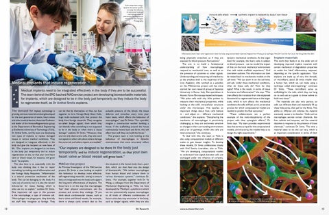
TU/e researchers using a dynamic bioreactor system to study the influence of mechanical loads on macrophage-material interactions. Photograph by Vincent van den Hoogen Photography.
Inflammation-driven heart valve regeneration inside the body using bioresorbable materials. Adapted from Wissing et al. npj Regen. Med. 2017 and De Kort et al. Adv. Drug Deliv. Rev. 2021.
Medical implants that induce regeneration Medical implants need to be integrated effectively in the body if they are to be successful. The team behind the ERC-backed MACxercise project are developing bioresorbable materials for implants, which are designed to be in the body just temporarily as they induce the body to regenerate itself, as Dr Anthal Smits explains. The demand for implant technology is growing, and new materials are being developed for the next generation of stents, heart valves and other medical devices. Associate Professor and leader of the ImmunoRegeneration group at the Department of Biomedical Engineering at Eindhoven University of Technology (TU/e), Dr Anthal Smits, and his team are developing new types of implants to replace damaged tissues in various medical conditions. They are designed to integrate effectively in the body and give the recipient an new lease of life. “Our implants are designed to be there in the body just temporarily and to induce regeneration on site, so that your own heart valve or blood vessel, for instance, will grow back,” he explains. The idea here is to essentially trick the body into thinking that it has to repair something, by making use of inflammation and the Foreign Body Response. “Inflammation is a natural protective mechanism of the body. This can be damaging to the body if it runs out of control, but it is also the natural kick-starter for tissue healing, which is what we try to exploit,” outlines Dr Smits. One important cell type in this process is the macrophage, a type of immune cell. “Macrophages are phagocytes, they basically eat stuff they recognise as foreign. They
12
can do that by themselves or they can fuse together to form Foreign Body Giant Cells, huge multi-nucleated cells that protect the body from foreign materials. They recognise so-called ‘danger signals’, from which they can identify when something is not supposed to be in the body or when there is tissue damage,” explains Dr Smits. “However, they are not only destructive cells, they also send out signals to other cells about where damage has occurred, and where repairs are needed.”
pulsatile pressure of the blood, the tissue deforms and stretches every time your heart beats, which affects the behaviour of macrophages,” says Dr Smits. “For a parallel, imagine a construction worker building a house. If the scaffolding around the house continuously moves back and forth, this will affect how well they can build the house.” The project team is now looking at the behaviour of macrophages in dynamic environments that more accurately reflect
“Our implants are designed to be there in the body just temporarily and to induce regeneration, so that your own heart valve or blood vessel will grow back.” MACxercise project As Principal Investigator of the MACxercise project, Dr Smits is now looking to exploit this behaviour to develop more effective, self-regenerating materials, aiming to ensure they are integrated into the body and support the long-term effectiveness of implants. The focus here is on the way that macrophages ‘feel’ their physical environment, and the stresses and strains they undergo. “If you think about cardiovascular tissues, such as heart valves and blood vessels, for example, there is always cyclic stretch due to the
the situation in the human body than a petri dish, which can then feed into the design of biomaterials. “We isolate macrophages from human blood and culture them in various bioreactor systems,” continues Dr Smits. “For example, together with Dr Ye Wang, a colleague from the Department of Mechanical Engineering at TU/e, we have developed the ‘MiniGym’, a platform in which we can systematically expose macrophages to all kinds of different combinations of factors that they may encounter in the body, such as danger signals, while they are also
EU Research
being physically stretched, as if they are exposed to blood pressure fluctuations.” The aim is to build a fundamental understanding of how macrophages respond to mechanical cues, as well as to the presence of cytokines or other signals. Understanding and measuring cell mechanics at the smallest level is the expertise of Dr Livia Angeloni, who worked as a postdoc on the MACxercise project and has since started her own research group at Sapienza University in Rome, Italy. She specialises in Atomic Force Microscopy-based techniques. “We poke cells with tiny little probes to measure their mechanical properties, while looking at the cells’ intracellular structure under the microscope. This teaches us important things about how cells behave in - and adjust to - different mechanical conditions,” she explains. “Deciphering the mechanics of macrophages is particularly challenging as they are extremely sensitive to changes in their surrounding environment and a lot of pathways within the cells are interconnected,” she continues. To deal with this, the team at TU/e is also using computational models at different scales to gain deeper insights. To develop these models, Dr Smits collaborates closely with Prof Sandra Loerakker, also at TU/e. “We are developing computational models to understand how signals between cells are exchanged under the influence of complex,
Human Foreign Body Giant Cells (fused macrophages) cultured under cyclic stretch. Image courtesy of Dr Bente de Kort and Ir Thijs Conner.
www.euresearcher.com
dynamic mechanical conditions. At the organ level, for example, the heart valve is subject to blood pressure - we can model the impact of that on the local mechanical environment that cells inside scaffolds experience,” Prof Loerakker outlines. This information can then be related back to mechanistic models on the cell level. “We can zoom in on the cell level, and ask; ‘under these mechanical conditions, what do these cells do? How do they signal? What is the result, in terms of tissue formation and inflammation?” she says. “This then affects the transition from the implanted biomaterial to regenerated tissue at the larger scale, which in turn affects the mechanical conditions the cells will feel, so it’s an iterative process for which computational models are needed to predict the final outcomes.” “These collaborations are a perfect example of the multi-disciplinarity of the project with clear synergistic effects,” Dr Smits says. “My team provides experimental data that serves as input for the computational models, and vice versa; the models help us to design the right experiments.”
Implant materials This work then feeds in to the wider aim of developing improved implant materials with certain mechanical or degradation properties to evoke the ‘ideal’ inflammatory response, depending on the specific application. “Our implants are made up of very thin threads, or microfibers, about 20 times smaller than a human hair, which we can make using a technique called electrospinning,” continues Dr Smits, “These microfibers serve as scaffolding for the cells, which they can hang on to and use as a guide to know where to build new tissue.” The materials are also very porous, so cells can infiltrate them and essentially fill up these little pores, then pull on the fibres. This ability to infiltrate and colonise the material eventually causes it to dissolve. “Cells such as macrophages secrete certain chemicals, like free radicals and enzymes, and the material dissolves in response to the presence of those chemicals,” continues Dr Smits. The time a material takes to do this can vary, which is an important consideration in terms of their
Schematic representation of macrophages in a microfibrous biomaterial, exposed to mechanical loads. Created by the ICMS Animation Studio.
13
MACxercise Dissecting Macrophage Mechanobiology to Engineer Immuno-Regenerative Biomaterials
Project Objectives
The ERC funded MACxercise program uses advanced engineering tools to systematically dissect macrophage responses to dynamic mechanical cues and their impact on biomaterial driven tissue regeneration.This work aims to improve prediction of macrophage behaviour and enable the rational design of regenerative implants.
Project Funding
The MACxercise program is financially supported by the European Research Council, ERC Starting Grant, Project 101042538 * The work is further supported by the Gravitation Program “Materials Driven Regeneration” and the Summit Program “DRIVE-RM”, both funded by the Netherlands Organization for Scientific Research (024.003.013 and SUMMIT.1.027, respectively).
Project Partners
Soft Tissue Engineering and Mechanobiology group at TU/e: https://www.tue.nl/en/research/research-groups/ soft-tissue-biomechanics-and-tissue-engineering/
Contact Details
Principal Investigator, Dr A.I.P.M. (Anthal) Smits Eindhoven University of Technology Department of Biomedical Engineering Eindhoven, The Netherlands T: +31 402474738 E: a.i.p.m.smits@tue.nl W: https://www.tue.nl/en/research/ researchers/anthal-smits/ Materials-Driven Regeneration program: https://mdrresearch.nl/ DRIVE-RM program: https://drive-rm.nl/ *Views and opinions expressed are however those of the author(s) only and do not necessarily reflect those of the European Union or the European Research Council. Neither the European Union nor the granting authority can be held responsible for them.
Dr Anthal Smits, Dr Livia Angeloni, and Dr Sandra Loerakker (left to right)
Dr Anthal Smits is an Associate Professor in the Department of Biomedical Engineering and the Institute for Complex Molecular Systems at Eindhoven University of Technology (TU/e). Dr Livia Angeloni is a Tenure-Track Assistant Professor in the Department of Basic and Applied Sciences for Engineering at Sapienza University of Rome. Dr Sandra Loerakker is Professor of Modeling in Mechanobiology at the Department of Biomedical Engineering at TU/e.
14
potential application, an issue that Dr Smits and his colleagues are investigating in collaboration with the project. “No two patients are the same and a lot of patients may be on medication that affects their inflammatory response, how the immune cells respond to the material,” he outlines. “It may be that patient A needs additional medication to slow down or accelerate material degradation for example, while patient B doesn’t, and so patient A may degrade the material faster. We don’t know yet if - and how - that affects the longterm outcome.” These questions are topic of active investigation in several other research consortia, alongside the MACxercise project, including the Materials-Driven Regeneration and the DRIVE-RM programs.
Trials A number of clinical trials are being conducted by spin-off companies that originate from the overarching research group at TU/e. One trial centres around using a stent device called a Resorbable Fibrillated Scaffold (RFS) to treat Chronic Limb Threatening Ischemia (CLTI), a fairly common condition which affects the lower legs and feet. “This trial is being conducted by STENTiT, a spin-off company from our Department. So far it’s progressing well, but it’s a fairly small patient cohort until now,” acknowledges Dr Smits. Further trials are ongoing, looking at the effectiveness of certain materials in different applications. “The team at Xeltis - another spin-off - are carrying out a trial looking at self-healing blood vessels for kidney dialysis for example. They’ve been working in this area for a relatively long time, and we have a long-standing collaboration with them,” says Dr Smits. “These clinical experiences are very important to us, as they give us important insights, for example into what immune cells are present at which time and if - and how this may differ between patients.” The main aim with these types of selfhealing implants is to improve quality of life
for patients. “By having a living replacement that can adapt and heal itself, just like your own tissues and organs do, we hope to avoid complications that can occur with non-living implants,” says Dr Smits. “One important aspect of the technology we develop is that the implants are relatively simple devices; they don’t require any human or animal tissue or complicated and expensive cultures in the lab,” he continues. “This means that these implants have the potential to be broadly accessible. They are a sustainable and affordable solution.” Dr Smits is keen to translate the research at his lab into societal benefits, yet at the same time he is motivated by fundamental questions. “I find immune cells fascinating,” he says. “It’s incredible to see these cells doing what they do under the microscope, to learn what they are capable of and how intricately it is all regulated. When you realise that all of that is continuously happening inside your body while you are sitting there, it is truly mind-blowing”. This work requires close collaboration between different disciplines, and Dr Smits is keen to stress that these projects are very much a team effort. “We’ve been able to develop these technologies because we build on the pioneering work of our mentors and because we work as a team with complementary expertise from different disciplines, including chemists, immunologists and clinicians,” he outlines. “What makes our work innovative is that we study immune cells with our ‘engineering glasses’ on. This means that we ask different sometimes seemingly stupid - questions than an immunologist would do for example,” he outlines. “By closely collaborating with experts from different disciplines we are able to share information effectively as we look to translate our findings into tangible progress. This complementarity enables us to tackle scientific challenges in a different way which will, hopefully, lead to new solutions.”
Dr Anthal Smits showing a self-regenerating blood vessel graft based on biodegradable material. Photograph by Leonie Voets.
EU Research