28 minute read
national and regional FMD control


Species
Confirmed transmission to species
Advertisement
Exposure and serotype Clinical signs
Antibody tested positive Virus positive
Sambar deer (Rusa unicolor) Nat (O) Y Y
Sheep (Ovis aries) Nat/Exp Y Y Y Sika deer (Cervus nippon) Cattle Sheep Exp (C) Mild Y Y Spotted deer (Axis axis) Nat (O) Y Y
Springbok (Antidorcas marsupialis) Squirrel, Grey (Sciurus carolinensis) Nat (SAT-1, 2, 3) Y N
Exp (A, C, O) Y Y
Squirrel, Indian (Funambulus pennanti) Steenbok (Rhaphicerus campestris) Swine (Sus scrofa domesticus) Tapir, Asian (Tapirus indicus) Tapir, South American (Tapirus terrestris) Thomson’s gazelle (Eudorcas thomsonii) No transmission Exp (A, Asia-1, C, O) Severe Y
Nat Y
Nat/Exp Y Y Y Nat Y
Nat Y
Nat (A, C, O) Y
Topi (Damaliscus korrigum) Nat (O) Y Tsessebe (Damaliscus lunatus) Nat (SAT-1, 2, 3) Y Vicugna (Vicugna vicugna) Exp Vole, European water (Arvicola amphibius amphibius) Water Vole Exp (A, C, O) Y Y Y Vole, Field/Short-tailed (Microtus agrestis) No transmission Exp Y Y Warthog (Phacochoerus aethiopicus) Nat (SAT-1, 2, 3) Exp (SAT-2) Severe Y Y Y
Waterbuck (Kobus ellipsiprymnus) Water rat (Hydromys chrysogaster) Nat (SAT-1, 2, 3) Y
Exp (A, SAT-1) Mild Y Y
Watussi/Wild buffalo (Bos taurus) Nat Nat (A) Y Y Y
White-tailed deer (Odocoilleus virginianus)
Wildebeest, Black (Connochaetes gnou) Cattle Exp (O) Exp (O) Nat (O) Nat Y Y Y
Y Y Y Y
Y
Wildebeest, Common (Connochaetes taurinus) Nat (A, SAT-1, 2) Nat (O, SAT-1, 2) Exp (O)
Y Mild Y Y Y Y
Species
Confirmed transmission to species
Exposure and serotype Clinical signs
Antibody tested positive Virus positive
Wild boar (Sus scrofa) Nat Nat (O) Exp (A) Exp (O) Y Y Y Y Y Y Y Y
Wombat (Vanitatus hirsutas) No transmission Exp (A, O, SAT-1) N Y Y
Yak, Domestic (Bos grunniens/ Poephagus grunniens) Nat (O) Y Y
Yak, Wild (Bos mutus) Nat (O) Y Y
Antelope species may serve to propagate FMD. Impala, particularly in the KNP and possibly elsewhere in subSaharan Africa, have been associated with outbreaks in cattle (43). Ninety per cent of infections in impala occur from June to November. This is the same time in which buffalo calves are losing their maternal antibodies and becoming infected with FMDV (5). Since impala have not been shown to become carriers, it is suspected that impala are an intermediary species and become acutely infected and spread the virus to cattle outside KNP by jumping over fences (43). Foot and mouth disease outbreaks are seen regularly in the countries of Central Asia. There are several important wildlife species that are impacted by FMD. Mongolian gazelles (Procapra gutturosa) from the Eastern Steppe of Mongolia have been infected by FMDV; however, several studies indicate that it is the continued circulation of FMDV in the domestic livestock of the Mongolian Eastern Steppe that results in the virus entering the susceptible gazelle population (7, 29). Furthermore, there is no evidence for the persistence of the virus in the gazelle population between outbreaks (38), and actions such as culling of Mongolian gazelles and fencing do not appear to have any impact on the disease in livestock (7). In Kazakhstan, Saiga antelope (Saiga tatarica) are susceptible to FMD and suffer from more severe disease than what is seen in domestic ruminants. Saiga populations have declined dramatically due to excessive hunting in the 1990s. Mortality can be high (as much as 75% in experimentally infected animals) and past outbreaks have resulted in a loss of 10% of a population (27). Foot and mouth disease has been found in wildlife in the Middle East. FMD has infected captive populations of Arabian oryx (Oryx leucoryx) in Bahrain and the United Arab Emirates (UAE) with high mortality, and seropositivity has been found in at least two captive individuals in Saudi Arabia. However, FMDV has not been detected in wild Arabian oryx populations in Saudi Arabia (14). Antibodies to all seven serotypes of FMDV have been found in dromedary camels (Camelus dromedaries) (47). They are believed to be mostly resistant to clinical FMD and virus isolation is sometimes difficult; however, natural and experimental infections have occurred. They are not believed to play a significant role in the transmission to livestock (45). Outbreaks among mountain gazelles in Israel have resulted in about 10% to 15% of the population becoming acutely infected with a 50% mortality rate (35). Domestic water buffalo (Bubalus bubalis) are common throughout Asia and are susceptible to FMD. In India, water buffalo are frequently kept with both cattle and sheep (23). Maroudam et al. demonstrated that water buffalo can transmit FMDV to cattle and to each other (24). Clinical signs in water buffalo tend to be more covert than the clinical signs in cattle. In addition, water buffalo appear to have a longer incubation period and are infective prior to exhibiting any lesions. Water buffalo can become acutely infected as well as become a carrier for the virus (23, 24). Persistence of infection was found from one to two years after exposure to the virus (4). Feral swine, wild boar (Sus scrofa) and feral water buffalo (B. bubalis) are often found in proximity to livestock and could play an important role in the epidemiology of FMD. In Sri Lanka, FMD outbreaks frequently occur in areas in close proximity to national parks where there are significant populations of feral water buffalo and wild boar (32). Severe FMD has been reported in mithun (Bos frontalis), yak (Bos grunniens) and gaur (Bos gaurus). Frequently, migratory cattle come into contact with these species and transmission occurs. Asian elephants suffer a moderately severe disease particularly in the younger animals, and a serosurvey at an Indian zoo found positive antibodies
to FMD despite no history of clinical disease in many elephants (18). Other species documented to be infected are provided in Table I. We found only one published experimental study conducted on the susceptibility of various Australian fauna (36). Clinical disease was not apparent for most of the infected animals, and only the red kangaroo (Megaleia rufa), tree kangaroo (Dendrolagus matschiei), water rat (Hydromys chrysogaster) and echidna exhibited mild clinical symptoms. Others are listed in Table I. Despite experimental infections of several European cervids (13, 16), reports of natural infection in captive animals at European zoos (34), and the recent report of infected roe deer (Capreolus capreolus) and wild boar (Sus scrofa) in Bulgaria, there is no evidence for the maintenance of FMDV in wildlife in Europe (10). Severe natural and experimental infections have been reported in reindeer (Rangifer tarandus) (39). Experimental infections have been demonstrated in many species (Table I). A survey during and after the 2011 outbreak of FMD serotype O among livestock in Bulgaria found a low seroprevalence and clustered distribution of positive roe deer and wild boar, indicating that FMD failed to become established in the wild populations. This suggests that wildlife populations are not able to maintain FMDV in the absence of FMD infection in livestock (10). The first and only known outbreak of FMD in North American wildlife occurred in mule deer (Odocoileus hemionus) after an outbreak among cattle in California in 1924. Ten per cent of a population of mule deer exhibited clinical FMD. A mathematical model created by Ward et al. (44) found that wild deer and feral pigs have the potential to amplify disease spread and form a possible reservoir of FMD virus infection in Texas, but this has never been found in real outbreaks anywhere. Experimental data indicate that there are many North American species that are susceptible to FMD and are capable of transmitting the disease to cattle. North American bison (Bison bison) and elk (Cervus elaphus nelsonii) have been infected with FMDV experimentally. Others are listed in Table I. There has been no evidence of transmission from wildlife to livestock in South America and no history of outbreaks of disease despite the fact that many South American wild animal species are susceptible to FMDV (31). New World camelids can become infected with FMDV; however, they are not highly susceptible and appear to be unable to transmit the disease (32). Llamas were difficult to infect under experimental conditions and, when infected, they developed only very mild disease (12). Other reports of susceptible species in South America are presented in Table I. Despite conjecture on the role of South American wildlife as a possible reservoir for FMDV, there is no evidence to date to support that claim.
Efforts to manage wildlife and feral domestic animals
The feral animal situation should be considered separately from what is occurring with native wildlife. Feral animals are more similar to domestic animals than truly wild, native animals. They technically derive from domesticated genetic stock and so retain many of the physiological and behavioural qualities of domesticated animals. They are also more likely to be in contact with domestic livestock and humans. Although there has not been any sufficient evidence for the propagation of FMDV within feral populations (32), there does exist a greater risk than the risk presented from native wildlife. A wild boar found near the Turkish border was found with FMD lesions just prior to an outbreak among domestic livestock in Bulgaria (10). Feral animals have not been studied extensively with regard to FMD; however, several studies suggest that there is the possibility that feral swine have the ability to become carriers of the virus in their pharynx at least 33–35 days post inoculation (10, 26). Another report from an FMD outbreak in Israel suggested aerosol transmission from feral swine in Jordan. Mongolian gazelles, as mentioned previously, have not been found to be a reservoir of FMD but are passively infected with FMDV when outbreaks occur among livestock (7, 29, 38). Modified stamping out, as was conducted in 2010 in Mongolia for Mongolian gazelles and livestock, involved the culling of clinically diseased animals. This method does not further decrease the spread of FMD, as not all infected animals will demonstrate clinical illness and often FMD is infectious prior to any lesions. The controlled movement of people and livestock as well as vaccinating before and during outbreaks are more effective means of handling an outbreak of FMD (38). Veterinary cordon fences are commonly used to separate livestock from wildlife in southern Africa. This method is accepted by the OIE as one of the methods of establishing FMD disease-free zones in southern Africa (22,
39). However, the reliance of FMD exclusion provided by fences is problematic. The high cost of construction, maintenance and patrolling of the fences may be cost prohibitive in certain countries (37). Fences are frequently subject to various environmental and human pressures, including flooding, breaks due to wildlife movement, particularly elephants, and damage due to theft (22). The sheer magnitude of fencing that exists in some parts of southern Africa (for example, the perimeter fence surrounding the Kruger National Park is 750 km long) makes it difficult for fences to be maintained in a timely manner (21). Compromised fences allow cattle to move into reserves and wildlife areas.
The additional impact to wildlife populations and local people cannot be ignored. Fences interfere with normal migration patterns and in times of water scarcity block wildlife from critical water sources. Fences also prevent genetic exchange among populations of various wild species, thus potentially resulting in inbreeding and loss of genetic diversity. Fenced wildlife populations may remain small and capped, which could have significant effects on long-term survival or can exceed the carrying capacity of the land available, resulting in overcrowding, malnutrition, increased infectious disease problems and the need for population control efforts.
Recommendations
The diversity of this virus and its behaviour make it problematic to generalise any one strategy. The actual method used to control FMD will ultimately be regionally specific. Eradication is not feasible and may not be needed everywhere. Sub-Saharan Africa has the unique consideration of a significant reservoir of FMD with a wild species that may never truly result in disease freedom defined geographically. Efforts should be directed at finding opportunities for Africa to participate in international trade and to improve the economic situation that do not require complete eradication of FMD. Mechanisms such as commodity-based trading, where there is a negligible risk for transmission of FMD, provide one such opportunity. Vaccine programmes should be adapted to improve efficacy through the incorporation of appropriate strains based on the circulating viruses, increasing the frequency of vaccination using currently available vaccines, ensuring proper vaccine handling and conducting post-vaccination monitoring. Improved vaccines are needed. In addition, mixed land-use scenarios, such as those envisioned for the Transfrontier Conservation Areas (TCFAs), where there is added utilisation of wildlife resources, could be highly beneficial both economically and politically.
Conclusion
To date, the scientific evidence indicates that outside the sub-Saharan Africa situation, with SAT types of FMD adapted to African buffalo, effective control of FMD among domestic livestock will result in the protection of both livestock and wildlife without requiring direct management or interventional activities directed at wildlife. Control of feral domestic animals may be required. In parts of southern Africa, revenues from tourism now exceed the total revenues of agriculture, fisheries and forestry combined (1), and thus strategies selected for FMD control need to ensure that the costs of controlling the disease are put into context with the revenues that are or could be generated by other land-use choices, selection of species managed for meat, trophies, hides or tourism, or other means of ensuring safe and FMD-free meat such as that provided by compartmentalisation and commoditybased trade mechanisms. This broader view of animal health and human livelihoods and well-being provides an opportunity for the veterinary profession to make a significant contribution to the global good. The protection of wildlife should be a priority in the Global FMD Control Strategy.
References
1. Albertson A. (2010). – The Scott Wilson ‘fencing impacts’ report: Ten years on. In Fencing impacts: A review of the environmental, social and economic impacts of game and veterinary fencing in Africa with particular reference to the Great Limpopo and Kavango-Zambezi Transfrontier Conservation Areas (Ferguson K. & Hanks J., eds). Mammal Research Institute, Pretoria, 83–98. 2. Anderson E.C., Doughty W.J., Anderson J. & Paling R. (1979). – The pathogenesis of foot-and-mouth disease in the African bu alo (S ncerus ca er) and the role of this species in the epidemiology of the disease in Kenya. J. Comp. Pathol., 89 (4), 541–549.
3. Arzt J., Baxt B., Grubman M.J., Jackson T., Jule N., Rhyan J., Rieder E. Waters R. & Rodriguez L.L. (2011). – The pathogenesis of foot-and-mouth disease II: Viral pathways in swine, small ruminants, and wildlife; myotropism, chronic syndromes, and molecular virus-host interactions. Transbound. Emerg. Dis., 58 (4), 305–326. 4. Barros J.J., Malirat V., Rebello M.A., Costa E.V. & Bergmann I.E. (2007). – Genetic variation of foot-and-mouth disease virus isolates recovered from persistently infected water bu alo (Bubalus bubalis). Vet. Microbiol., 120 (1–2), 50–62. 5. Bastos A.D., Bosho C.I., Keet D.F., Bengis R.G. & Thomson G.R. (2000). – Natural transmission of foot-andmouth disease virus between African bu alo (S ncerus ca er) and impala (Aepyceros melampus) in the Kruger National Park, South Africa. Epidemiol. Infect., 124 (3), 591–598. 6. Bengis R.G., Thomson G.R., Hedger R.S., De Vos V. & Pini A. (1986). – Foot-and-mouth disease and the African bu alo (Syncerus ca er). 1. carriers as a source of infection for cattle. Onderstepoort J. Vet. Res., 53 (2), 69–73. 7. Bolortsetseg S., Enkhtuvshin S., Nyamsuren D., Weisman W., Fine A., Yang A. & Joly D.O. (2012). – Serosurveillance for foot-and-mouth disease in Mongolian gazelles (Procapra gutturosa) and livestock on the eastern steppe of Mongolia. J. Wildl. Dis., 48 (1), 33–38. 8. Condy J.B. & Hedger R.S. (1974). – The survival of foot-and-mouth disease virus in African bu alo with nontransference of infection to domestic cattle. Res. Vet. Sci., 16 (2), 182–185. 9. Condy J.B., Hedger R.S., Hamblin C. & Barnett I.T. (1985). – The duration of the foot-and-mouth disease virus carrier state in African bu alo (i) in the individual animal and (ii) in a free-living herd. Comp. Immunol. Microbiol. Infect. Dis., 8 (3–4), 259–265. 10. European Food Safety Authority (2012). – Scienti c opinion on foot-and-mouth disease in Thrace. EFSA J., 10 (4), 2635. 11. Ferris N.P., Condy J.B., Barnett I.T. & Armstrong R.M. (1989). – Experimental infection of eland (Taurotrages oryx), sable antelope (Ozanna grandicomis) and buffalo (Syncerus caffer) with foot-and-mouth disease virus. J. Comp. Pathol., 101 (3), 307–316. 12. Fondevila N.A., Marcoveccio F.J., Blanco Viera J.B., O’Donnell V.K., Carrillo B.J., Schudel A.A., David M., Torres A. & Mebus C.A. (1995). – Susceptibility of llamas (Lama glama) to infection with foot-and-mouth-disease virus. Zentralbl. Veterinärmed., B, 42 (10), 595–599. 13. Forman A.J., Gibbs E.P., Baber D.J., Herniman K.A. & Barnett I.T. (1974). – Studies with foot-and-mouth disease virus in British deer (red, fallow and roe). II. Recovery of virus and serological response. J. Comp. Pathol., 84 (2), 221–229. 14. Frolich K., Hamblin C., Jung S., Ostrowski S., Mwanzia J., Streich W.J., Anderson J., Armstrong R.M. & Anajariyah S. (2005). – Serologic surveillance for selected viral agents in captive and free-ranging populations of Arabian oryx (Oryx leucoryx) from Saudi Arabia and the United Arab Emirates. J. Wildl. Dis., 41 (1), 67–79. 15. Gainaru M.D., Thomson G.R., Bengis R.G., Esterhuysen J.J., Bruce W. & Pini A. (1986). – Foot-and-mouth disease and the African buffalo (Syncerus caffer). II. Virus excretion and transmission during acute infection. Onderstepoort J. Vet. Res., 53 (2), 75–85. 16. Gibbs E.P., Herniman K.A. & Lawman M.J. (1975). – Studies with foot-and-mouth disease virus in British deer (muntjac and sika). Clinical disease, recovery of virus and serological response. J. Comp. Pathol., 85 (3), 361–366. 17. Grubman M.J. & Baxt B. (2004). Foot-and-mouth disease. Clin. Microbiol. Rev., 17 (2), 465–493. 18. Hedge R., Gomes A.R., Giridhar P., Venkatesth M.D., Sudarshan K.J., Shivshankar & Renukaprasad C. (2010). – Screening of elephants (Elephas maximus) for foot and mouth disease virus antibodies by liquid phase block ELISA. Zoos Print, 25 (11), 27. 19. Hedger R.S., Condy J.B. & Golding S.M. (1972). – Infection of some species of African wild life with foot-andmouth disease virus. J. Comp. Pathol., 82 (4), 455–461. 20. Howell P.G., Young E. & Hedger R.S. (1973). – Foot-and-mouth disease in the African elephant (Loxodonta africana). Onderstepoort J. vet. Res., 40 (2), 41–43, 45, 47–52. 21. Jori F., Brahmbhatt D., Fosgate G.T., Thompson P.N., Budke C., Ward M.P., Ferguson K. & Gummow B. (2011). – A questionnaire-based evaluation of the veterinary cordon fence separating wildlife and livestock along the boundary of the Kruger National Park, South Africa. Prev. Vet. Med., 100, 210–220. 22. Jori F., Vosloo W., Du Plessis B., Bengis R., Brahmbhatt D., Gummow B. & Thomson G.R. (2009). – A qualitative risk assessment of factors contributing to foot and mouth disease outbreaks in cattle along the western boundary of the Kruger National Park. Rev. Sci. Tech. Off. Int. Epiz., 28 (3), 917–931. 23. Maddur M.S., Kishore S., Gopalakrishna S., Singh N., Suryanarayana V.V. & Gajendragad M.R. (2009). – Immune response and viral persistence in Indian buffaloes (Bubalus bubalis) infected with foot-and-mouth disease virus serotype Asia 1. Clin. Vaccine Immunol., 16 (12), 1832–1836.
24. Maroudam V., Nagendrakumar S.B., Madhanmohan M., Santhakumar P., Thiagarajan D. & Srinivasan V.A. (2008). – Experimental transmission of foot-and-mouth disease among Indian buffalo (Bubalus bubalis) and from buffalo to cattle. J. Comp. Pathol., 139 (2–3), 81–85. 25. McVicar J.W., Sutmoller P., Ferris D.H. & Campbell C.H. (1974). – Foot-and-mouth disease in white-tailed deer: Clinical signs and transmission in the laboratory. Proc. Annu. Meet. U.S. Anim. Hlth Assoc., 78, 169–180. 26. Mohamed F., Swafford S., Petrowski H., Bracht A., Schmit B., Fabian A., Pacheco J.M., Hartwig E., Berninger M., Carrillo C., Mayr G., Moran K., Kavanaugh D., Leibrecht H., White W. & Metwally S. (2011). – Foot-and-mouth disease in feral swine: Susceptibility and transmission. Transbound. Emerg. Dis., 58 (4), 358–371. 27. Morgan E.R., Lundervold M., Medley G.F., Shaikenov B.S., Torgerson P.R. & Milner-Gulland E.J. (2006). – Assessing risks of disease transmission between wildlife and livestock: The saiga antelope as a case study. Biol. Conserv., 131 (2), 244–254. 28. Moussa A.A.M., Daoud A., Tawfik S., Omar A., Azab A. & Hassan N.A. (1979). – Susceptibility of water-buffaloes to infection with foot-and-mouth disease virus. J. Egypt. Vet. Med. Assoc., 39 (1), 65–83. 29. Nyamsuren D., Joly D.O., Enkhtuvshin S., Odonkhuu D., Olson K.A., Draisma M. & Karesh W.B. (2006). – Exposure of Mongolian gazelles (Procapra gutturosa) to foot and mouth disease virus. J. Wildl. Dis., 42 (1), 154–158. 30. Perl S., Yadin H., Yakobson B., Zuckerman E. & Orgad U. (1989). – Pathological changes in mountain gazelles challenged with FMD virus with special reference to pancreatic lesions. Rev. Sci. Tech. Off. Int. Epiz., 8 (3), 765–769. 31. Pinto A.A. (2004). – Foot-and-mouth disease in tropical wildlife. Ann. N.Y. Acad. Sci., 1026 (Oct), 65–72. 32. Roeder P.L. (2009). – Opportunities and constraints posed by wildlife in the diagnosis and epidemiological analysis of foot and mouth disease virus infection. Proceedings from the first OIE/FAO global conference on foot and mouth disease: the way towards global control, 24-26 June 2009, Asunción, Paraguay, 101–106. 33. Salt J.S. (1993). – The carrier state in foot and mouth disease – an immunological review. Br. Vet. J., 149 (3), 207–223. 34. Schaftenaar W. (2002). – Use of vaccination against foot and mouth disease in zoo animals, endangered species and exceptionally valuable animals. In Foot and mouth disease: facing the new dilemmas (G.R. Thomson, ed.). Rev. Sci. Tech. Off. Int. Epiz., 21 (3), 613–623. 35. Shimshony A., Orgad U., Baharav D., Prudovsky S., Yakobson B., Bar Moshe B. & Dagan D. (1986). – Malignant foot-and-mouth disease in mountain gazelles. Vet. Rec., 119 (8), 175–176. 36. Snowdon W.A. (1968). – The susceptibility of some Australian fauna to infection with foot and mouth disease virus. Aust. J. Experim. Biol. Med. Sci., 46 (6), 667–687. 37. Sutmoller P., Thomson G.R., Hargreaves S.K., Foggin C.M & Anderson E.C. (2000). – The foot-and-mouth disease risk posed by African buffalo within wildlife conservancies to the cattle industry of Zimbabwe. Prev. Vet. Med., 44 (1–2), 43–60. 38. Thomson G.R. (2011). – Current disease control policies and ‘knowledge gaps’ in the epidemiology of foot and mouth disease on Mongolia’s eastern steppe. Report on a consultancy conducted on behalf of the Wildlife Conservation Society, 25 February 2011, 1–30. 39. Thomson G.R., Vosloo W. & Bastos A.D. (2003). – Foot and mouth disease in wildlife. Virus Res., 91 (1), 145–161. 40. Vosloo W., Bastos A.D.S., Sahle M., Sangare O. & Dwarka R.M. (2005). – Virus topotype and the role of wildlife in foot and mouth disease in Africa. In Conservation and development interventions at the wildlife/livestock interface: implications for wildlife, livestock and human health (Osofsky S.A., Cleavland S., Karesh W.B., Kock M.D., Nyhus P.J., Starr L. & Yang A., eds). IUCN, Gland, Switzerland and Cambridge, United Kingdom, 67–80. 41. Vosloo W., Bastos A.D., Sangare O., Hargreaves S.K. & Thomson G.R. (2002). – Review of the status and control of foot and mouth disease in sub-Saharan Africa. In Foot and mouth disease: facing the new dilemmas (G.R. Thomson, ed. Rev. Sci. Tech. Off. Int. Epiz., 21 (3), 437–449. 42. Vosloo W., de Klerk L.M., Boshoff, Botha B., Dwarka R.M., Keet D. & Haydon D.T. (2007). – Characterisation of a SAT-1 outbreak of foot-and-mouth disease in captive African buffalo (Syncerus caffer): Clinical symptoms, genetic characterisation and phylogenetic comparison of outbreak isolates. Vet. Microbiol., 120 (3–4), 226–240. 43. Vosloo W., Thompson P.N., Botha B., Bengis R.G. & Thomson G.R. (2009). – Longitudinal study to investigate the role of impala (Aepyceros melampus) in foot-and-mouth disease maintenance in the Kruger National Park, South Africa. Transbound. Emerg. Dis., 56 (1–2), 18–30. 44. Ward M.P., Laffan S.W. & Highfield L.D. (2007). – The potential role of wild and feral animals as reservoirs of footand-mouth disease. Prev. Vet. Med., 80 (1), 9–23. 45. Wernery U. & Kaaden O.R. (2004). – Foot-and-mouth disease in camelids: A review. Vet. J., 168 (2), 134–142.
46. Woodbury E.L. (1995). – A review of the possible mechanisms for the persistence of foot-and-mouth disease virus. Epidemiol. Infect., 114 (1), 1–13. 47. Yousef M.R., Mazloum K.S. & Al-Nakhli H.M. (2012). – Serological evidence of natural exposure of camels (Camelus dromedaries) to foot and mouth disease virus. Vet. World, 5 (4), 197–200.
The Progressive Control Pathway for FMD (PCP-FMD): a tool for developing sustainable long-term national and regional FMD control
K.J. Sumption (1)*, M. McLaws (1)*, C.J. Bartels (1)*, P.W. De Leeuw (3)* , J. Domenech (2), J. Lubroth (3)* & G. Ferrari (3)*
(1) Food and Agriculture Organization of the United Nations (FAO), European Commission for the Control of Foot-and-Mouth Disease (EuFMD), Viale delle Terme di Caracalla, 00153 Rome, Italy (2) World Organisation for Animal Health (OIE), 12 rue de Prony, Paris, France (3) Food and Agriculture Organization of the United Nations (FAO), Viale delle Terme di Caracalla, 00153 Rome, Italy
* The views expressed in this publication are those of the authors and do not necessarily reflect the views of the Food and Agriculture Organization of the United Nations. Also, the designations employed and the presentation of material in this information product do not imply the expression of opinion whatsoever on the part of the Food and Agriculture Organization of the United Nations concerning the legal status of any country, territory, city or area or of its authorities, or concerning the delimitation of its frontiers or boundaries. Content and errors are exclusively the responsibility of the authors. Correspondence: Keith.Sumption@fao.org
Summary
The Progressive Control Pathway for FMD (PCP-FMD) was designed to assist countries where foot and mouth disease (FMD) is endemic to develop sustainable national FMD control policies appropriate to their livestock sectors, and medium- to long-term national strategies for progressive reduction of the disease impact. First developed by the European Commission for the Control of Foot-and-Mouth Disease (EuFMD) and Food and Agriculture Organization of the United Nations (FAO), further development of the PCP was recommended by the World Organisation for Animal Health (OIE)/FAO Global Conference in 2009 in Paraguay and, since 2011, adopted by both FAO and the OIE as a joint tool. The PCP-FMD is composed of five stages of increasing level of control, to the point where an application to the OIE for official recognition of freedom from FMD (with or without vaccination) may be successful and sustainable. The first stage, appropriate for all countries not officially free of from FMD, involves a low cost but comprehensive assessment of FMD risk and control options, considering the capacity and drivers for public and private investment and the benefits to sectors, culminating in a revised national policy and longer term strategy. The PCP processes assist the identification of gaps to be addressed and the development of national or regional support projects. National PCP progress underpins regional progress, and 10- to 15-year regional roadmaps have been developed in Asia and Africa, complementing those existing in South-East Asia and China and in South America. The PCP is thus an important tool in the global strategy for FMD, based on self-sustaining national investments backed by regional efforts to address transboundary issues.
Keywords
Europe – Foot and mouth disease – Global Strategy – Progressive Control Pathway (PCP) – Regional Roadmap – Vaccination – Virus pools – West Eurasia.
Introduction
Foot and mouth disease (FMD) is recognised as one of the most important, if not the most important, diseases of animals. In endemic countries, it is among the most common diseases of livestock, and recent surveys have indicated that exposure in the first year of life may approach 50% of ruminants (and pigs), and multiple episodes of FMD occur during the productive life of an animal. FMD distribution roughly mirrors economic development; wealthy countries have, through enormous effort, mostly eradicated FMD and the setback of an incursion can cost millions or billions of dollars to control, whereas many less developed countries have continuous circulation of FMD infection and experience major epidemics every 2–4 years. Sustained FMD freedom has been achieved in
over 60 countries and, possibly, one of the seven FMD serotypes (serotype C) has disappeared from circulation, in part resulting from the general control efforts in Europe and South America. Given the burden of infection and frequency of epidemics, and the number of less developed countries that do not have publicly funded vaccination programmes, the prospects for achieving the national or zonal freedom from FMD in most cases appear so far out of reach that they cannot realistically be considered within the near future. The scale of investment and competencies required are daunting for any country. In countries already applying preventive vaccination, the high recurrent cost of such programmes and unclear or unrealistic expectations of these programmes have raised questions about the extent of stakeholder support, responsibilities and benefits, and sustainability. For these reasons, the Progressive Control Pathway for FMD (PCP-FMD) was developed as a framework for the development of sustainable FMD management, and regionally co-ordinated ‘roadmaps’ have been developed for countries in most of Eurasia and Africa using the PCP-FMD approach and assessment tools. The initial take-up of the approach at national level has been encouraging, but sustained support will be needed to ensure that the benefits of the approach are translated into national policy and practice, and a mature capacity to evaluate progress is achieved at national level.
PCP-FMD principles and application
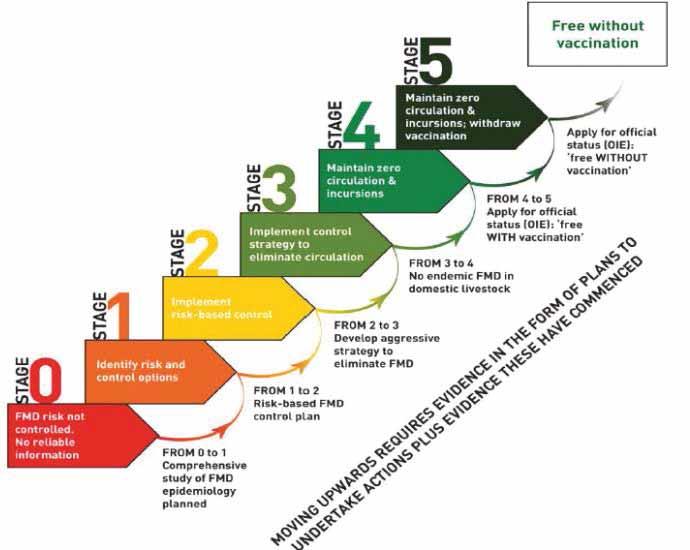
The PCP-FMD has been developed by the European Commission for the Control of Foot-and-Mouth Disease (EuFMD) and the Food and Agriculture Organization of the United Nations (FAO) in 2008 (2) to assist countries where FMD is still endemic to progressively reduce the impact of FMD and the load of FMD virus. The PCP-FMD was adopted by FAO, EuFMD and the World Organisation for Animal Health (OIE) in 2011 as a joint tool, creating for the first time a ‘single framework’ that includes all possible levels of FMD control, from uncontrolled virus circulation to rigorously maintained freedom from infection without vaccination. The development of this single and comprehensive framework is an important step in international FMD management, enabling the possibility of setting national, regional and even global targets for progression. The PCP-FMD is composed of five stages that guide the planning and management of efforts to increase the level of control to the point where an application to the OIE for endorsement of the national control programme or, at higher levels, official recognition of freedom from FMD (with or without vaccination) may be successful and sustainable (Fig. 1). The stages and requirements have been published in detail (1). Stage 1 assists identifying appropriate control options, and Stage 2 involves the implementation of the chosen policy. It is not expected in Stage 2 that control measures will reduce FMD incidence across the entire population; measures might be largely privately financed, or a balance of public funding (e.g. in border regions) and private funding (e.g livestock keeper-supported vaccination programmes). Stage 2 therefore does not imply large investments at the national level, but if the producer is to pay for preventive measures, he/she will expect adequate information and access to effective vaccines, and this in itself will create demand for information and results to guide vaccine selection. However, Stage 3, when progressive elimination of virus circulation is the objective, normally requires very significant national capacity and ongoing investment, including the ability to regulate internal trade and ensure sufficient immunity is maintained in critical populations to prevent virus circulation; evidence of progress within Stage 3 is important precursor for endorsement by the OIE (6) of the FMD control programme. Stages 4 and 5 involve the development of capacity and evidence needed for compliance with the official OIE status for freedom of FMD with or without vaccination, respectively. The PCP approach is based on the following four principles: 1. Active monitoring for FMD virus circulation and understanding the epidemiology of FMD virus transmission.
This underpins the foundation of any control programme, and therefore activities to meet these requirements are common in all stages. The improved information generated is of benefit nationally and regionally. 2. Activities in each PCP stage are appropriate to the required reduction in virus circulation and mitigation of disease risk to be achieved.
3. The optimisation of resource use for FMD control is achieved through the targeting of measures to the husbandry systems and critical risk points where the impact on disease control and/or virus circulation will be greatest.
4. Activities and their impacts are measurable at each stage, comparable between countries, and generate information and potential benefits to national as well as international stakeholders. The monitoring of outcomes (indicators of control effectiveness), within a national FMD management system, is included at the higher PCP stages.
Fig. 1 Stages in the Progressive Control Pathway for FMD (PCP-FMD)
Passage to a higher stage requires completion of the milestones indicated below the arrows, and initiating key actions of the higher stage. Stages 4 and 5 involve activities that lead to submission of applications for official recognition of freedom
Source : FAO/EuFMD
It should be noted that the second and third principles support a risk-based approach to national control strategies, and, through the focus on critical risk reduction points, avoid prescriptive requirements for capacities or activities that are not identified as important to risk reduction. This potentially reduces the level of investment at national level at Stage 2, but entry into Stage 3 requires a very thorough assessment of the costs and benefits since the competencies and level of investments required to reduce virus circulation are very much higher. As such, PCP-FMD can be viewed as a policy optimisation pathway, assisting countries to determine their optimal position after considering the national situation of risk, costs to control, and benefits to sectors and public and private stakeholders. Since PCP Stage 1 involves a comprehensive assessment of control options based on what is necessary, achievable and sustainable with national resources invested by public and private sectors, the importance of this initial stage should not be underestimated in the development of a national PCP roadmap where the optimal stage target may be different between zones of the country, for socio-economic as well as risk reasons. Although it may also be viewed as a pathway towards FMD eradication, it cannot be assumed that all countries will find economic advantages to proceed further than Stage 2. On the other hand, countries usually free of FMD that detect an incursion of the disease would normally not enter the pathway, but rather would act to eradicate the disease and reapply directly to the OIE for reinstatement of an officially recognised FMD-free status as soon as possible.


