10 minute read
3D Printers Move Beyond Curiosities


cines or other, less effective treatment methods.”
Transplanted donor valves have drawbacks, sometimes requiring antirejection medications and subsequent surgeries to replace the initial valve since it does not grow as the child ages. Davis and his team are developing child-sized valves using the patient’s own cells through Induced Pluripotent Stem Cells technology. “We want to fix the patient, using the patient, within the patient,” says Davis.
Stitches and staples are effective at closing wounds, but they can also be invasive and painful and leave visible scars. Felmont Eaves, professor of plastic and reconstructive surgery at Emory and director of the Emory Aesthetic Center, has spent six years using 3D printing to transform the way surface lesions and wounds are treated.
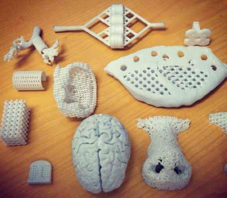
As part of the DIY revolution, Emory doctors are using 3D printers to make body parts, medical tools, training models, and prototypes.
At the Emory Orthopaedics & Spine Center, sports medicine surgeons like Matthew Pombo are using 3D printers to design custom knee replacements for elite athletes during their careers and after retirement.
“In aging athletes, you can see the impact of a lifetime of sports and activity,” says Pombo, who has performed about 500 customized 3D-knee replacements at Emory Johns Creek Hospital. “The patient-specific 3D-knee implant is ideal for those with arthritic knees who are considering surgery.”
Making patients’ lives easier at home is the aim of Steve Goudy, director of the Division of Pediatric Otolaryngology at Children’s Healthcare of Atlanta and associate professor at Emory’s School of
Medicine, who is using 3D printers to create medical devices that patients can take home with them. One of his current projects is a collaboration between Emory, Georgia Tech, and the Global Center of Medical Innovation. He is working to create a better infant nasal suction device. “As a parent, I remember nights where my kids could not breathe or eat,” says Goudy. “All parents want to give their kids the best medical attention possible. This device would allow them to be more self-sufficient at home.”
Tissues and cells are the inks that Michael Davis, associate professor of cardiology and biomedical engineering and director of the Children’s Heart Research and Outcomes (HeRO) Center, is using in his lab.
“We are printing patient-specific heart valves and heart patches for pediatric patients,” he says. “Children are often treated using repurposed adult medi-
“We created a very dynamic and flexible device that will pull a wound together and relieve its tension,” he says. “3D printing allowed us to cost-effectively make hundreds of models and prototypes to perfect the dimensions, shape, and thickness of our device design.”
Doctors were at a loss for ways to treat a baby with a windpipe so weak it often collapsed, so they approached biomedical engineer Scott Hollister, who used 3D printing to craft a custom, infant-sized tracheal splint to open the baby’s windpipe. Hollister, the Patsy and Alan Dorris Chair in Pediatric Technology at the Coulter Department of Biomedical Engineering at Emory and Georgia Tech, is now setting up the Center for 3D Medical Fabrication. “We want to support clinicians’ ideas by providing them with the capability to design and 3D-print patient-specific devices,” he says. “This new center makes it clear that the development of groundbreaking 3D-printed creations by Emory innovators will continue to grow, evolve, and proliferate.” Aspen Ono
Regulating A Racing Heart
In a pinch, epinephrine is a good thing. It prepares the body to respond to a threat. When sustained over time, however, the effects of epinephrine—also known as adrenaline—can be threatening to the heart.
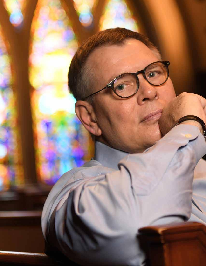
Brittany Martin, a children’s dance coach, experienced troubling cardiac symptoms over the course of a year, culminating in a visit to Emory University Hospital. “My body was in fight or flight all the time,” she says.
In 2015, Martin began seeing a doctor in McDonough for an elevated heart rate and was prescribed beta-blockers. She had been concerned about thyroid problems because of her family history. She attributed her heart’s racing to stress: Her dance team at the Platinum Peaches studio in Decatur was getting ready for a competition.
Then, one Monday last summer, she felt much worse. After putting her daughter on the bus to school, she grew concerned about an increasingly bad headache, chest pain, and heart palpitations.
Her husband drove her to the emergency room, where doctors ran tests to determine if she was having a heart attack. They performed an electrocardiogram and looked for troponin in her blood, a sign of damage to the heart muscle. Everything looked fine, which was puzzling.
She was sent for a head CT. The severity of her headache by that point made her kick and scream, she recalls. After an extra troponin test was in the suspicious range, Martin was sent to the cardiac catheterization lab. She remembers thinking: “I’m 30 years old—why am I having a heart cath?”
Doctors didn’t see any blockages that would cause her problems. But an ultrasound examination of her heart showed that the walls of the base were not moving, although the apex or lower conical tip was still contracting. And she had an ejection fraction (a measure of the heart’s pumping efficiency) of 30 percent, similar to someone with heart failure.
To supervising cardiologist Stam Lerakis, this pattern indicated a rare condition called reverse Takotsubo cardiomyopathy. The standard form of Takotsubo cardiomyopathy is known as stress-induced cardiomyopathy or “broken heart syndrome,” and occurs mainly in post-menopausal women.
“We do see [standard Takotsubo] regularly,” says Lerakis, recalling a case in which a man had a stroke and his wife, because of her distress, developed the syndrome.
In the reverse form, which Martin appeared to have, the apex still contracts. It is more common among younger women, possibly because of age-related changes in how the heart responds to epinephrine. Lerakis and his team sent Martin for a cardiac MRI to solidify the diagnosis.
“The catheterization results did not explain the weakness of the heart,” he says. “We needed to determine if there was a problem with the heart walls and see if the muscle was still alive. That’s how we found the tumor.”
A mass about the size of a quarter was discovered close to Martin’s spine. Based on her symptoms, the doctors suspected it was a paraganglioma, a tumor that produces epinephrine and its chemical relative, norepinephrine. Adding to doctors’ suspicions, her blood and urine showed high levels of normetanephrines, metabolic products of epinephrine and norepinephrine also known as catecholamines. “In Takotsubo, the heart is essentially stunned, but we expect that it will be able to recover,” Lerakis says. “Her heart was being bombarded by catecholamines.”
To learn more, Lerakis turned to endocrinologist Sol Jacobs, who called for an octreotide scan—a diagnostic technique used to find catecholamine-producing tumors. “Given her clinical presentation and biochemical data, we still suspected this was a paraganglioma,” Jacobs says.
Martin was prepared for surgery. To counteract the effects of the catecholamines, she was given alpha- and beta-blocker drugs, which restored her heart to normal pumping efficiency. But she wasn’t out of danger. “Any manipulation of the tumor could cause a sudden release of catecholamine, leading to severe increases in heart rate and blood pressure,” says thoracic surgeon Seth Force, who removed the tumor. “That’s why the blood pressure and heart rate have to be controlled by medicines prior to surgery, to avoid any cardiac issues.”
Genetic tests revealed that Martin has a mutation that increases the risk of developing a paraganglioma. One of her daughters has the genetic mutation as well, so they both will need to get periodic scans. As for now, she is planning more dance competitions. “I’m doing great,” she says. “My relatives can’t believe that I was in the hospital last year.”


Calming A Heart Storm
The first time Nils Ericson’s heart slipped out of its normal rhythm was on a Sunday. He was in church with his family when he suddenly slid out of his seat and started shaking, leading his wife to think he was having a seizure.
“I did not feel it coming,” says Ericson, a vice president at Olympus. “I felt a little warmth on the top of my head, and I just … went to sleep.”
This was the start of a medical mystery that took several unexpected twists and turns. In fact, Ericson’s diagnosis, treatment, and recovery were the basis of a lecture given to Emory first-year medical students this spring, which Ericson himself was able to attend.
Cardiac electrophysiologist Michael Lloyd used Ericson’s case as an example of a common heart disorder: ventricular tachycardia (fast heartbeats) that turned into cardiac arrest.
Later, at the hospital, Ericson experienced a type of arrhythmia called “refractory electrical storm,” which is rare and carries a high mortality rate.
He survived because, in the middle of the night, Lloyd thought to ask anesthesiologists to perform a procedure he had only read about—a stellate ganglion block, an injection of local anesthetic into the neck that can have a heart-calming effect.
Emory doctors have used the procedure to stabilize the heart of other patients with stubborn arrhythmias. For Ericson, it worked well enough that it gave doctors time to discover the source of his trouble and provided a clue as to how he should be treated long-term.
Still, two frightening days passed before that moment of inspiration.
After Ericson regained consciousness at church and sat up, he passed out again. Members of the congregation tried to revive him and were preparing to use an automated external defibrillator, but he was able to walk to an ambulance that took him to Emory Johns Creek Hospital.
There, doctors looked for a blockage of Ericson’s coronary arteries. When they found none, they decided to send him to Emory University Hospital. Kathryn Ericson remembers hearing staff shout “Code blue” when her husband needed to be resuscitated another time.
A nurse urged her to repeat words of encouragement to her husband during the ambulance ride in the rain. “I consider it a miracle he didn’t code on the way there,” she says.
At Emory University Hospital, doctors stabilized Ericson and told his wife to go home and rest. She returned the next day optimistic. But on Monday night, the electrical storm began.
On multiple occasions, Ericson’s heart began beating quickly and then stopped altogether, requiring an electrical shock to bring it back. This kept occurring, despite doctors’ interventions—administering anti-arrhythmic drugs, calming beta-blockers, and, eventually, propofol sedation.
“We were escalating with several kinds of drugs,” says cardiology fellow Matthew Crim, who was on duty along with attending cardiologist Maan Jokhadar. “But all those shocks send the heart into a spiral. He was deep in the spiral.”
By this time, Ericson’s sons had arrived at the hospital. His oldest, then a first-year student at Medical College of Georgia, knew enough about what was happening that every time he heard the “buzz—boom” of the defibrillator, he became upset.
“Dr. J cautioned us not to Google ‘electrical storm,’ ” says Kathryn Ericson. “But we did anyway. We must have had a dozen people in there praying.”
Around 2 a.m. Tuesday, Lloyd called anesthesiologist Boris Spektor, who was on acute pain service, and asked him to try the stellate ganglion block.
The block involves injecting anesthetic into a star-like cluster of nerves in the neck, guided by ultrasound. Spektor says he performs the procedure around once per month but mostly to alleviate escalating chronic pain. The delicate procedure is relatively convenient because it can be performed at the bedside, he says.
Lloyd and Spektor’s aim was to temporarily disconnect Ericson’s heart from the stimulation it was getting from the sympathetic nervous system. The intended effect was similar to that of the beta-blockers doctors already had tried: to shield the heart from adrenaline.
“It was one of those situations when all other options had been exhausted,” Lloyd says. “It gave us a window of time to do imaging on his heart, which we couldn’t do before.”
To extend the duration of the anesthetic block, Spektor added dexamethasone, an anti-inflammatory steroid.
When Ericson’s heart calmed down in response to the stellate ganglion block, it was a clue that inflammation was part of what was causing his arrhythmia.
MRI and PET imaging showed a hot spot in the left ventricle, which doctors suspected was cardiac sarcoidosis. Usually found in the lungs, sarcoidosis is the body’s response to an unknown irritant.
“Nobody knows where sarcoid comes from,” Lloyd says. “It’s known as a great mimic of other disorders.”
Under the microscope, sarcoidosis looks like the lumps of immune cells, called granulomas, that surround tuberculosis bacteria. In Ericson’s heart, those lumps were interfering with the signals carried from the pacemaker region to the rest of the muscle, and triggering spikes of their own.
Ericson’s long-term treatment would need to include shrinking the sarcoidosis, so he started anti-inflammatory treatment with corticosteroids.
Since then, he has transitioned to a milder anti-inflammatory drug, methotrexate. For preventive purposes, he also has an ICD (implantable cardioverter-defibrillator), but it has not been triggered.
When questioned by medical students about his recovery during the lecture, Ericson explained that a revamp of his diet came out of discussions with Jokhadar about side effects of the corticosteroid treatment. He learned that the drugs often induce hunger and weight gain. Encouraged by his vegan mother-in-law, he resolved to eliminate meat as well as alcohol from his diet.
“I would eat cardboard if it meant staying alive,” Ericson says. “I don’t crave meat, but I did recently start eating fish because I found my legs were feeling weak.”
Before the electrical storm, Ericson was doing yoga and “P90X” workouts, but upon first coming home, he had difficulty just walking down the driveway. A complication of his long hospital stay was irritation of the sciatic nerve, which made walking or even sitting painful.
Ericson recalls that his father, after quadruple bypass surgery, was not able to return to normal activities.
He is working hard to recover and is now running a minute at a time with walking breaks. He has also returned to traveling for his job.
“I refuse to let this thing beat me,” he says.
Windows of Opportunity
Individual donors and organizations help Emory researchers’ efforts to develop therapies to improve blood flow, prevent inflammation, and encourage healing from cardiovascular disease.
To find out more, contact Gabrielle Stearns, director of development, at 404.727.2512 or gabrielle. stearns@emory.edu
Clockwise from top: Cardiothoracic surgeon Bradley Leshnower, assistant professor of surgery, (far left) shows Ebrima Bah a stent similar to the one that saved his life; Bah, pictured here after his surgery, was able to go home on Christmas Day; a post-operative CT scan shows the stent graft in the descending thoracic aorta with aortic remodeling.


