3 minute read
PERIODONTAL HOES SCALPELLI PARODONTALI

by elite dental

Advertisement
Titanium Curettes And Scalers Curettes E Scalers In Titanio
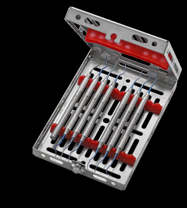
Kit Perimplantite
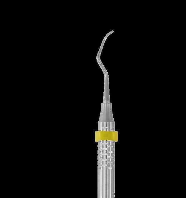
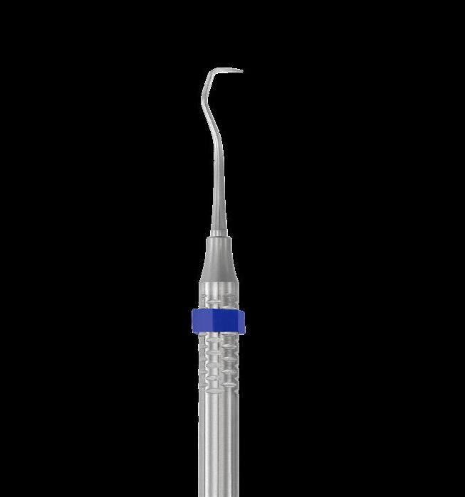
The bacterial aggression observed on the neck portion of the implants is similar to the bacterial aggression observable at the muco-gingival junction of the tooth. The stainless steel instruments may contaminate the titanium made implants while plastic instruments do not fully remove the failing tissues around the implant. Hence the need of using instruments made of titanium, the unique solution for a real efficient and fully safe handling on and close to the implants sites. Blue colour for a simple and fast identification of titanium tips, from standard gray of stainless steel. L’aggressione batterica osservata nel collo degli impianti è simile a quella presente a livello della giunzione muco gengivale del dente. Gli strumenti in acciaio possono contaminare il titanio degli impianti, mentre quelli in plastica non sono in grado di rimuovere in modo efficiente la parte in oggetto. Da qui la necessità di usare strumenti in titanio, la soluzione sicuramente più adeguata per una gestione del problema efficiente ed in piena sicurezza in prossimità degli impianti. Il colore blu delle punte, serve per identificare in modo semplice e sicuro il titanio impiegato, rispetto al tradizionale acciaio.
548/4 PT Probe CP15
627/4Ti.HL8 Curette Columbia 4R/4L Titanium
651/11Ti.HL8 Scaler H6-H7 Titanium
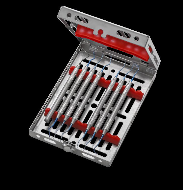
669/5-6Ti.HL8 Gracey short type Titanium
669/7-8Ti.HL8 Gracey short type Titanium
669/11-12Ti.HL8 Gracey short type Titanium
669/13-14Ti.HL8 Gracey short type Titanium
980/9 Gammafix tray Doi
Tunnel Technique Instruments
Clinical Case Caso Clinico
TUNNEL TECHNIQUE, DR. STABLUM
Gingival recession is defined as the apical displacement of the soft tissue margin from its normal position on the crown of the tooth to levels on the root surface beyond the cemento-enamel junction. It has a high incidence among the population and often leads to severe aesthetic problems, tooth sensitivity and /or cervical carious lesions. In the last years several surgical techniques have been developed aiming at performing good root coverage. The unfolding of the “Tunneling technique” represent an interesting improvement for the correction and management of this kind of mucogingival problems. The technique can be performed for single recessions or Miller Class and Class II multiple recessions, it has been shown to be efficacious combination with subepithelial connective tissue graft (SCTG) or with acellular dermal matrices (ADMS).
TECNICA TUNNELING, DR. STABLUM
Le recessioni gengivali sono definite come uno spostamento in senso apicale del margine gengivale, hanno un’alta incidenza nella popolazione e spesso creano grossi problemi di estetica, sensibilità dentinale e/o carie cervicali. Negli anni sono state sviluppate numerose tecniche finalizzate alla copertura radicolare. Lo sviluppo della “Tecnica a Tunnel” rappresenta un interessante miglioramento nella gestione di queste problematiche; può essere utilizzata sia per recessioni singole sia per quelle multiple di grado e II di Miller e può essere eseguita in associazione ad innesti di tessuto connettivo e/o matrici dermiche acellulari.
1 Multiple recession Miller class and II (21/22/23) Recessioni multiple classe e II di Miller (21/22/23)
2 Sulcular incision at T bone depth crest cutting the connective tissue fibers with blade 3638/69
Incisione sulculare fino alla cresta ossea sezionando le fibre connettivali con lama 3638/69
3 Papilla elevation performed according to the tunneling technique (698/1.HL8)
Elevazione della papilla secondo tecnica a tunnel (698/1.HL8)
4 Tunnel elevation at full thickness in the coronal part for preserving integral tissue volume (698/2.HL8) Scollamento del tunnel a spessore totale nella parte più coronale del lembo per mantenere inalterato lo spessore (698/2.HL8)
5 Moving beyond the mucogingival line at split thickness in order to increase the flap mobility in the coronal direction (698/2.HL8) Si oltrepassa la linea mucogengivale mantenendo il lembo a spessore parziale per aumentare la mobilità in senso coronale (698/2.HL8)
6 Checking the mobility with periodontal probe
Si verifica la mobilità del tunnel con sonda parodontale
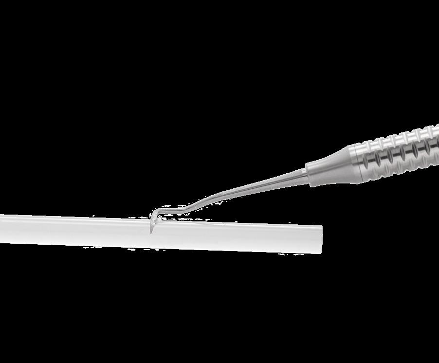
7 Root scaling of the coronal part of recession in order to remove the contaminated cement Rimozione del cemento contaminato con curette nella parte coronale delle recessioni
8 Insertion of the connective tissue graft in the tunnel Inserimento dell’innesto connettivale all’interno del tunnel
9 Graft suturing in order to stabilize it with the overlying coronal gingival flap Innesto ancorato al tunnel e posizionato coronalmente tramite suture
Inspection (3 months) Controllo (3 mesi)
Inspection (3 years) Controllo (3 anni)
Graduated in 1993, University of Verona, Faculty of Dentistry and Prosthodontics. Active member of SIO/IAO (Italian Society for Osteointegration/ Italian Academy of Osteointegration), active member of Florence Perio Group, active member of AIIP (Italian Academy of Implant Prosthesis), active member of GIR (group Implant Research), member of SIDP. From 1999 to 2001, he cooperated with Dr. Giano Ricci in Florence and active as tutor at biennial courses. In 2013 he co-authored the SIO book: IMPLANT SUCCESS: DIAGNOSIS, TREATMENT PLANNING AND OPERATIVE PROTOCOLS. Dr. Stablum currently writes for National and International Dental magazines and is engaged as regular speaker at congresses, in Italy and abroad. He manages his own dental practice in Borgo Valsugana (TN) as specialist in periodontics and implantology.
Laurea in Odontoiatria e Protesi Dentaria nel 1993 conseguita presso l’Università di Verona. Socio attivo SIO/IAO (Società Italiana Osteointegrazione/ltalian Academy of Osteointegration), Socio attivo del FLORENCE PERIO GROUP socio attivo AIIP (Academia Italiana Impianto Protesi), socio attivo GIR (Group lmplant Research), socio SIDP. Dal 1999 al 2001 collabora presso lo studio del Dott. Giano Ricci a Firenze dove svolge anche attività di tutor ai corsi biennali. Nel 2013 Coautore nel libro della Società Italiana di Osteointegrazione: “IL SUCCESSO IN IMPLANTOLOGIA: DIAGNOSI PIANO DI TRATTAMENTO E PROTOCOLLI OPERATIVI. Pubblica su riviste nazionali ed internazionali, relatore a congressi in Italia e all’estero e svolge la libera professione a Borgo Valsugana (TN) occupandosi prevalentemente di parodontologia e implantologia.


