
7 minute read
Unilateral Absence of Pulmonary Artery in an Adult Patient Presenting with Haemoptysis ACase Report with Brief Review of Literature

from August 2020
by Ejima

JOURNAL OF THE INDIAN MEDICAL ASSOCIATION, VOL 118, NO 08, AUGUST 2020
Case Report Unilateral Absence of Pulmonary Artery in an Adult Patient Presenting with Haemoptysis : A Case Report with Brief Review of Literature
Advertisement
Tony Ete 1 , Swapan Saha 2 , Vanlalmalsawmdawngliana Fanai 3 , Arun Kumar 3 , Habung Mobing 4 , Shakeel Ahamad Khan 3 , Utpal Kumar 3 , Narang Naku 5 , Animesh Mishra 6
Unilateral absence of pulmonary artery should be considered in patients presenting with haemoptysis and recurrent respiratory infections. Usually it is a diagnosis of exclusion. Patient may present with non specific symptoms. A high index of suspicion with proper investigations, non invasive as well as invasive, are required for diagnosis and management. The diagnosis is usually confirmed by CT and MRI. Angiography is done only for patients who require embolisation or revascularisation surgery.
[J Indian Med Assoc 2020; 118(8): 62-3]
Key words : Hemoptysis, pulmonary artery, angiography.
Unilateral absence of a pulmonary artery (UAPA), a rare condition, usually occurs in combination with other cardiovascular conditions like tetralogy of Fallot (TOF) or septal defects. Patients with isolated absence of one pulmonary artery often present with dyspnoea, chest pain, hemoptysis or recurrent chest infections but may be asymptomatic till late adulthood. About 20% of the patients develop inconsequential hemoptysis, although massive hemoptysis is very rare 1 . Therefore, diagnosis may be difficult due to these nonspecific presentation 2 , sometimes diagnosed incidentally on chest radiographs. Here we are presenting a case with isolated absent right pulmonary artery with haemoptysis, as clinical presentation.
CASE REPORT
A 37-year gentle man, non hypertensive, non diabetic and non smoker, presented with recurrent episodes of hemoptysis for the last six months, scanty in amount. There was no history of fever, breathing difficulty or recurrent chest infection. No history of pulmonary tuberculosis in the past. Physical examination revealed body temperature of 36.6 o C, pulse rate of 90 beats per minute, respiration rate of 16 per minute and blood pressure of 100/70 mmHg. Clinically there were no signs of cyanosis, edema, or clubbing of the fingers. Cardiac and chest auscultation was normal. The electrocardiogram revealed normal sinus rhythm.
Department of Cardiology, North Eastern Indira Gandhi Regional
Institute of Health and Medical Sciences, Shillong 793018 1 MBBS, MD, DM (Cardiology), Assistant Professor and Corresponding Author 2 MBBS, MD, DM (Cardiology), Consultant, Department of Cardiology, Desun Hospital Siliguri 3 MBBS, MD, Senior Resident 4 MBBS, MD, Senior Resident, Department of Medicine 5 MBBS, MS, Assistant Professor, Department of General Surgery 6 MBBS, MD, DM (Cardiology), Professor and Head
Received on : 25/06/2020 Accepted on : 02/07/2020 Editor's Comment :
Unilateral absence of pulmonary artery (UAPA) should
be considered in patients presenting with hemoptysis and recurrent respiratory tract infections.
A high index of suspicion is required for diagnosis. It
is usually a diagnosis of exclusion. Chest radiograph may suggest the diagnosis whereas echocardiography can be used for the evaluation of possible associated cardiac anomalies and assessment of pulmonary hypertension.
The diagnosis is usually confirmed by CT scan and
MRI. Angiography is done for patients who require embolisation or revascularization surgery.
Echocardiogram was normal with normal pulmonary artery pressure except for absent right pulmonary artery. A chest radiograph (postero-anterior view) revealed dilated artery (?Main pulmonary) in the right hilum with alveolar infiltrates in right lung field. The right hemi diaphragm was elevated without any cardiac and mediastinal displacement (Fig 1). Computed tomography (CT) angiogram revealed the absence of the right main pulmonary artery. A focal vascular dilation was detected in right lung possibly representing an aneurysmatic formation or an arteriovenuous fistula (Fig 2 A). Blood was supplied to the right lung by tortuous, dilated arterial branches of indeterminate origin (Fig 2 B).
In view of patient’s symptom and to look for source of blood supply to the right lung, patient was taken for catheterization and pulmonary angio and aortogram was done with an intention to proper decision making for the management. Pulmonary angiogram showed absent right pulmonary artery (Fig 3 A). Selective angiogram of right subclavian artery showed upper zone of right lung is supplied by right vertebral artery and lower zone is supplied by right internal mammary artery (Fig 3 B).
As the patient’s symptom improved thereafter, he was kept under close follow-up on an outpatient basis, with symptomatic
62
JOURNAL OF THE INDIAN MEDICAL ASSOCIATION, VOL 118, NO 08, AUGUST 2020
Fig 2 — Transaxial CT angiogram with intravenous contrast in a soft tissue window (A) shows an absent left pulmonary artery (B) tortuous, dilated arterial branches supplying right lung
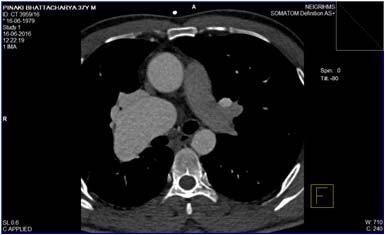

Fig 1 — Chest radiograph (postero-anterior view) shows dilated artery in the right hilum with alveolar infiltrates in right lung field with elevated right hemi diaphragm and supportive treatment for recurrent haemoptysis. Fig 3 — (A) Pulmonary angiogram showed absent right pulmonary artery Selective angiogram of right subclavian artery showed upper zone of right DISCUSSION lung is supplied by right vertebral artery and lower zone is supplied by right
The prevalence of UAPA is 1 in 200,000 young internal mammary artery adults 2 and it can occur in an isolated manner revascularization of peripheral branches to the pulmonary although most cases are associated with other hilum can be attempted 9 . Hemoptysis can be managed cardiovascular anomalies like tetralogy of fallot 2,3 . In about with embolization, lobectomy or pneumonectomy 10 . two third of cases, isolated UAPA involves the right lung 1 . In conclusion, UAPA should be considered in patients Developmental alteration of ventral bud of the ipsilateral 6th presenting with haemoptysis and recurrent respiratory aortic arch is thought to be the embryological basis of infections. Chest radiograph may suggest the diagnosis UAPA 4 . In the affected artery, generally the distal whereas echocardiography can be used for the evaluation of intrapulmonary branches remain intact which can be possible associated cardiac anomalies and assessment of supplied by collaterals from bronchial, intercostal, internal pulmonary hypertension. The diagnosis is usually confirmed mammary, subdiaphragmatic, subclavian or even coronary by CT and MRI. Angiography is done only for patients who arteries 5 . Clinical course of many patients with isolated UAPA require embolisation or revascularisation surgery. is benign and a diagnosis is not made until adulthood 2 . In REFERENCES symptomatic patients, one study showed, chest pain, pleural 1 Harkel DJT, Blom NA, Ottenkamp J — Isolated unilateral effusion and recurrent infections to be present in 37% of absence of a pulmonary artery: a case report and review of patients, while dyspnea or exercise intolerance in 40% of the literature. Chest 2002; 122: 1471-7. patients. Pulmonary hypertension was present in 44% of 2 Bouros D, Pare P, Panagou P, Tsintiris K, Siafakas N — Th e patients. Hemoptysis occurred in about 20% of patients varied manifestation of pulmonary artery agenesis in whereas high-altitude pulmonary edema was seen in approximately 10% of patients 1 . Hemoptysis is caused by collateral circulations that create high pressures in venous adulthood. Chest 1995; 108(3): 670-6. 3 Presbitero P, Bull C, Haworth SG, de Leval MR – Absent or occult pulmonaryartery. Br Heart J 1984; 52(2): 178-85. 4 Thomas P, Reynaud-Gaubert M, Bartoli J–M, Augé A, Garbe system 5 . The systemic collaterals usually arise from the L, Giudicelli R, et al — Exsanguinating hemoptysis revealing bronchial, intercostals, subclavian or subdiaphragmatic the absence of left pulmonary artery in an adult. Ann Thorac arteries 1 . The diagnosis of UAPA is, in generally, based on Surg 2001; 72: 1748-50. history, physical examination and findings on chest 5 Kadir IS, Th ekudan J, Dheodar A, Jones MT, Carroll KB — radiographs. Pulmonary function test in patients with UAPA Congenital unilateral pulmonary artery agenesis and is usually unremarkable 6 . CT thorax with contrast enhancement confirms the absence of the affected pulmonary artery. Echocardiography is helpful for exclusion aspergilloma. Ann Th orac Surg 2002; 74(6): 2169-71. 6 Werber J, Ramilo JL, London R, Harris VJ — Unilateral absence of a pulmonary artery. Chest 1983; 84: 729-32. 7 Hayek H, Palomino J, Thammasitboon S— Right pulmonary of other cardiac anomalies and pulmonary hypertension. artery agenesis presenting with uncontrolled asthma in an Pulmonary angiography is the gold standard and is usually adult: a case report. J Med Case Rep 2011; 5: 353. reserved for patients requiring embolisation or 8 Turner DR, Vincent JA, Epstein ML — Isolated right pulmonary revascularisation surgery 7 . artery discontinuity. Images Paediatr Cardiol 2000; 4: 24-30.
At present there is no consensus regarding 9 Welch K, Hanley F, Johnston T, Cailes C, Shah MJ — Isolated management of patients with UAPA. Some recommends serial echocardiography of asymptomatic patients for the development of pulmonary hypertension 8 . On the other hand, unilateral absence of right proximal pulmonary artery: surgical repair and follow-up. Ann Th orac Surg 2005; 79(4):1399- 402.


63


