Journal of Engineering Technology and Sciences
Volume 01, Issue 01, September 2024
www.jets.org.in
Journal of Engineering Technology and Sciences
Volume 01, Issue 01, September 2024
www.jets.org.in
K.Surya Department of Chemistry, Velammal Engineering College, Chennai
To Cite this Article
K.Surya, “Efficacy of Nanomaterials and Nanotechnology In Diagnosis and Treatment of Heart Disease”, Journal of Engineering Technology and Sciences, Vol. 01, Issue 01, September 2024, pp.-1520
Abstract: In the current era of equipment, a number of potential point care devices have been developed and these have paved the way for the next generation point care test. Heart disease requires a global average of life expectancy. Heart disease factors are on the rise today due to a poor diet high in saturated fat, salt and sugar, but people have a poor lifestyle. Factors combined with complementary and advanced methods provide information that clearly shows the increasing trend of cardiovascular disease prevalence. It is very important that advanced health management is achieved by making appropriate decisions based on rapid diagnosis, intelligence data analysis and information analysis. On-site monitoring ensures that early detection of diagnoses facilitates better diagnosis, control and management of the disease. It also facilitates rapid medical decisions as the disease can be diagnosed at an early stage, leading to better health outcomes for patients, prompting them to begin treatment sooner. Currently, oral medications or invasive surgery are prescribed for the treatment of vascular disease. Large gap of drugs with new therapies for better patient outcomes. Nanotechnology provides more effective therapeutic solutions with better prognosis and reduced side effects
Keywords: Cardiovascular diseases (CVD), Nano particles, Nanotechnology, Pharmacological therapies, medical implants biocompatibility, biomedical implantable devices.
Cardiovascular disease (CVD) is commonly defined as cardiovascular disease (Edgardo et al., 2020). This includes cardiovascular and cardiovascular diseases such as peripheral vascular disease, coronary heart disease, and stroke. According to the World Health Organization (WHO), cardiovascular disease is one of the leading causes of death in the world. Therefore, we need to improve the management of Geoparks. Nanotechnology is commonly used to use nototonic technology to prevent, diagnose, treat, or repair damaged tissue in the body (Khalidetal.,2014).Today,nanotechnologyhasmadegreatstridesintheworldbyimprovingtheunderstanding of diseases at the molecular level and expanding nanotechnology. It is becoming more and more important for CVD management.Nanoparticles, particles in the nanometer range, show great promise in a wide range of cardiovascular applications. Nanoparticles are ideal for targeted delivery of therapeutic and contrast agents because they are mobile within the system and extravascular system (Patrick and Bertrand, 2011). The main features of Nanotite are drug targeting and strategic goal setting, and further development of medical imaging technology (Syed and Ayman, 2018). This technique can be applied to dental implants, heart shadows, and bone grafts (Priyadarshini et al., 2019).
In addition, antibacterial nanoparticles can be used in wound healing, coatings and medical textiles (Federica and Mauro, 2019). Various types of target ligases, such as peptides, can be attached to nanotubes to identify hidden sites. Cellular nanoparticles enable new applications for intracellular DNA delivery and RNA
Published by: Surya Publications www.jets.org.in 15 | Page
detection. Nanoparticles can be introduced into cells by processes such as endocytosis, phagocytosis, and pinocytosis. In this sense, the mechanism of absorption into different cell types can be determined in different ways. The intracellular and phagocytosis are specific internal pathways that are first initiated by proteins or receptors.
Pinocytosis, on the other hand, is a non-specific pathogen that occurs when cells absorb the surrounding body fluids. These specific and non-specific cell proliferation pathways indicate that most natural pathways can enter cells. Absorption efficiency of nanotoxin cells depends on several factors, including size, shape, charge, molecular surface area, density, decoration, and compression of nanocells (Soyeon et al., 2018). Over the last few decades, various nanotechnology systems have been designed for drug delivery and bioimplantation. Nanomaterials are a remarkable first step in overcoming the limitations of classical biological raw materials. In addition, the development of nanoparticles has the potential to advance many new therapies that have the potential to modernize the treatment of cardiovascular disease. Heart valves, blood transfusions, vascular grafts, tacometers, and stressers are examples of cardiovascular surgery commonly used in medicine. This review focuses on the latest trends in the application of nanotechnology in cardiovascular therapy. It also covers the control and manufacture of embedded devices based on the concepts of nanomethods and drug delivery.
Nanostructures have long been known to have unique physicochemical and surface properties such as surface area to volume ratio, surface energy, surface roughness, wet ability, and reflectance. The response can be tuned to facilitate improved mechanical and biological performance of the biomaterial. Surfaces can be classified into "ordered nanostructures" and "random nanostructures". In addition to the above advantages, the placed surface nanostructures have a stronger effect on cell activity than random nanostructures. For example, cell association is improved based on exposure guidelines. Incorporation of nanostructured surfaces into conventional materials has been applied to many implantable devices and other devices approved by regulatory intervention, and large scale to be approved for the development of new materials and therapeutics. Long-term examination is required. However, whether nanodispersion in approved materials can bring nanocharacteristics to the surface in nanomaterials is a complex process. The surface of the biomaterial allows for an interface between the device and the human body. The nanoscale features of the surface of the material are similar in size and can affect the biological response at the cellular and tissue levels, and thus are direct to proteins and biomolecules in the body. The interface is improved (Figure: 1).
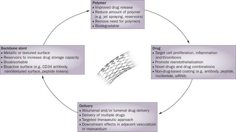
Figure: 1. Nano-features on materials surface can affect the biological responses at the cellular and tissue level because of similar dimension, thereby improving the direct interactions with proteins and biological molecules in the body
Specially modified nanosurfaces can be used to derive suitable bioreactors for the device. The physicochemical properties and the chemical composition of the surrounding environment play a very important role in the interaction of cells and substances. In the body, the extracellular matrix is the main environment for cells. When nanomaterials with nanostructured surfaces are introduced into an organism, their unique characteristics can affect proteins and can also affect cell adhesion, proliferation, differentiation, and phenotype (Marie-Claude Hofmann., 2014). These features may include surface energy, surface area, surface roughness, wettability, charge, topography, morphology, chemical composition, and mechanical properties. Therefore, in the case of cardiovascular stents, it can lead to an increase. Adhesion of endothelial cells is a desirable event for rapid endothelialization, or promotes smooth muscle cell adhesion and growth by promoting neointima formation and blocking the lumen.
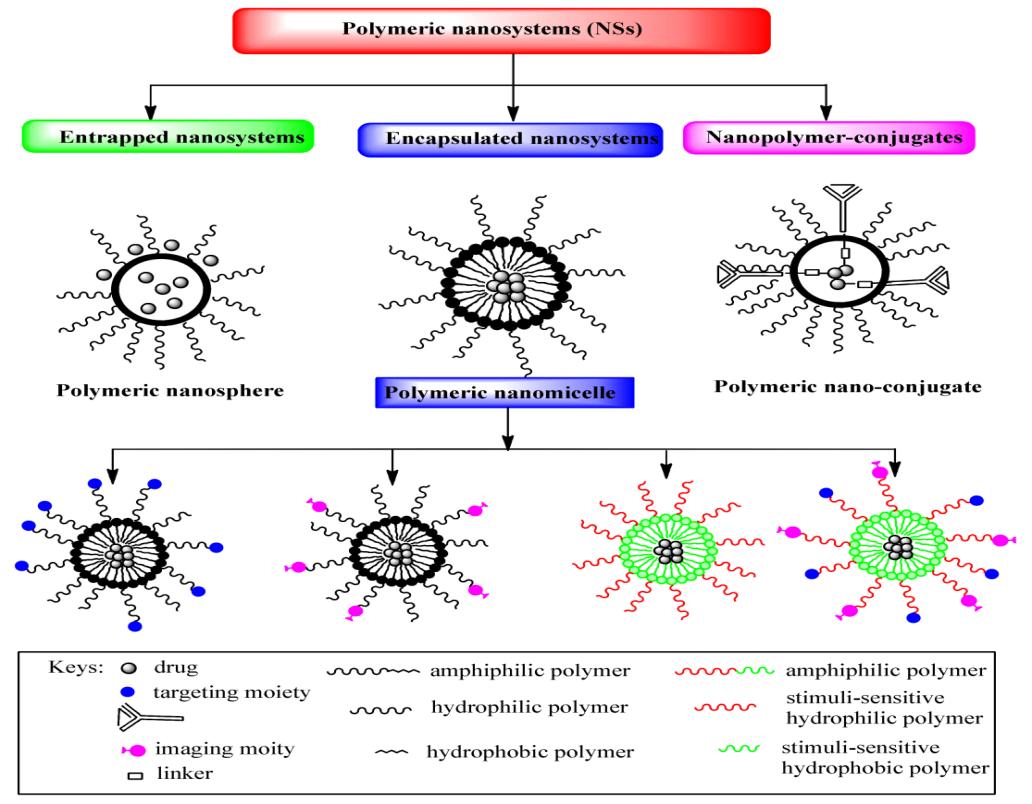
Nanoparticles show considerable potential in providing a platform for targeted drug delivery due to their unique multifunctional properties. Delivering and targeting the drug to the site of interest is the most important feature of nanoparticles. Nanomaterials for CVD Drug delivery Biodegradable polymers such as poly (lactide) (PLA), poly (lactide-coglycolide) copolymer (PLGA), poly (ε-caprolactone) (PCL), poly (acid) amine , Types of polymeric nanomaterials for drug delivery applications that have been used in various preparations (Figure: 2).
Poly (lactide-coglycolide) polymeric nanoparticles (PLGA) were prepared and their surface modified with a cationic agent. The study claims that the absorption of coated nanoparticles in the artery is 10 times higher than that of uncovered nanoparticles. A new formulation of high molecular weight anti-restaurant nanoparticles (Eleonora et al., 2018). They found that arterial absorption depends on size because particles with a small diameter (approximately 100 nm) entered the ex-vivo model of the canine carotid artery more frequently than particles with a larger diameter that is mere (200 nm). In comparison, this shows the ability of the reduced nanoparticles to be taken up in the in vivo model. In addition, the projected nanoparticles were preferentially located in the cell cytoplasm, with smaller
17 | Page
nanoparticles (70 nm) havinga higher absorption capacity (27-fold) than smaller nanoparticles and its diameter of 200 nm. Quercetin as an antioxidant has been reported to have a protective effect against cardiovascular disease.
Explain the potential of these particles in vitro and in vivo. Nitric acid (NO) can mediate endothelial cells and vascular smooth muscle function. It has been reported that DNA synthesis in smooth muscle cells can be inhibited by NO-releasing polymers. NO has been functionalized for the ligand. Based on their report, a single layer protective gold group (MPC) was prepared using both ligands to significantly increase the NO payload. More research is needed to modify cationic primary amine functional groups with charge neutralizing groups to eliminate potential aminerelated toxicity and / or deterioration of nitrosamine formation after NO release.
In addition to serving as a platform for functional groups, drugs can also be loaded with nanoparticles. The involvement of platelets in the pathogenesis of CVD, such as atherosclerosis, myocardial infarction and thrombosis, has made platelets an important target in drug delivery systems. Liposomes to address the mentioned disorders. Intermittent peripheralarterydisorders andotherperipheral artery disorders canalso be treated byapplying liposomes. Encapsulation of PGE-1 with lipid nanoparticles reduces drug degradation and enhances the therapeutic effect of drugs. Thrombosis therapy has been reported by applying liposomes. Then there is the blockage of blood vessels, which is closely related to myocardial infarction. Recent studies have reported surface functionalized peptides of arginyl-glycyl-aspartic acid (cRGD) containing antithrombotic urokinase in liposomal carriers. The cRGD peptide selectively binds to the active platelet receptor GPIIb // IIIa. The use of nanoparticles in the early detection of atherosclerosis is a good example. They used monocrystalline magnetic nanoparticles (MNPs) for the non-invasive detection of vascular cell adhesion molecule (1), a flash point. Nanoparticles have been used to improve magnetic resonance imaging (MRI). Due to their targeting function, it can be used as a complete contrast medium for MRI. A successful application of this technique is the imaging of post-implantation rejection sites in mouse heart transplantation. Signals produced by macrophages successfully labeled with magnetic beads indicate the extent and location of rejection. With the successful identification of macrophages, inflammatory responses and foreign bodies could be evaluated in the future in several other cardiovascular diseases using similar techniques.
The hydrophobic core of the micelle regulates the encapsulation of pharmaceutical compounds, and the hydrophilicity of the shell increases the circulation time of the micelle. Due to their small size, micelles can pass through biofilms and enter tissues. High molecular weight micelles are more stable compared to liposomes and low molecular weight surfactants, due to the significantly lower excipient ratio than the drug. Therefore, these means of transport not only present a very low risk of systemic toxicity, but are also cost-effective. In addition, the synthesis of micelles is extremely controllable and can be easily modified to obtain more effective treatments by adding targeting or tracking molecules. The most common polymers used in the preparation of micelles include poly (amino acid) or polyester derivatives to form hydrophobic regions (Yasser and Mohamed, 2018).In a recent study, the micelles were a block copolymer of polycaprolactone-b-poly (2- (dimethylamino) ethyl methacrylate) (PCL-PDMAEMA) and a mixture of polycaprolactone methoxypolicaprolactone-b (mPEG-PCL). Target micelles loaded with ramokinase (LK) using polycaprolactone-b- block copolymer
Nanocoating can be used for dental implants, stent coatings for cardiovascular implants, and orthopedic joint replacements. The surface interacts with the negative charge of the cell membrane and the intracellular absorption of the nanoparticles' attractiveness. In vivo experiments have shown that isothiocyanate-coated fluorescent nanoparticles (FITC) -coated stents improve fluorescence in the environmental layer and tumors compared to stents containing only polymers (FITC). This happens partially when the human body detects it. The surface of the stent as a foreign body
| Page
and / or an embedded polymer can cause inflammation, ignoring its biodegradability. The use of nanomaterials to mimic natural structures is another trend that leads to improved physical, mechanical and biological properties of implants.
This approach provides a solution to potential problems by promoting the proliferation of endothelial cells and suppressing the proliferation of vascular smooth muscle cells. The nanoparticles bound to the stent leave the surface of the stent, enter the damaged epithelium, and are inserted into the arterial tissue (Mahdi et al., 2016). However, this system did not deliver the drug to the arterial layer, as opposed to nanoparticles. However, in another study, this problem was solved using a layer of polymer stents by choosing a composite layer of nanotubes. Another approach is to use nanodrinting to replace the nanotube coating on the surface of the metal stent. The main complication of these stents is the formation of polymer-related blood clots that can be suppressed using polymer-free surfaces.
In addition, hollow metal stents showed a slower thrombus formation rate than drug-eluting stents. The design of nanostructured wires from all metals has been the goal of many studies. Preferred technical properties of the surface obtained by a chemically corrosive surface can be achieved by chemical vapor deposition at high temperatures. IPad frequency plasmas, capable of producing radial-based metal nanostructures on the stent surface, have been used in recent years to provide porous, high-density structures that affect endothelial cells. The interaction between the stent and the endothelial cells, and the effect of the stent on endothelialization, are important factors before deciding how to design and synthesize the stent. The effect of nanostainless stents on the mechanism of endothelial processes has been elucidated. These stents are filled with paclitaxel. The results of in vivo studies have shown that the above stents accelerate reendothelialization, improve vascular healing and significantly reduce inflammatory flow compared to sirolimus polymer stents or individual types.
In this appraisal, we focused in principle on nanostructured surfaces, nanoparticles and nanocomposites and discussed important substances in nanomaterials for cardiovascular applications. The evidence obtained in this review showsthatnanotechnologyhas thepotentialfortherapeuticapplicationofCVD,asdetermined bytranslationalclinical trials. With growing speculation in nanotechnology and related infrastructure around the world, nanomedicines, nanomaterials and other related technologies will be supplemented and guided by extensive clinical trial processes.
It's just a matter of time. What is certain is that nanotechnology is committed to improving patients' health and well-being, and current advances in treatment are having a positive impact on the lives of patients around the world. To increase the effect on the drug, more in vivo studies should be performed and clinical trials ordered to fully understand the systemic behavior of nanoparticles, when the future of therapy turns to personal medicine, nanotechnologymay beinanoptimalpositiontoachievetheproposed goaloftailoringtreatmenttoindividualmedical conditions. On the other hand, there is considerable evidence to support the idea that nanotechnology has not yet had the full effect of revolutionary medicine.
Nanotechnology as a new type of science gives clinicians a clear perspective and hopes that they will achieve goals that were previously considered unattainable, but still deepen knowledge and apply nanotechnology. It is mandatory to increase. Nanomedicines have great potential for the treatment of CAD. Efficient systems for the delivery of various drugs are being urbanized. On the other hand, the challenges for their performance are many, such as determining the age of nanomaterials in living cells, sufficient information on biological safety for NP at the mobile level and obvious in living cells. In general, research and clinical translation in the application of nanotechnology have a long way to go. Major advances in nanomedicine have significantly improved the current treatment of CVD. With the decline in treatment and the growing scientific popularity, the future of CVD treatment is really very exciting.
References
[1] Edgardo Olvera Lopez, Brian D, Ballard, Arif Jan., 2020. Treasure Island (FL): StatPearls Publishing;
[2] Khalid , KhalafAlharbi.,Yazeed A.Al-sheikh.,2014. Saudi Journal of Biological Sciences,Volume 21, Issue 2: Pages 109-117
| Page
[3] Patrick Boisseau .,and Bertrand Loubaton.,2011.C. R. Physique,12,PP.No:620-636
[4] Syed A.A. Rizvi., and Ayman M. Saleh, 2018. Saudi Pharm J. Jan; 26(1): 64–70. doi: 10.1016/j.jsps.2017.10.012
[5] B., Rama,Chetan M., & Vijayalakshmi U.,2019. Journal of Asian Ceramic Societies, Volume 7, Issue 4
[6] Federica Paladini and Mauro Pollini 2019. Materials (Basel). Aug; 12(16): 2540. doi: 10.3390/ma12162540
[7] Soyeon Jeon, Jessica Clavadetscher, Dong-Keun Lee, Sunay V. Chankeshwara, Mark Bradley, and Wan-Seob Cho.,2018.Nanomaterials (Basel). Dec; 8(12):028.. doi: 10.3390/nano8121028
Marie-Claude Hofmann.,2014. Adv Exp Med Biol. 2014; 811: 255–275.doi: 10.1007/978-94-017-8739-0_13
[8] Eleonora Calzoni ., Alessio Cesaretti ., Alice Polchi ., Alessandro Di Michele.,Brunella Tancini .,and Carla Emiliani.,2018. Journal of Functional Biomaterials
[9] Yasser H. A. Hussein., and Mohamed Youssry ,2018.Materials (Basel) ; 11(5): 688. doi: 10.3390/ma11050688
[10] Mahdi Karimi ,Hossein Zare , Amirala Bakhshian Nik , Narges Yazdani ,Mohammad Hamrang ,Elmira Mohamed ,Parham [11] SahandiZangabad , SeyedMasoudMoosaviBasri ,LeilaBakhtiari ,and MichaelRHamblin.,2016.Nanomedicine(Lond).Mar; 11(5): 513–530. doi: 10.2217/nnm.16.3
| Page