4 minute read
Partial restorations on anterior teeth

from ESTRATTO DERCHI
by Grupo Asís

We have described how it is possible to comply with the principle of minimally invasive intervention in posterior teeth. At this point, it is important to evaluate the possibility of acting in the same way with the anterior teeth that need restoration to correct their shape and position, close diastemas, replace old composite restorations, repair and correct teeth, incisal abrasions, and erosions, and hide discolorations.31
The ceramic materials available on the market in recent years have drastically shifted the options for professionals in the restoration of anterior teeth. As indicated by Gurel32 , the “old” full coverage crowns are the first form of aesthetic restoration for anterior teeth, however they implied the need of sacrificing the enamel and a high risk of damaging the periodontal tissues and pulp.
Advertisement
It has been proven that the aesthetic goals can be achieved with stratified ceramic veneers without sacrificing the enamel and the entire tooth structure; therefore, these veneers have become the best choice possible in this regard. This procedure comprises creating a veneer prepared as a thin shell covering the buccal surface. Several authors33-36 have suggested that the minimum thickness of the preparation for ceramic veneers should not exceed 0.5 mm. However, the thickness of the current ceramic veneers is between 0.4 and 0.7 mm; these values are close to the thickness of the natural dental enamel. Due to the evolution of ceramic materials, today clinicians can show their skills by creating restorations that allow preserving enamel and promoting a better and longer adhesive cementation.
Regarding the preparation required to accommodate the veneer, there are different indications in the literature from the classical preparations of only the buccal surface, to those that require the prosthesis to cover the incisal ridge and the so-called “no-prep veneer”.
What should always be the mantra for these preparations was defined by Friedman37 in 2001, who supported and recommended the importance of preserving the enamel as much as possible because a better retention of ceramic veneers, in the long run, can be obtained by preserving at least 50% of the enamel surface by positioning all the finishing margins in the enamel. The reason for this choice, shared by most professionals, lies in the possibility of guaranteeing the resistance and duration offered by the adhesion on the enamel, as shown in previous studies on this topic.
Clinical case 5
Protocol for producing aesthetic veneers
E. Manca
The restoration of the front group with veneers has the purpose of establishing a fresh smile for patients. The images below show the operating sequence.
$ 1 Radiographic check. $ 2 Initial situation. $ 3 Detail of old restorations (buccal view). $ 4 Detail of old fillings (palatal view).
$ 5 Diagnostic wax-up. $ 6 Preparation of buccal surface. $ 7 Finishing of margins. $ 8 Elimination of unsustained enamel prisms.
$ 9 Assessment of occlusal margin reduction by means of mask. $ 10 Check of residual enamel thickness after preparation. $ 11 Impression detection using DSR. $ 12 Preparation of elements that receive temporary materials.
$ 13 Cementation of temporary materials. $ 14 Plaster base for removable stumps. $ 16 Wax model on plaster cast.
$ 17 Plaster stumps.
$ 21 Artifact test.
$ 24 Acid etching.
$ 28 Photopolymerization of cement. $ 18 Artifact test on plaster cast. $ 19 Check of thickness. $ 20 Four-tenth thickness.
$ 22 Start of cementation phase. $ 23 Surface cleaning.
$ 25 Adhesive distribution. $ 26 Acid etching of the artifact. $ 27 Etched veneers.
$ 29 Finishing of margins before dam removal. $ 30 Finishing of cervical margin after rubber dam removal.
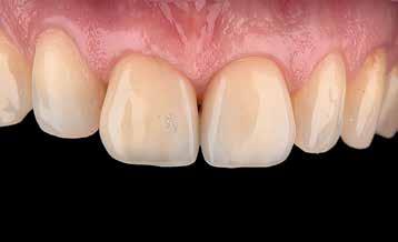
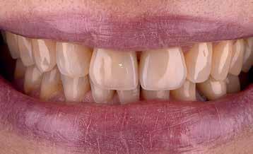
$ 31 Final result. $ 32 Smile with the integration of reconstructed elements.
The veneers can be made from different materials as follows, which affect cementation thickness and protocol:38 Ä resin composites; Ä fused glass-ceramics; Ä traditional feldspar-based ceramics; Ä heat-pressed ceramics (leucite-reinforced ceramics; one of the most commonly used); Ä CAD/CAM digital production.
resin composites
They are the cheapest option and can be stratified by the technician with a traditional sequence or be milled via the CAD/CAM systems. We will be able to evaluate the differences in Chapter 3. These veneers do not guarantee a stable aesthetic result because they tend to opacify, change color, and frequently fracture along the margins. Furthermore, the intrinsic porosity of the composite material represents a good substrate for plaque accumulation (! 1.25 and 1.26).
fuseD glass-ceramics
They are the first ceramic materials for dental use. Derived from Chinese porcelain, they are made of a glass phase and crystalline fillers. Baking of the modeled powders leads to the fusion of the glass portion that wraps and blocks the crystalline matrix. This ceramic, correctly termed porcelain, is chemically stable, provides excellent aesthetic results, and is long-lasting. Moreover, it shows a high resistance to compression (350-550 MPa), but an inadequate resistance to traction (20-60 MPa). As it is made mostly of glass, it has an insufficient hardness. This results in surface microcracks, which have severely limited its use.
traDitional felDspar-baseD ceramics
It was present in the market in the mid-60s of the 20th century, thanks to the studies of McLean and Hughes who defined it as “reinforced ceramic core system” as reinforced with feldspar glass and alumina (PJC, alumina – reinforced Porcelain Jacket Crown).
! 1.25 Characterization after stratification on model (courtesy of Andrea Piacentini, dental technician).
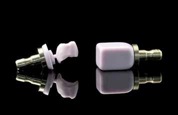
! 1.26 Block-milled composite element.


