11 minute read
WET MOUNT PREPARATION

by Grupo Asís

There are several types of wet mount, depending on the site of collection of the specimen: • cervico-vaginal wet mount • urinary wet mount or urocytogram • oral wet mount • cutaneous wet mount • rectal wet mount.
Material
Advertisement
The following materials are needed to prepare a wet mount: • microscope glasses • cover glasses (coverslips) • sterile saline solution (0,9% sodium chloride solution) • 10% potassium hydroxide solution • dropper for transferring saline solution onto the microscope slide • vaseline or clear nail polish.
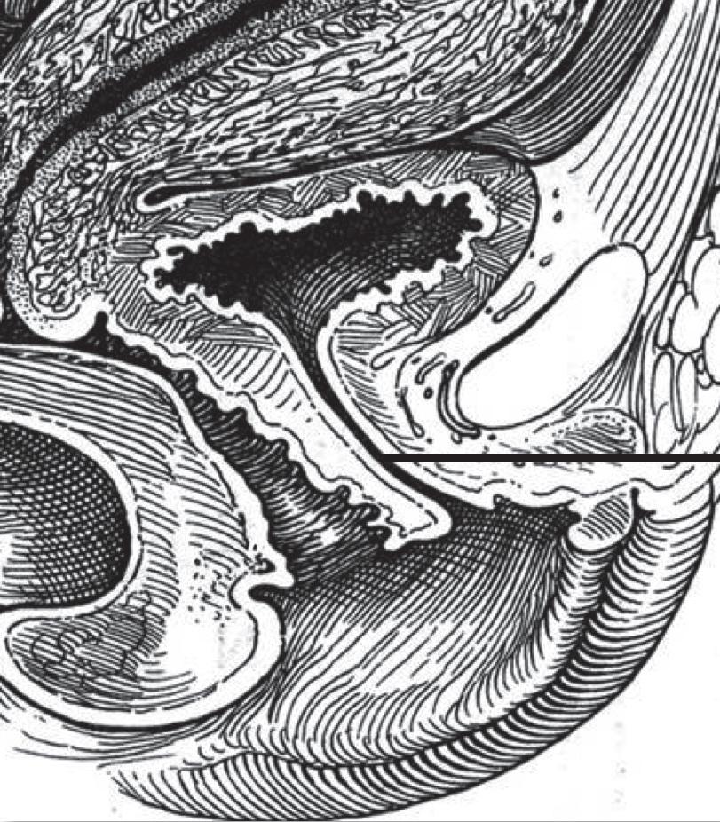
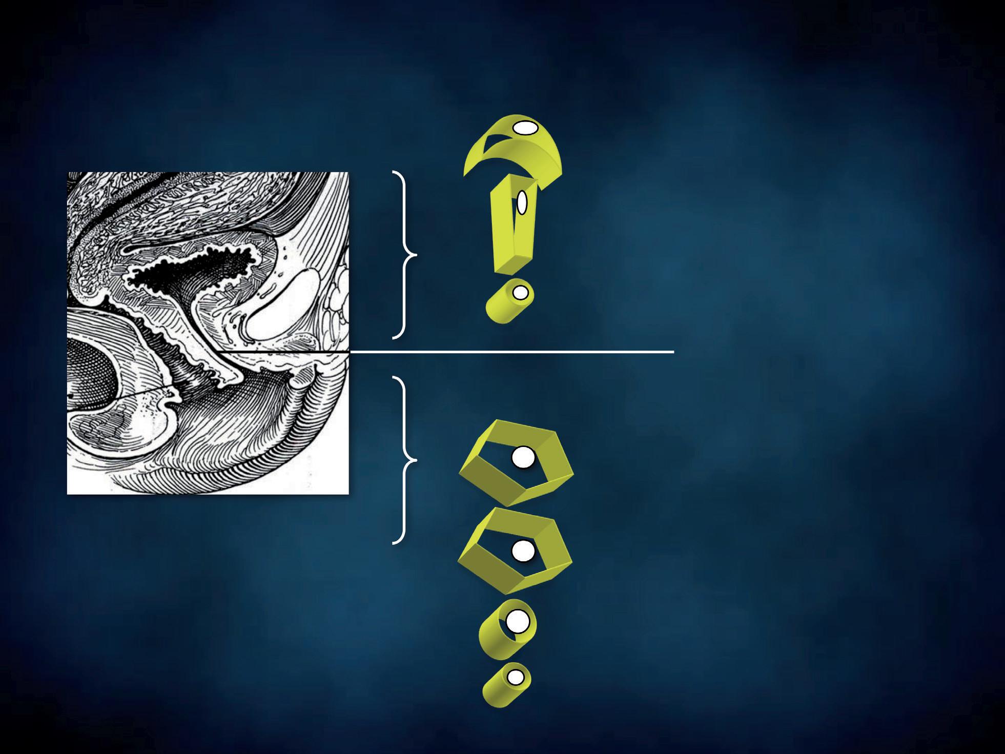
Cervico-vaginal wet mount technique
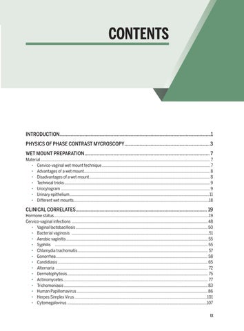
The patient should not have douched and used vaginal suppositories, creams, gels or foams for at least 48 hours prior to sampling. With the patient lying down in gynaecological position, a non lubricated bivalve self retaining speculum is gently inserted into the vagina, so that vaginal walls, fornices and cervix can be thoroughly visualized. A cotton swab or a spatula are used to collect discharge from the vaginal walls, the esocervix and the vaginal posterior fornix (Figure 5); a second cotton swab or an endocervical device can be rotated in the cervical canal. The swabs, the spatula or the endocervical device are immediately and lightly dabbed (and not rubbed) in a drop of sterile normal saline, previously applied on a microscope slide (Figure 6a). Alternatively, 10% potassium hydroxide (KOH) solution is suggested when vaginal fungal infection is suspected. In fact, use of 10% KOH in wet preparations improves the visualization of yeasts and mycelia by disrupting cellular material that might obscure yeasts, pseudohyphae or hyphae. Applying this type of solution between the slide and the coverslip is not recommended. In fact, despite the presence of a vaginal fungal infection and irritative symptoms, blastospores, hyphae and pseudohyphae may be not visible under direct microscopic examination and cultures give false negative results. In such cases, Candida uses protease to penetrate the vaginal epithelium, thus leaving a specific track on the cells, that the screener may easily detect. The use of KOH solution destroys the cells, thus hindering the recognition of the specific cytopathy induced by the fungal hyphae.
vaginal sampling
endocervical sampling
Figure 5 - Cervico-vaginal sampling.
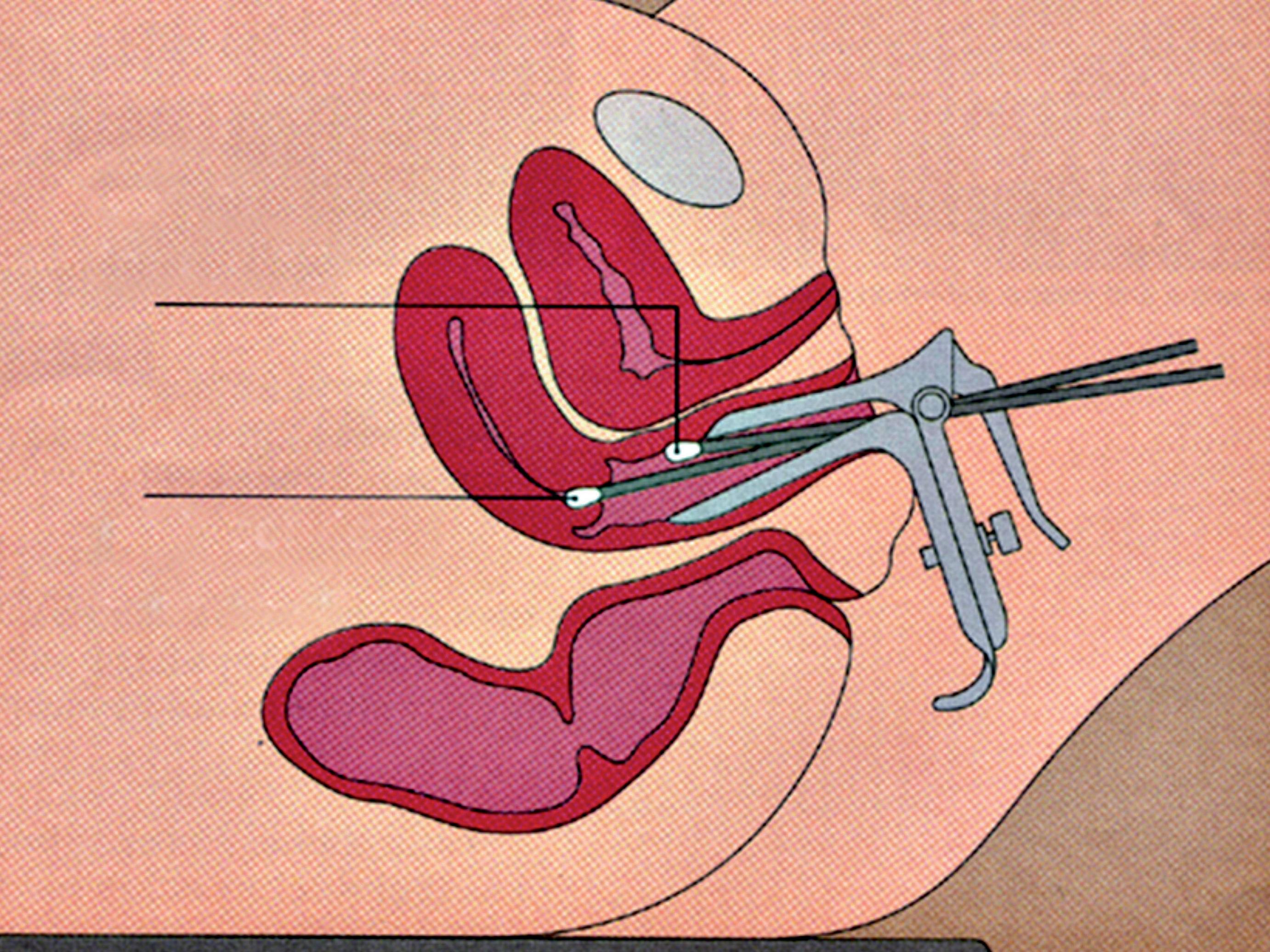
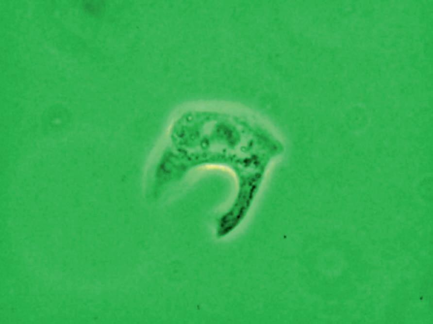
The specimen is mounted with a cover glass (Figure 6b) immediately after. Be careful to lower the coverslip so that it forms an angle of 45 degrees with the microscope glass and one edge touches the slide. This precaution will prevent air bubbles from being entrapped under the coverslip, interfering with the viewing and hampering organisms’ movements. The sample is thus ready to be examined under the Ph microscope, without any artefact due to fixation and staining. Each photomicrograph was taken through a Ph objective lens (x40) and an eyepiece (x10) connected with a camera, obtaining a 400x total magnification. Some images were magnified to highlight particular structures of cells or microorganisms. The green shade of the pictures is due to the green filter placed on the filter holder. When necessary, our microscopic findings had confirmatory cultures and the causative agents are indicated in brackets.
Advantages of a wet mount
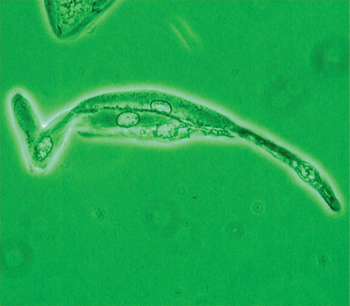
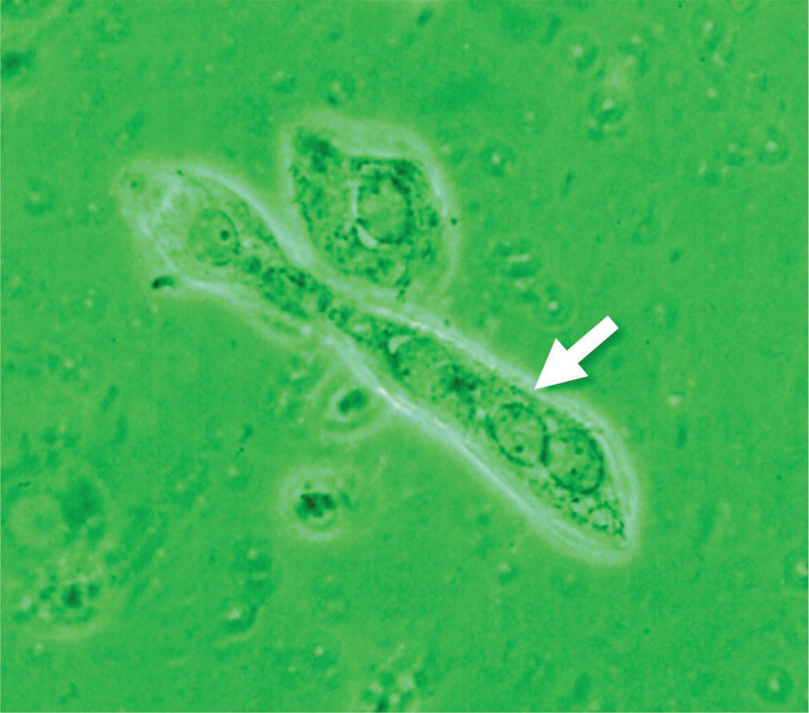
• No artefact: direct microscopy makes living organisms and biological structures clearly and immediately visible, without artifacts due to dehydration, fixation and staining – used for processing permanent mounts – that may alter the morphological characteristics and modify the physicochemical properties of the matter. • Quick preparation. • Observation of movements of protozoas and cell organelles such as cilia in ciliated endocervical cells.
Disadvantages of a wet mount
• Storage: wet mounts cannot be stored over a long time because the specimen dries up. This problem may be easily solved by taking photos of the microscopic images using a digital camera system applied to the phase contrast microscope.
a
b
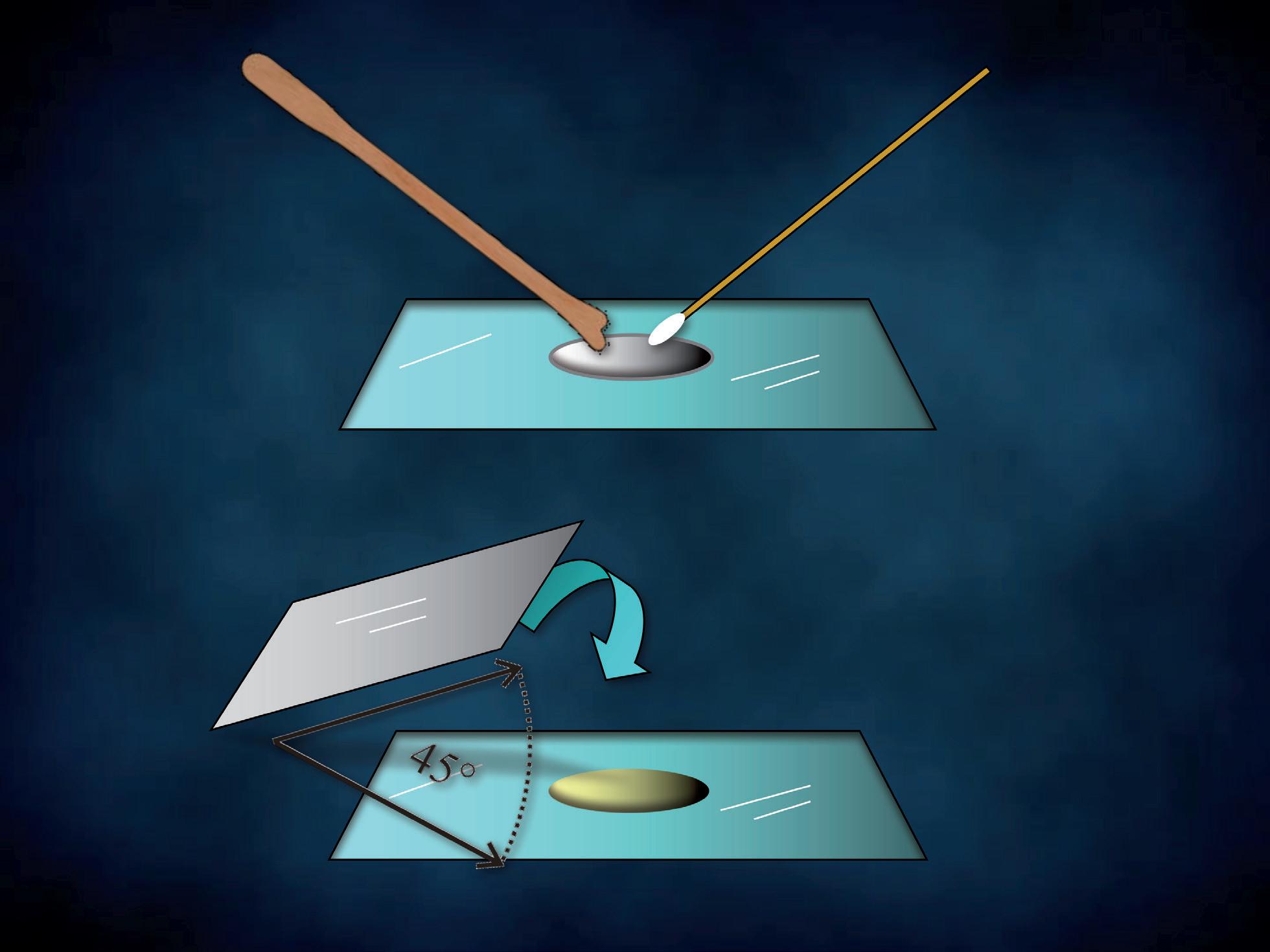
Figure 6 - Cervico-vaginal wet mount preparation.
• Difficulty in taking pictures due to the movement of protozoas or ciliated endocervical cells. However it is possible to film the moving organisms using the digital camera system or wait a few minutes for the movements to fade or disappear.
Technical tricks
• Your aim is to have sufficient water to fill up the space between coverslip and slide. The coverslip may float in case of excessive water. In such case, hold a paper towel close to one edge of the coverslip. This will draw out some water. • If the wet mount is too dry, air bubbles may form and the specimen may be squeezed between the coverslip and the slide. This would interfere with the viewing and hamper the organisms’ movements. In this case you can add a drop of water near the coverslip. • Bringing the objective lens too near the edge of the slide will result in the saline smearing the objective; this may interfere with the viewing and can damage the lens. • Touch the cover glass on the sides to avoid leaving fingerprints. • Air-drying, a common artifact with wet mounts, causes artificial nuclear enlargement. When a longer observation of the wet mount is necessary, a vaseline chamber can be prepared to prevent that the specimen dries up due to the heat of the lamp.
It is sufficient to gently rub the tip of a swab dibbed in vaseline on each edge of the coverslip, to form a thin and continuous vaseline ledge. The same result may be obtained by using a clear nail polish.
Urocytogram
With the patient’s external genitalia exposed, the urethral meatus is thoroughly cleaned with sterile saline solution.
URINARY wet mount
a
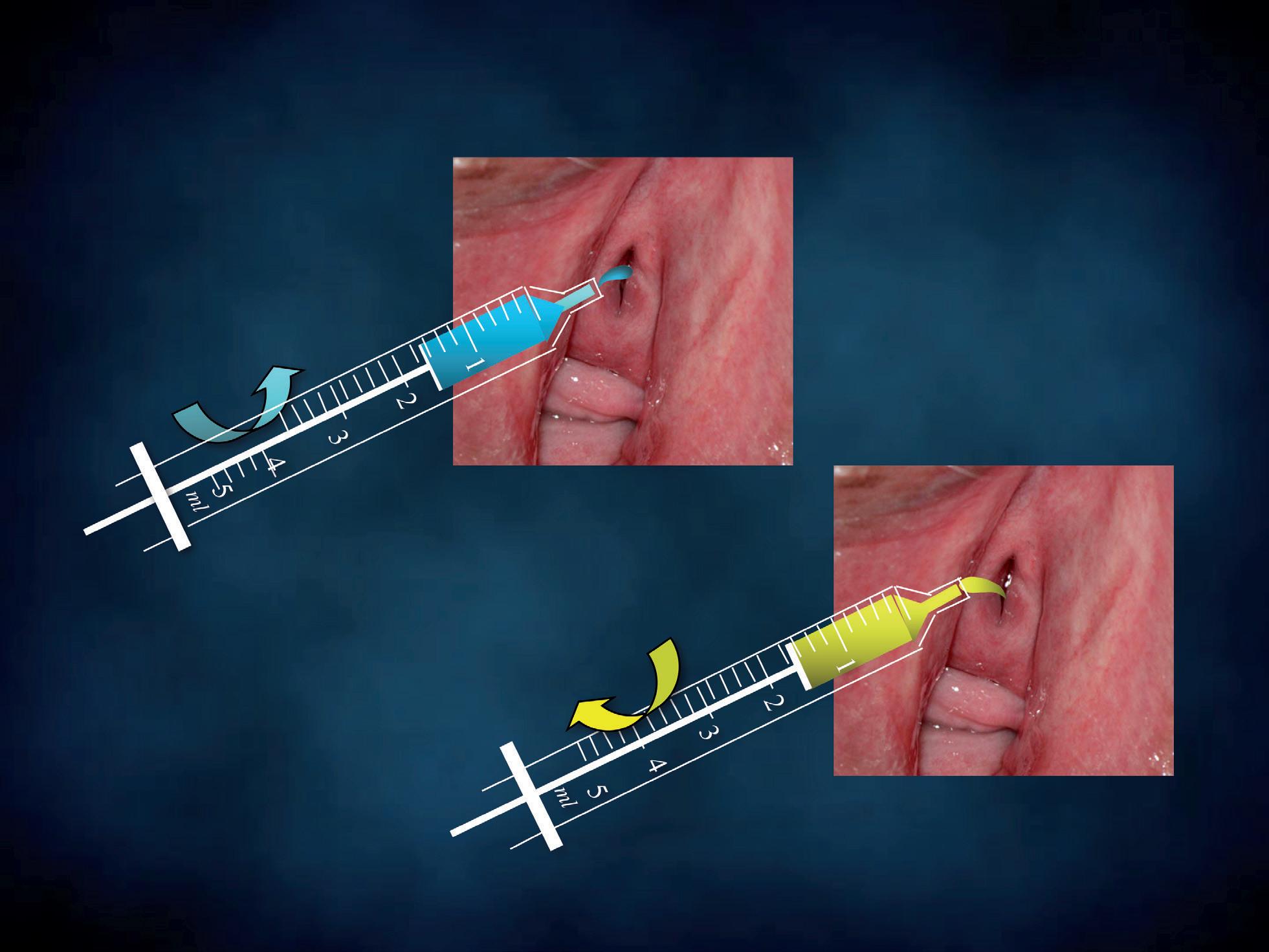
Figure 7 - Urinary wet mount sampling.
b
URINARY wet mount
Figure 8 - Urinary wet mount preparation.

Using a disposable needless syringe, we introduce 5-10 ml of saline into the urethra and the bladder (Figure 7a). Following this injection, a few drops of the urethral washing fluid are collected using the same syringe (Figure 7b). Soon after, one or two drops are poured onto the glass (Figure 8). The following procedures correspond to the ones used to process a cervico-vaginal wet mount.
Urinary epithelium
Two types of epithelia can be observed in the female urethra (Figure 9): • transitional epithelium • squamous epithelium.
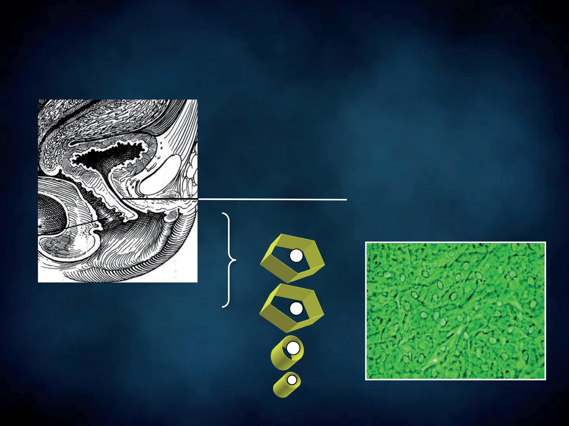
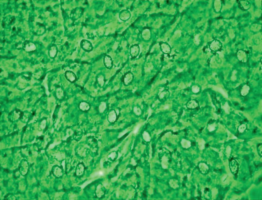
All the cells of both epithelia show a pale nucleus with finely granular chromatin. Transitional epithelium consists of multiple layers of epithelial cells able to contract and expand. It is called “transitional” because of this function in the transition from different degrees of distension. This highly specialized and unique epithelium, known as urothelium, lines the proximal 2/3rds of the urethra. Transitional epithelium also lines the renal pelvis, the ureters and the bladder. Urothelium is composed of three types of cells, proceeding from the basement membrane towards the lumen (Figures 9, 10): • basal cells • club-shaped cells • umbrella cells.
A basal layer of cuboidal cells rests on the basement membrane. The intermediate layer is composed of club-shaped or columnar cells, whose expanded end lays in the concavity of umbrella cells which vary in
URINARY EPITHELIA transitional
umbrella c
club-shaped c proximal 2/3rds
basal c
squamous
distal 1/3rd
Figure 9 - Urinary epithelia.
URINARY EPITHELIA transitional umbrella c
club-shaped c
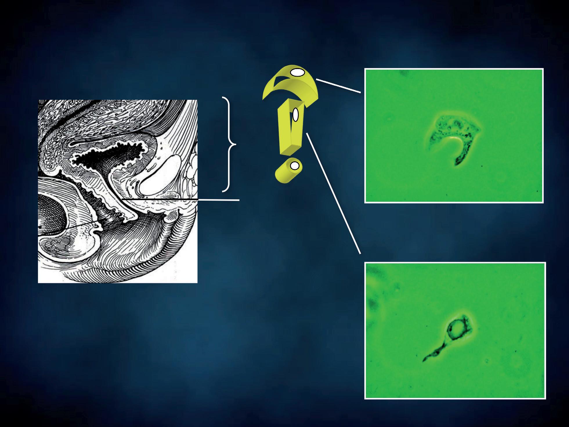
Figure 10 - Urinary transitional epithelium.

appearance, depending on the degree of distension. The superficial cells are referred to as umbrella cells with a domed apex when the organ or the tube in which they reside is not stretched. They are fairly large when released and may show multiple nuclei (Figure 11). The distal 1/3rd of urethra is lined by a nonkeratinized stratified squamous epithelium (Figure 9). Urethral squamous cells differ from squamous cells lining the vagina only for the presence of pale nuclei (Figure 12). A gradual change from squamous to columnar epithelium can be observed with advancing age. More squamous epithelium is present at the distal end of the urethra. There are small mucus-secreting urethral glands, that help protect the epithelium from the corrosive urine. The urocytogram is an essential diagnostic tool to observe bacterial adherence to urinary cells (Figures 13, 14), since it is not detectable by cultures and may cause recurrent urinary tract infections (UTIs). When urethral glands are interested by bacterial colonization or infection, bacteria can appear adhered to mucus filaments and the observation of clean filaments reveals the success of the pharmacological treatment (Figure 15). Even in presence of negative urine culture and in absence of dysuric symptoms, the microscopic evidence of bacterial adherence enables to plan prophylactic antimicrobial regimens, such as (i) the use of longterm, low-dose prophylactic antimicrobials taken at bedtime; (ii) intermittent single-dose treatment with antimicrobials; (iii) post-coital prophylaxis for women whose episodes of infection are associated with sexual intercourse. These antimicrobial regimens have all been demonstrated to be effective in managing recurrent uncomplicated UTIs in women. Furthermore, the screener can immediately recognize the same cell changes induced by pathogens, detectable in vaginal cells, like Candida cytopathy in urinary fungal infection (Figures 16, 17).
When fungal infection is detected in the male sexual partner by urocytogram, it is advisable to treat him as well, in order to avoid recurrent vaginal candidiasis.

Different wet mounts
We use spatulas to collect the specimens for the preparation of cutaneous (Figure 18), oral (Figure 19) and rectal wet mounts (Figure 20). The following procedures correspond to the ones described to process a cervico-vaginal wet mounts.
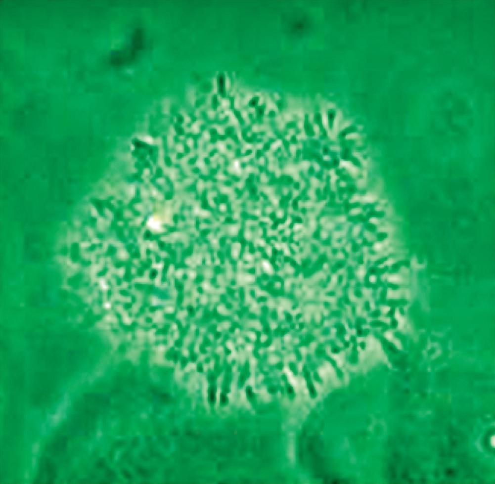
Figure 11 - Transitional superficial cells.
URINARY EPITHELIA transitional superficial cells
Flat and released superficial cells Multinucleated superficial cell
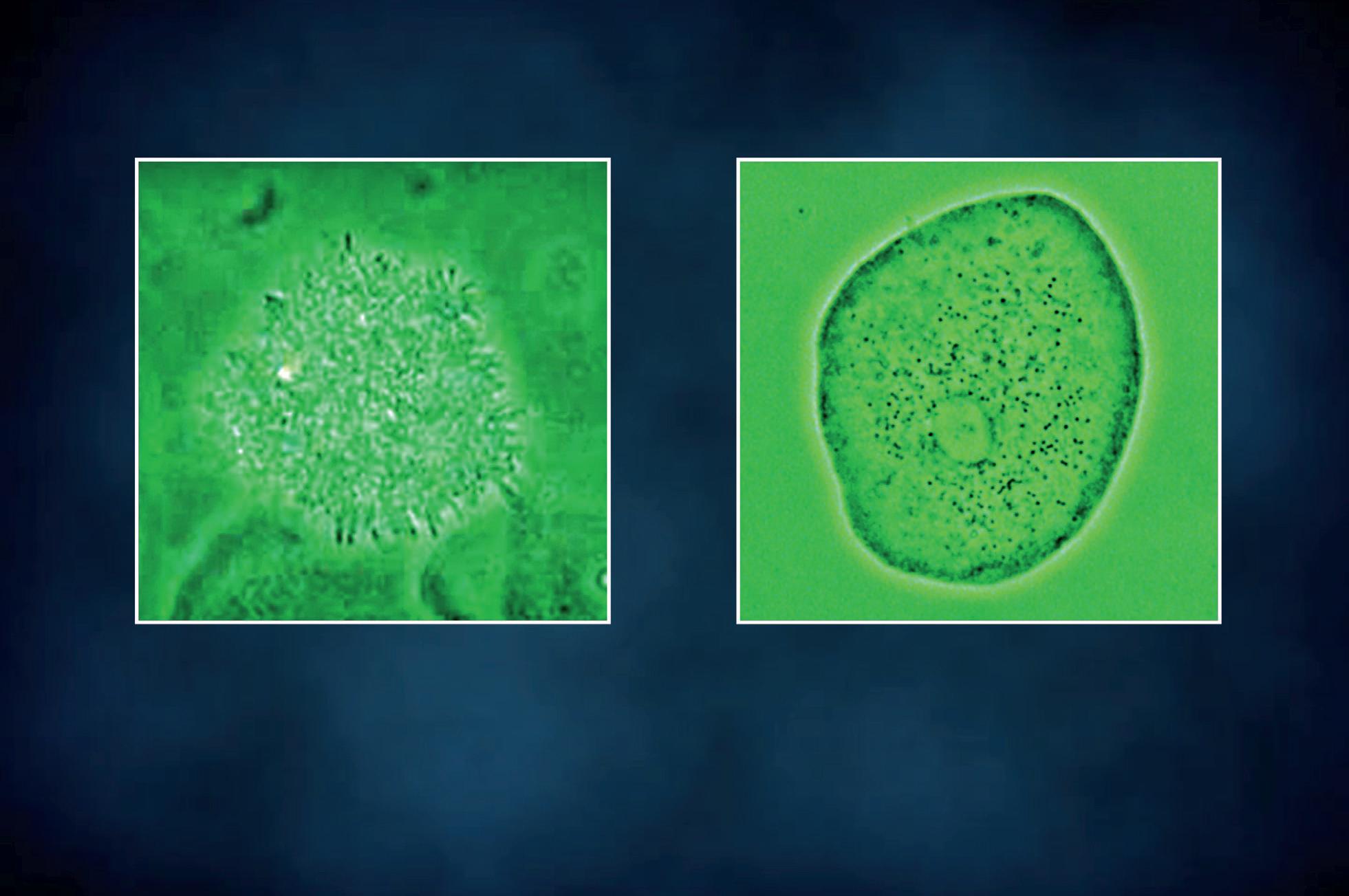
squamous squamous c
Figure 12 - Urinary squamous epithelium.
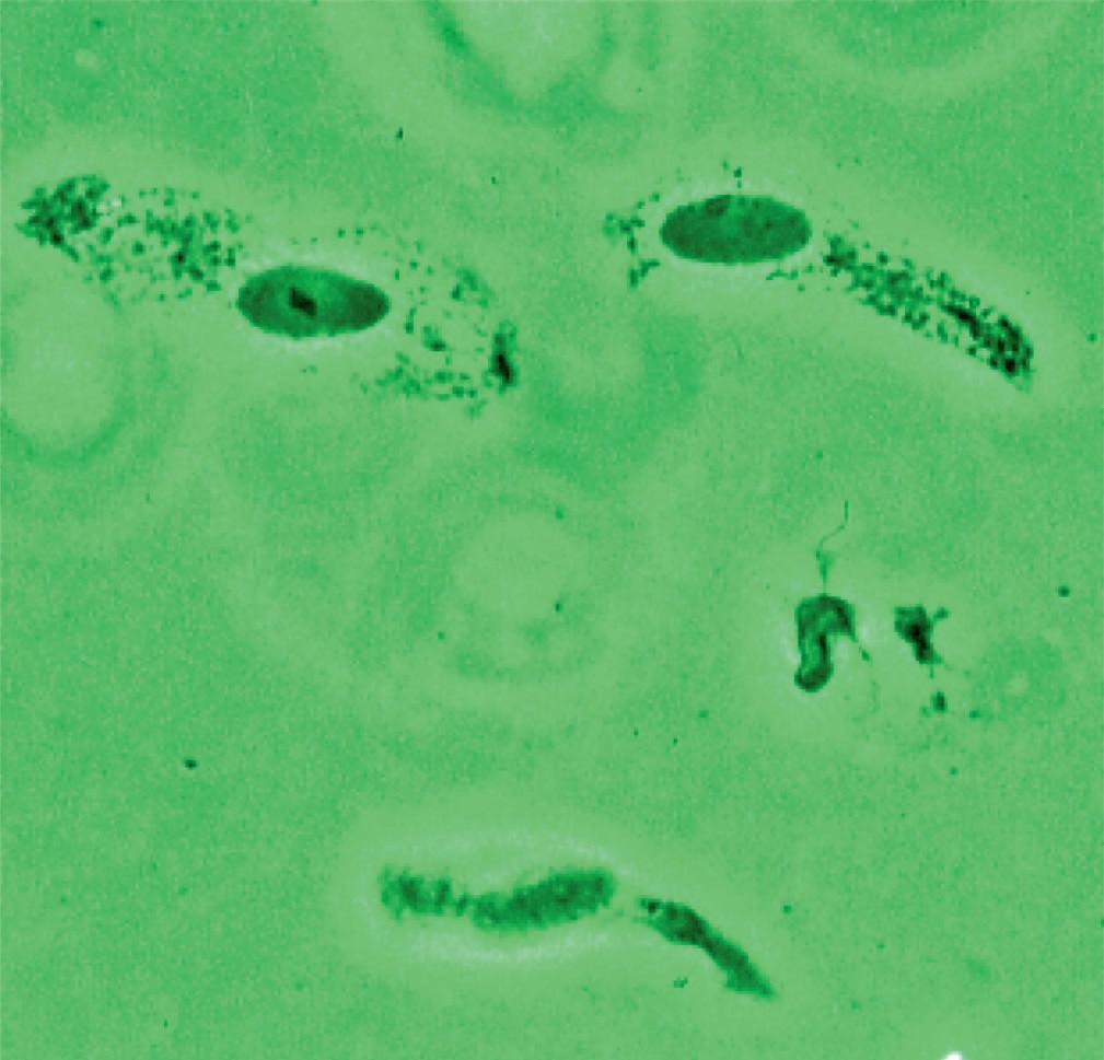
URINARY wet mount
Short bacilli adhered to a squamous cell
Small cocci adhered to a squamous cell
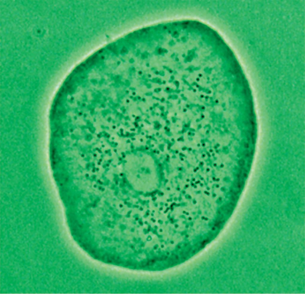
Figure 13 - Urinary wet mount: bacterial adhesion to squamous cells.
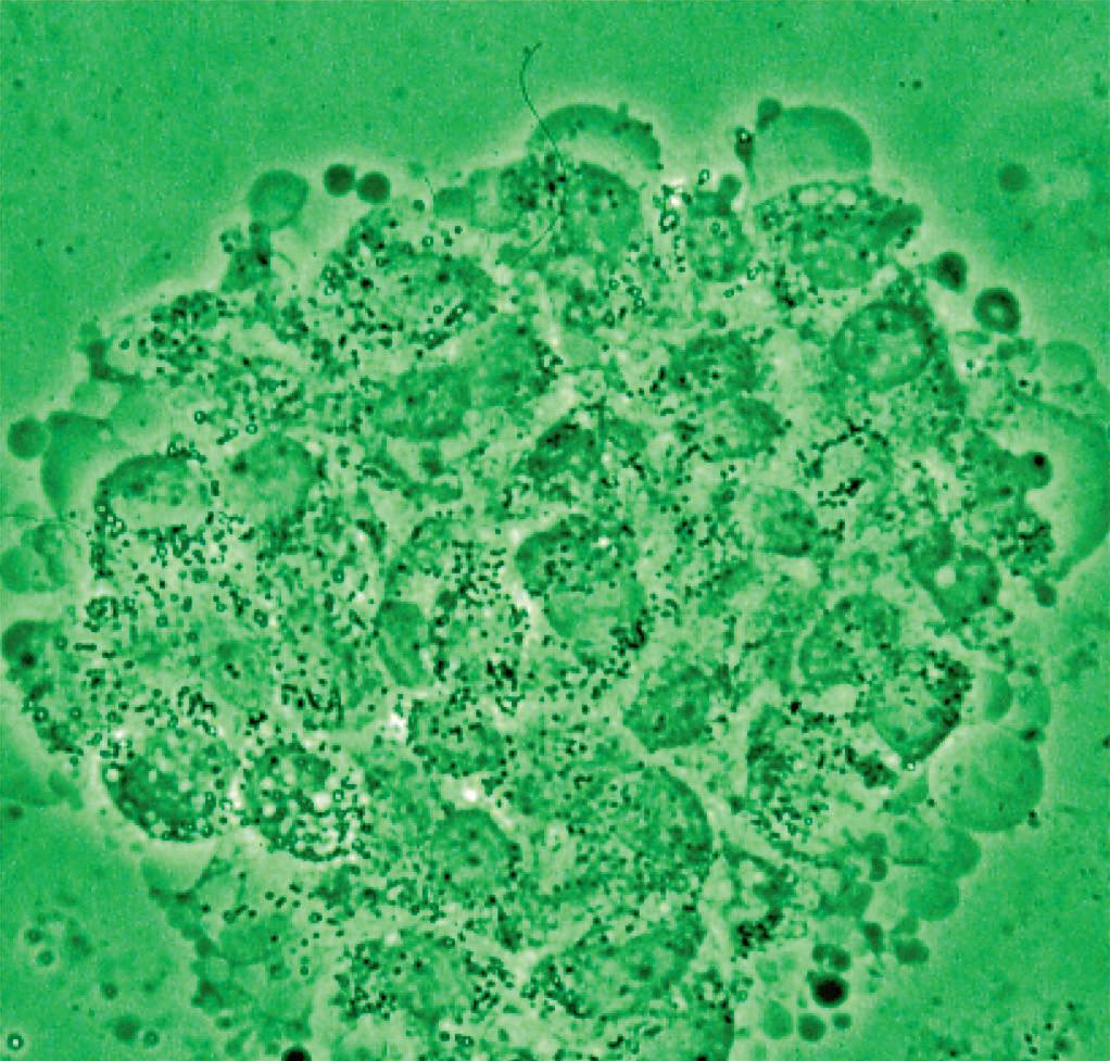
URINARY wet mount
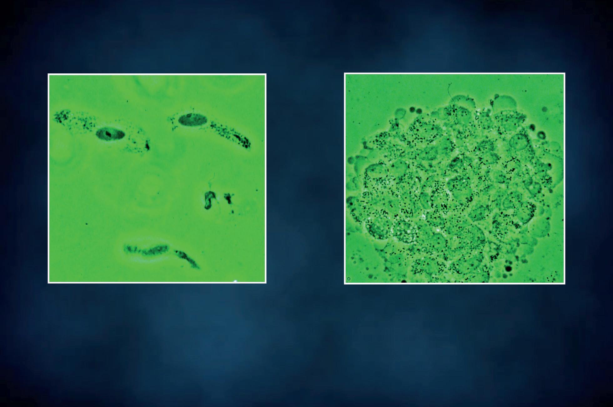
Pleomorphic bacteria adhered to transitional intermediate cells Pleomorphic bacteria adhered to transitional superficial cells
URINARY wet mount
Adhered bacteria to mucus filaments Clean mucus filaments after treatment

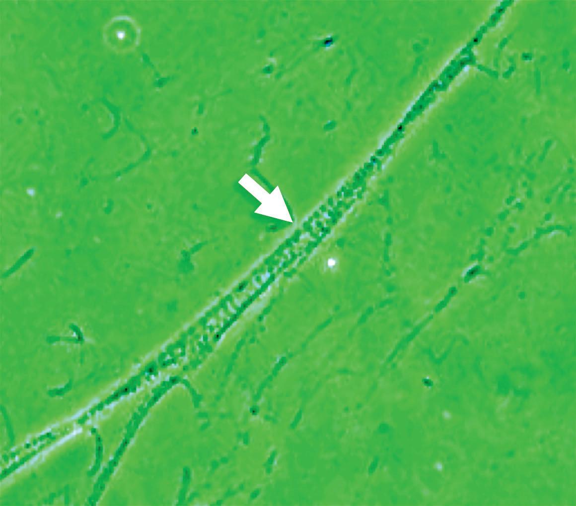

Figure 15 - Urinary wet mount: bacterial adhesion to mucus filaments.
URINARY wet mount
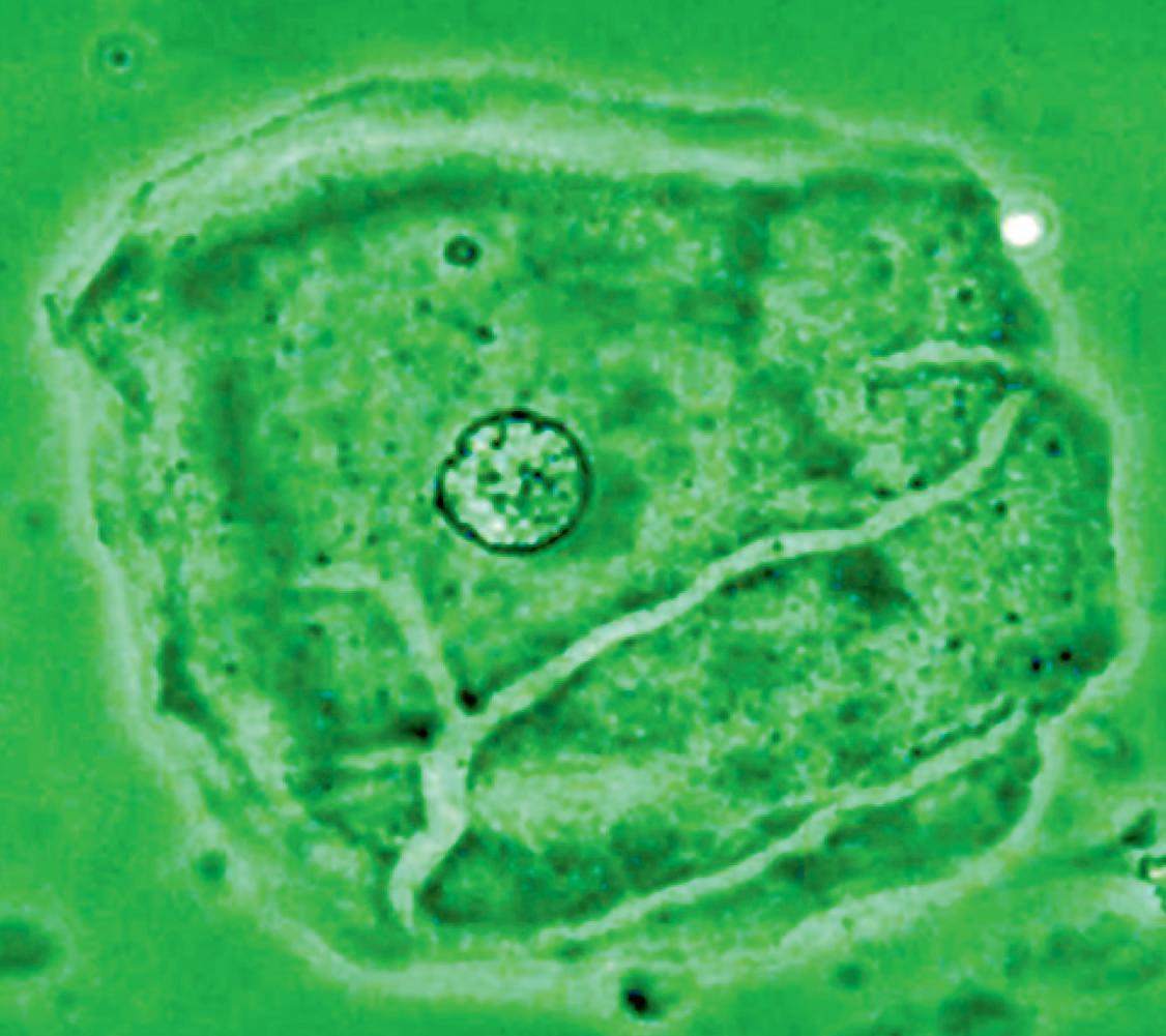

Figure 16 - Urinary wet mount: Candida cytopathy in a squamous cell.
URINARY wet mount
Candida cythopathy: cytoplasmic hole in in umbrella cell


Figure 17 - Urinary wet mount: Candida cytopathy in a transitional cell.
CUTANEOUS wet mount
Candida cytopathy: cytoplasmic hole and marginal erosion in horny cell Fungal indurated erythema
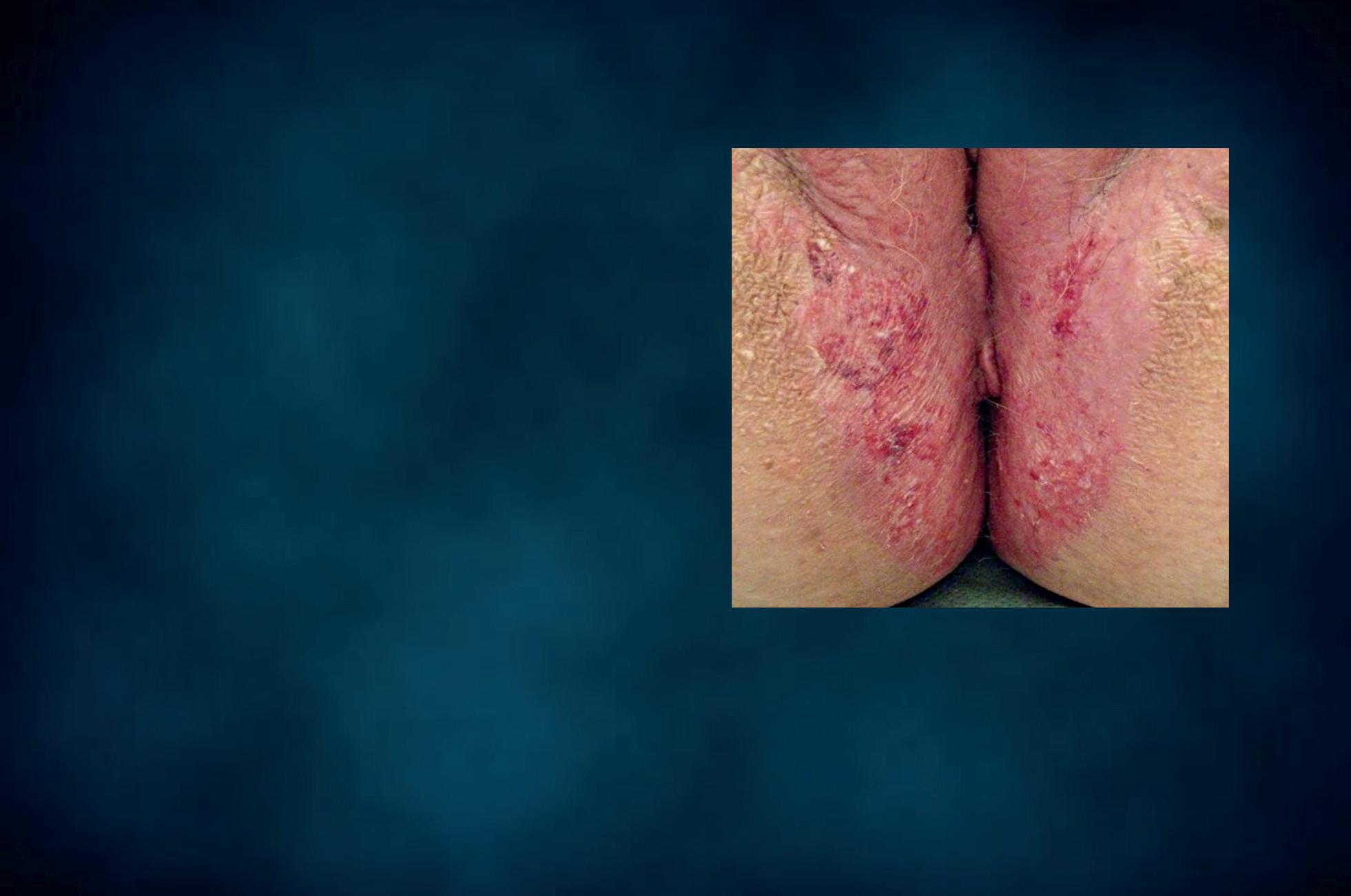
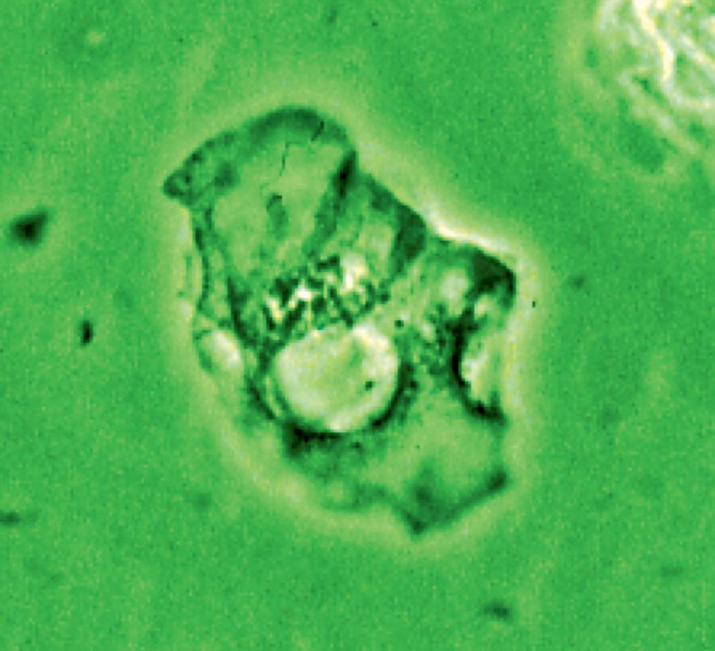


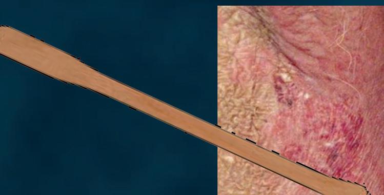
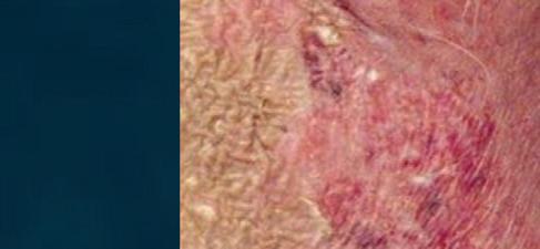
Figure 18 - Cutaneous wet mount: Candida cytopathy in a horny cell.
ORAL wet mount
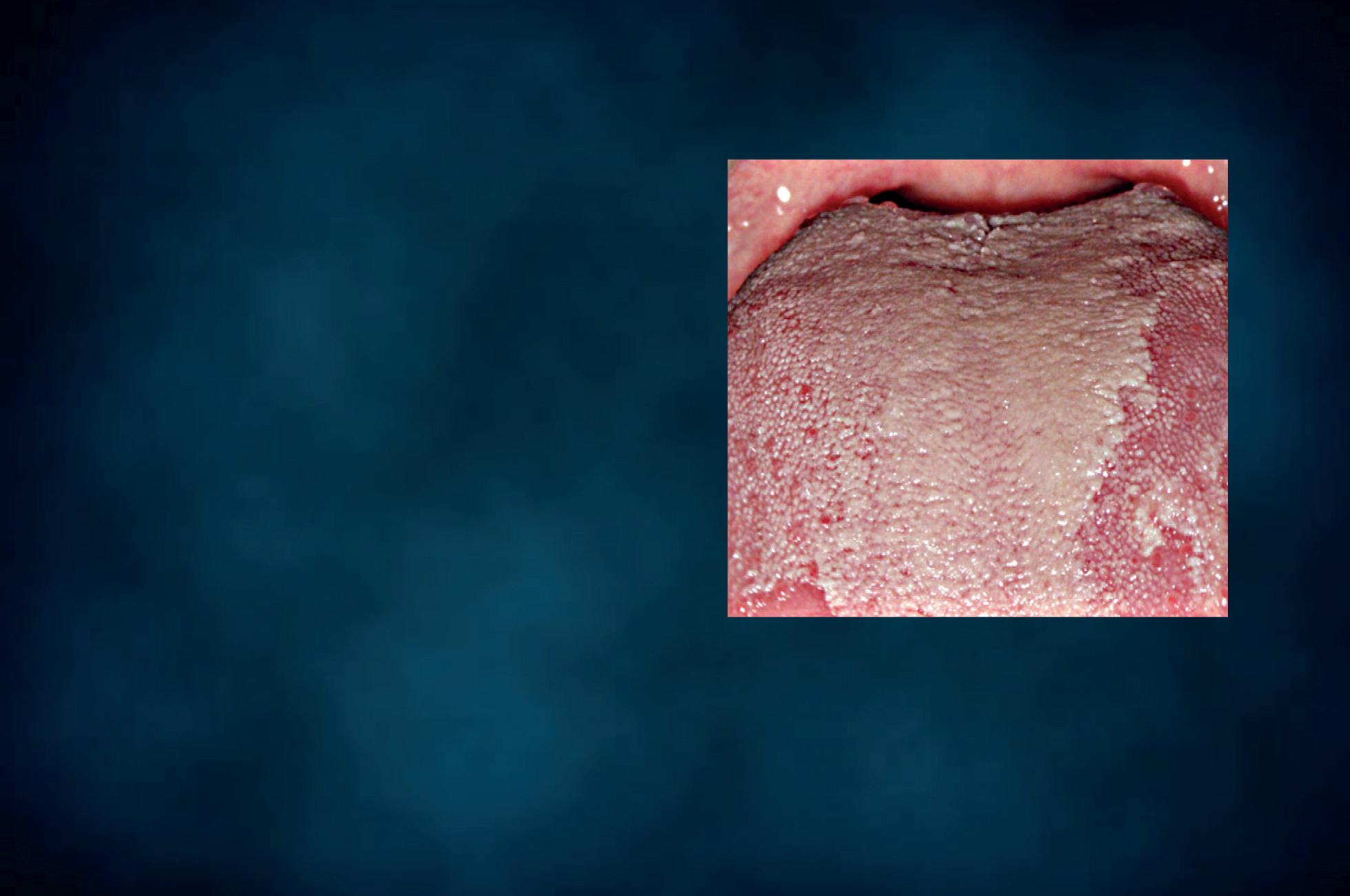
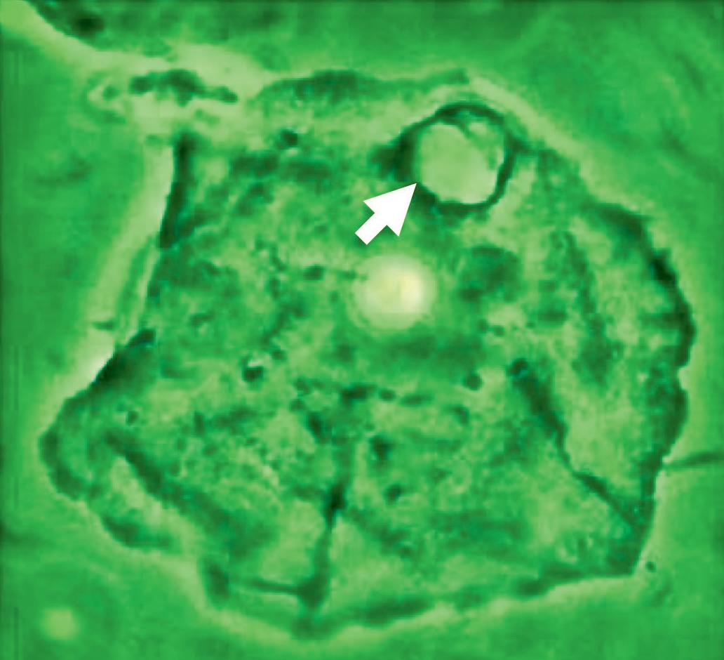

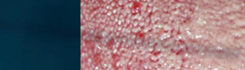
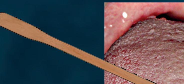

Figure 19 - Oral wet mount: Candida cytopathy in a squamous cell.
RECTAL wet mount
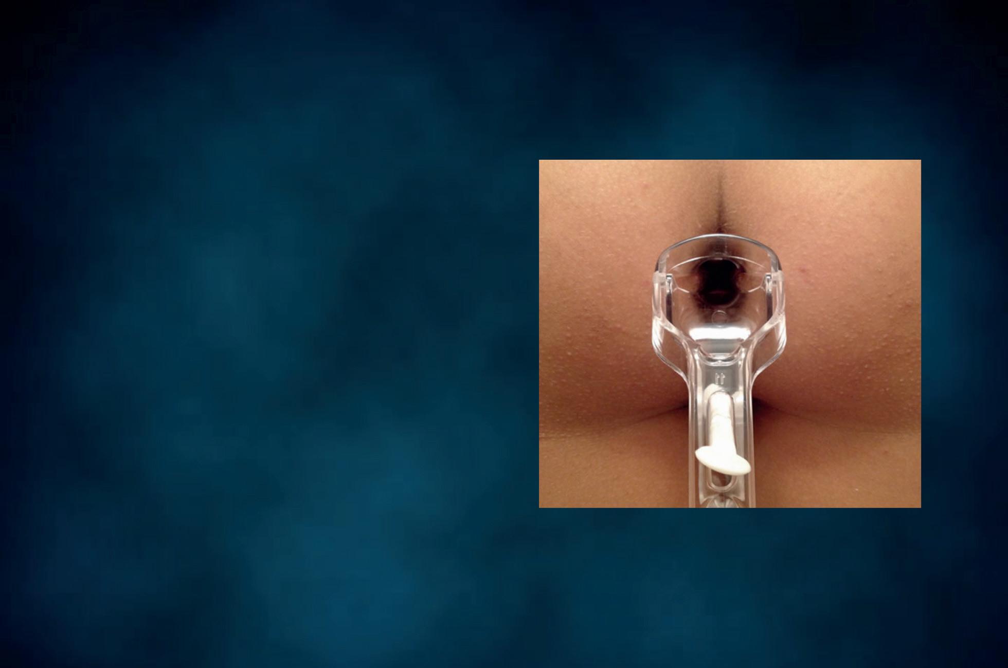
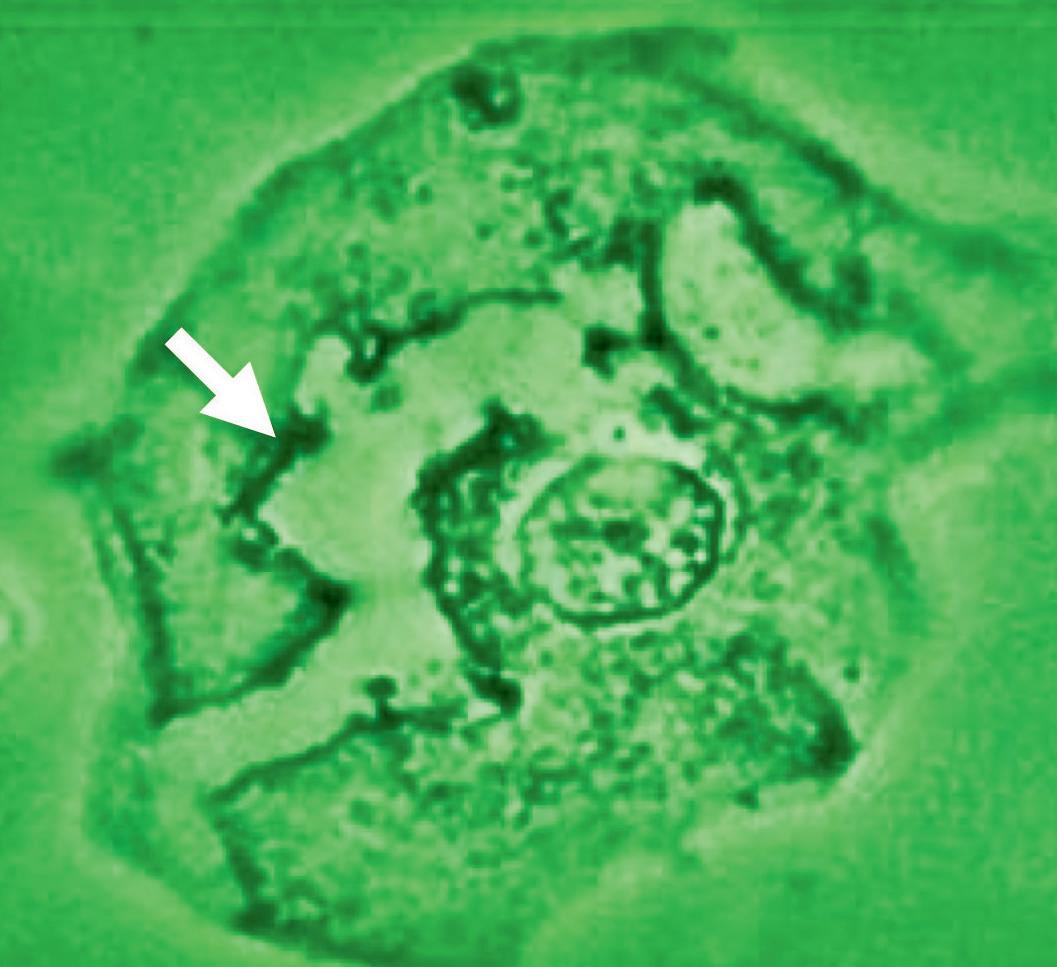
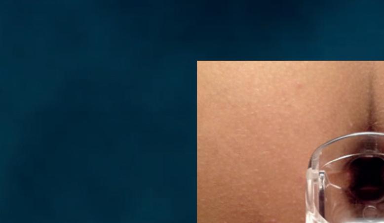

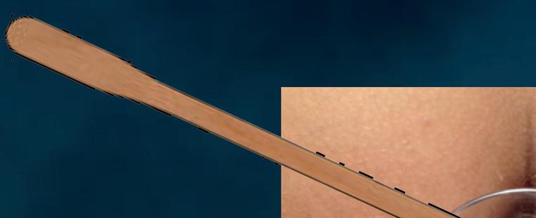

Figure 20 - Rectal wet mount: Candida cytopathy in a squamous cell.
When fungal infection is detected in the male sexual partner by urocytogram, it is advisable to treat him as well, in order to avoid recurrent vaginal candidiasis.
Different wet mounts
We use spatulas to collect the specimens for the preparation of cutaneous (Figure 18), oral (Figure 19) and rectal wet mounts (Figure 20). The following procedures correspond to the ones described to process a cervico-vaginal wet mount.