8 minute read
Surprises Every Day: Edward Bossen, MD
SURPRISES EVERY DAY
Dr. Edward Bossen, In His Own Words
Advertisement
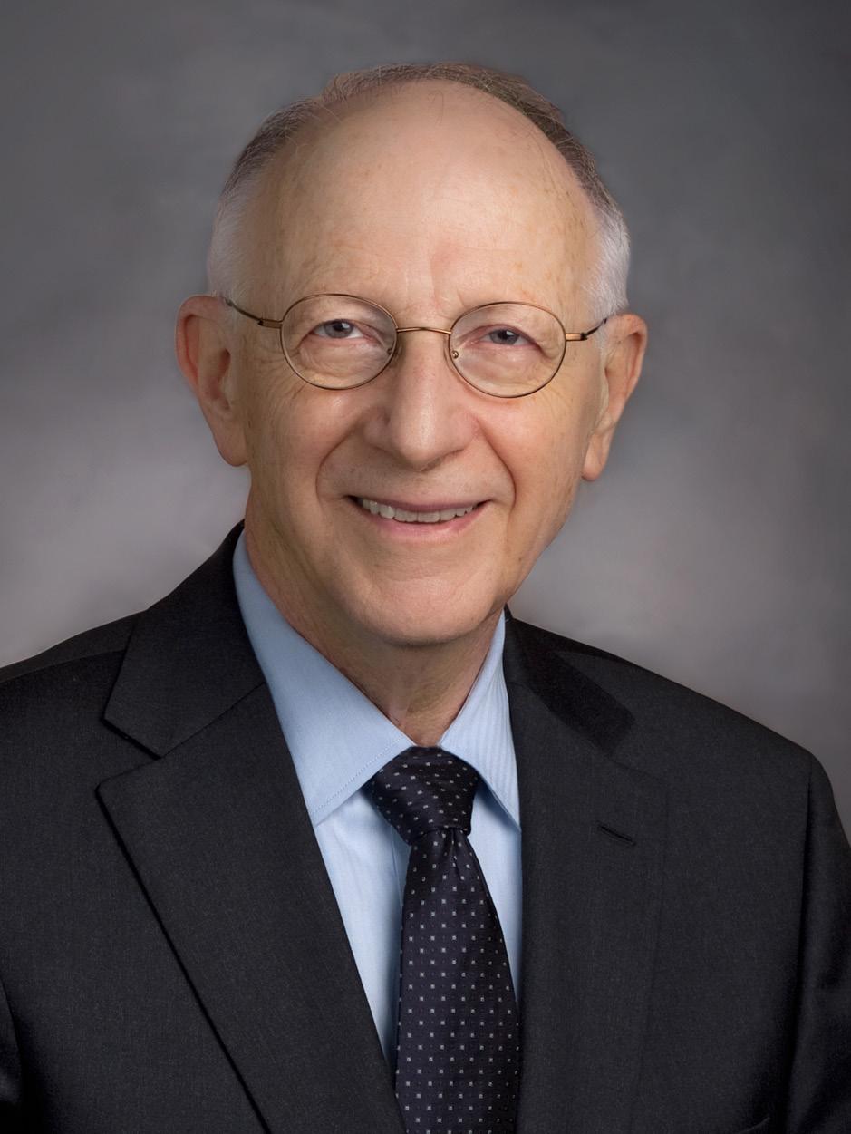
PROFESSOR EDWARD BOSSEN, MD has retired after an illustrious 47-year career in Duke Pathology. His teaching abilities and calm, patient, and genial manner are legendary, and his influence over the decades, including on the medical school, is still strongly felt. A master of muscle pathology and electron microscopy, he literally has written the course book on “Diseases of Skeletal Muscle,” accessible on our website.
(https://pathology.duke.edu/education/duke-pathology-instructional-material)
I arrived at Duke for medical school in 1961 planning to be a family practitioner. I had never heard of Pathology until my second year in medical school when I took my Pathology course. It was a surprise to me that you could make a career of something as interesting as diagnostic pathology. I decided Pathology was the career for me, and Duke the best place for my training.
I was blessed to have as mentors during residency Drs. Hackel, Vogel, Smith, Johnston, and, of course, Fetter. Current residents might be aghast at a typical autopsy case sign-out with Dr. Fetter. You didn’t count hours, because the sign-out could last days. We had around 800 autopsies a year between the VA and Duke (2019: 260 at Duke). Duke had 550 beds and the VA had about 250 beds (Duke: ~979). There was a requirement that hospitals autopsy 65% of deaths. Recently, I read that reinstituting a similar rule has been discussed because the age-old figure of 33% of autopsies having an unexpected finding has once again been acknowledged.

Prepping an IV in 1965 as a senior medical student
My first weekend on autopsy call was a July 4th long weekend. Another resident (I believe he was in Orthopedics—surgeons rotated through Pathology) and I covered both Duke and the VA and performed 10 autopsies, which was quite a feat considering we were neophytes. It took 4-6 hours per case. table, so one of us would dissect organs from a previous autopsy between the legs of the current autopsy. The surgical pathology grossing room at Duke was primitive, and probably illegal by today’s standards. The room was on the 4th floor and was basically a closet with no ventilation. There was a small oscillating fan. You would gross for 15 minutes or so and then leave the room to recover from the formaldehyde fumes. Fortunately, after a couple of years, grossing was moved to a room fitted with a hood.
Surgical pathology sign-out was also quite different from today. The only separate service was Neuropathology, so we dealt with all other specimens, e.g., skin, bone marrows, lymph nodes. The annual case load was about 10,000 cases. You reviewed your cases, wrote out in long hand your description and diagnoses and then took your cases to the lone senior resident on Surgical Pathology who reviewed the cases before you took them to the attending. This meant late nights for the poor senior resident.

Attending the Residents Luncheon in 1978, next to Dr. Dana Copeland
There were no multi-headed microscopes during my early years of training. You sat quietly in a chair while the attending reviewed your cases, occasionally asking you questions. You were expected to have reviewed the patient’s chart if the case was particularly challenging (this was before the days of computer records which meant you had to make a trip to the ward). Sign-outs could last hours or even days. One attending, however, was famous for saying, “Show me the key slide.” Fortunately, he did this only with autopsies and with senior residents. The idea was to see if you knew what was really important.
When you finished signing out a case you gave it to secretaries who typed up the report. Turnaround time was in the 7-10-day range. No one cared about the lag time. There was no rush to discharge patients because the payment system did not demand it, so no one complained. A prostatectomy patient, for example, would remain in the hospital until his catheter was removed, which was about 2 weeks, not the 24-48 hours given now.
I started doing research in my second year of residency, and in my fourth and fifth years became a fellow in immunopathology under Dr. David Rowlands, later the Chair at Pennsylvania and South Florida. One research focus was renal transplantation which had just begun at Duke, and there is a record of my involvement with the third transplant at Duke. I was invited to pursue a PhD in Immunology, but that was not possible because of my military commitment.
In those days all male doctors—and over 90% of doctors were males—were required to serve in the military or public health service, signing up in the last year of medical school. You could be called to active duty any time after your first year of postgraduate training, but pathologists were usually not activated until they finished training.

In his office in 2004
I was very fortunate. The chair, Dr. Kinney, was on the Board of the Armed Forces Institute of Pathology (AFIP), which was the premier referral site for military and civilian pathology cases at that time (no charge!). I had received a list of possible duty posts from the Army with instructions to select my preferences. This I knew was a hoax: the Army would put you where they wanted. I went to Dr. Kinney and asked his advice. He turned around in his chair, picked up the phone, and called the officer in charge of assignments for pathologists, a Duke medical graduate. Dr. Kinney turned around to face me and said I was going to the AFIP, the prime assignment in those days. Furthermore, I was to be in the Laboratory of the Deputy Director, Colonel James Hansen, another ex-Duke graduate. The Lab was known as the Laboratory of Skeletal Muscle Research, but was absorbed into the Neuropathology Branch, so I picked up some general neuropathology as well. We consulted on patients at Walter Reed and performed open muscle biopsies on adults. Children’s biopsies were performed by surgeons because of general anesthesia requirements.
My second year at the AFIP, I was seated at my desk one day with my back to the door. I heard it open and a voice asked: “Want a job?” I turned around, saw it was Dr. Kinney, and said yes. The door closed. A few weeks later I received a letter announcing that I was to be an Assistant Professor at Duke.
In July 1972, I returned to Duke, and practiced Surgical Pathology and Cytopathology. I was appointed Associate Director of the latter and in the late 1970s became Director of Surgical Pathology. I was also the Director of the Residency Training program for 3 years in the mid-1970s. The renal pathology transplant faculty I had worked with had left, so I applied my new-found knowledge of skeletal muscle to create a muscle pathology service, which expanded to provide consultations to regional and national practitioners, and it still does now. I also renewed my lecture series to the ENT residents which I had begun as a resident.
I was also fortunate to add cardiac muscle research with Joe Sommer. Some of that research involved finches, and occasionally the research subject would escape and fly around the lab. We wisely kept the doors closed so the bird couldn’t escape. Finch hearts beat at 450 beats per minute resting and up to 1000 a minute when exercising. This meant that the finch could only be airborne a few minutes before needing to rest on a lab bench where it could be recaptured.

Delivering his last lectures to medical students in February 2017 at the Trent Semans Great Hall classroom
I dropped my duties in Cytopathology in the mid-1980s when my duties as Director of Surgical Pathology and the growing muscle biopsy service took up most of my time. Eventually I became Director of Anatomic Pathology.
I owe much to the past chairs, Drs. Kinney, Jennings, and Pizzo for their support. I also had the good fortune to know Dr. Forbus, though he was then no longer chairman. I must also mention Dr. John Shelburne who became acting chair after Dr. Jennings retired. This was a very difficult time for the department and John handled the situation with wisdom and grace. I know Dr. Huang will continue the tradition of great leadership.
I have fond memories of many of the technical and clerical staff. There is not room to list everyone who helped me. I must, however, mention Susan Watson, Terrie Harris, and Bonnie Lynch, who did their best to keep me out of trouble. Wayne Terrell and Bert Dotson were essential to the muscle service. Of course, the PhotoPath folks, Susan Reeves and Steve Conlon, whose function is vital to this visually-oriented department, are owed a debt of gratitude for their excellent work. I cannot mention PhotoPath without remembering Carl Bishop who started out as the first autopsy diener in the Department in the early 1930s and later became the department’s superb photographer. He was extremely compulsive, known to thoroughly clean the room before every new photography session. Documenting publication photomicrographs together at the Zeiss Ultraphot microscope was a process passed through from Mr. Bishop to Bill Boyarsky and then to Susan, and was always a highlight.
I am happy to say that I picked the right career and institution for me. As a pathologist you are not bound to a single area of medicine. Every day there are surprises to be seen under the microscope that may spark a research interest or just make you want to explore the disease process you have observed. Every day is a learning experience. If you are a compulsive observer and learner, Pathology is the field for you.
In honor of Dr. Bossen, an award was initiated in 2016. The Edward H. Bossen Team Player Award is presented to the resident or fellow who has distinguished themselves in their commitment, values, work ethic, and contribution to team morale.

