1. Targeting antigen heterogeneous B cell malignancies with multi-specific CAR molecules
2. The CD19 CAR T hinge region influences avidity and cytotoxicity in CD19 SNP variants in B cell Lymphoma
3. Mapping specificity of de novo designed pMHC-targeting minibinders
4. In situ tumor-to-cDC1 reprogramming improves expansion and anti-tumor activity of TILs for adoptive cell therapy
5. Dissecting the Impact of RAS Pathway Inhibition on TIL Expansion and Therapeutic E icacy
6. Neoantigen Identification using Dendritic Cell Reprogramming
7. CRISPR-PD1 modified tumor infiltrating lymphocytes for adoptive therapy for patients with metastatic melanoma
8. Enhancing anti-cancer immunity with combinatorial CRISPR perturbations in tumourinfiltrating lymphocytes
9. Modeling CAR T Cell Dysfunction in Solid Tumors Using 3D Heterospheroids
10.Optimizing tumor-infiltrating lymphocytes through nonviral CRISPR/Cas-engineering for enhanced adoptive T cell therapy
11.Deciphering the molecular signature of induced pluripotent stem cell to T-cell di erentiation using CRISPR activation screens
12.Bridging T Cell Immunotherapy and Stem Cell Engineering Advancing Allogeneic T Cell Manufacturing from Induced Pluripotent Stem Cells
13.Biomarkers of endothelial dysfunction for prediction of acute toxicities in patients treated with chi meric antigen receptor T-cells for hematological cancers
14.Single-cell based multiomics profiling of tumour infiltrating lymphocyte in adoptive cell therapy reveals signatures of relevance for clinical response
15.Tracking antigen specific T cell responses in rapidly expanded TIL-ACT products of cervical cancer patients
16.Nonviral CRISPR/Cas-engineered tumor-infiltrating lymphocytes with inducible cytokine release and checkpoint resistance for adoptive T cell therapy
17.Macrophage activation before lymphodepletion can predict CRS, ICANS, and late cytopenia after CD19 CAR-T cell therapy for hematological cancer
18.DAN-CART 1901: Preliminary results of safe and feasible administration of the fresh autologous anti-CD19 CAR-T product DK-CLIC-1901
19.GD2-targeting Tumor Infiltrating Lymphocytes for Pediatric Patients with CNS Tumors
20.Impaired function of CLL-derived CD19 CAR T cells in a 3D tumor microenvironment reveals CXCR4 and IL-10 as potential targets
21.Generation and Validation of HER2-targeting CAR-TILs for Ovarian Cancer: Investigating the potential as an adoptive cell therapy
22.T cell-targeted gene delivery for in vivo CAR-T cell therapy
23.Functional Dissection of Cytotoxic and Non-Cytotoxic Cellular States of a CD19 CAR T Infusion Product from the DAN-CART 1901 Trial Using Xdrop® Co-Encapsulation
24.FLIC - Flow Cytometry and Imaging Core
25.Adenoviral vectored full-length antigens for expansion of polyclonal CD4+ and CD8+ T cell responses in human dendritic cell and T cell co-culture
26.Harnessing Endogenous miRNAs for Targeted Activation of CRISPR/Cas Systems
27.Novel T-Cell Epitope Mapping and In-Depth Characterization of T-Cells in Respiratory Syncytial Virus Infection
28.Bispecific immunotherapy of Multiple Myeloma: understanding the clonal diversity of treatment response
29.Characterizing CD8+ KIR+ Regulatory T cell-mediated immunosuppression in cancer
30.Identification and characterization of splice-derived t-cell antigens in blood cancers
31.Develop and validate DNA-barcode labelled MHCII multimers for detection of antigenspecific CD4 T cells displayed towards large libraries of epitopes
32.In vivo Expansion of genetically engineered CAR T cells using antigen presenting polymer particles(T-Boost)
33.Human Endogenous Retroviruses as novel targets for Cancer Immunotherapy
34.CREATIC – Building a European Excellence Centre in Central Europe for ATMPs. From Capacity Building to Therapy Development to Patient Treatment
35.Immune signatures associated with clinical response to the personalized neoantigen vaccine EVX-01 and pembrolizumab in advanced melanoma
36.In-situ characterisation of T-cell reactivity
37.Cell avidity: the next-gen binding assay to advance cell-based and cell-engaging therapeutic development
38.Learning TCR–pMHC Binding from Designed TCRs
39.Improving CAR-T Cell Persistence and E icacy Through Epigenetic Reprogramming
40.Overcoming Tumor Apoptosis Resistance by Enhancing Chimeric Antigen Receptor Tcell Cytotoxicity by Delivery of Cathepsins through the Granzyme-perforin Pathway
41.Vaccine-induced PD-L1-specific T cells indirectly enhance antitumor immunity through myeloid cell modulation
42.TR-TIL Platform: Antigen-agnostic pan-cancer identification of polyclonal tumor-reactive CD4⁺ and CD8⁺ TILs for TCR discovery
43.Broad TGFβ-specific T cell immunity is associated with improved outcomes following radiotherapy and checkpoint blockade: rationale for multi-epitope TGFβ immunemodulatory vaccination in pancreatic cancer
44.MelTMG: A Tandem Minigene Platform Encoding Melanoma Minimal Epitopes for Detection of Tumor Antigen–Specific T Cell Reactivity in Melanoma
45.Identification and Characterization of MCP-1 (CCL2) Specific T cell Immunity
Title: Targeting antigen heterogeneous B cell malignancies with multi-specific CAR molecules
B-cell malignancies represent a major clinical challenge due to high relapse rates and limited efficacy of existing therapies. While CAR-T cell therapy has transformed the treatment landscape, long-term outcomes remain limited by antigen-loss-mediated escape and inadequate CAR-T cell persistence. Consequently, addressing these limitations becomes critical. The study focuses on the rational design and optimization of bispecific third-generation CAR-T cell constructs to effectively target antigen-heterogeneous B-cell malignancies. Bispecific CAR constructs targeting combinations of key B-cell antigens like CD19, CD20, and CD22 were designed and systematically evaluated tomitigateantigen escape. In silico structural analyses were integratedtoguidescFvorientationwithoptimalstructuralstability.Becauseaccesstopatient-derivedantigenloss samples is limited, clinically relevant antigen-loss model systems were generated using CRISPR-Cas9 engineered B-cell lymphoma cell lines with defined antigen knockouts to facilitate screening of bispecific CAR constructs. Functional evaluation in these models revealed significant differences among different bispecific combinations, demonstrating that not all bispecific CAR constructs perform equivalently under antigen-loss conditions. The study also aimed to enhance CAR-T cell persistence and functional efficacy using acomprehensive,multi-parametricscreeningofdual costimulatorydomaincombinations.Unlikeconventional approachesemphasizingprimarilyoncytotoxicityorproliferation,thisscreeningevaluatedmultiplefunctional parameters under repeated antigen exposure. This integrated analysis revealed that certain costimulatory combinations excelled in one parameter but underperformed in others, emphasizing the necessity of multiparameter evaluation for rational CAR design. Some costimulatory domains, however, demonstrated balanced performanceacross allfunctional parametersexamined.Overall,thisstudyunderscorestheimportanceofscFv pairing and rational costimulatory selection, providing a framework for developing effective and persistent CAR-T cells with translational potential in antigen-heterogeneous B-cell malignancies.
The CD19 CAR T hinge region influences avidity and cytotoxicity in CD19 SNP variants in Bcell Lymphoma
Ladina Hörtensteiner1,2,5*, Harpreet Mandhair1,5*, Sara Fischer1*, Katja Seipel1, Fanny S Krebs3,4, Vincent Zoete3,4, Urban Novak1,5, Thomas Pabst5, Marc Wehrli1,5#
1. Department for Biomedical Research (DBMR), Translational Cancer Research (TCR), University of Bern, 3010 Bern, Switzerland
2. Graduate School of Cellular and Biomedical Sciences (GCB), University of Bern, Bern, Switzerland
3. Computer-aided molecular engineering group, Department of Fundamental Oncology, Lausanne University, Ludwig Lausanne Branch, 1066 Epalinges, Switzerland
4. Molecular Modelling Group, SIB Swiss Institute of Bioinformatics, 1015 Lausanne, Switzerland
5. DepartmentofMedicalOncology, Inselspital,Bern UniversityHospitaland University ofBern, 3010Bern, Switzerland
# correspondence: marc.wehrli@insel.ch
Abstract
Outcomes after CD19-directed chimeric antigen receptor (CAR) T-cell therapy in relapsed/refractory diffuse large B-cell lymphoma (DLBCL) are heterogeneous, and biomarkers to guide product selection are lacking. The germline CD19 SNP rs2904880, encoding Val174 (V174) or Leu174 (L174), has been linked to differential responses to FMC63-based CAR T cells. We investigated how CAR hinge architecture modulates interactions with CD19 variants and relates to clinical outcomes.
In silico modeling of the FMC63–CD19 complex (PDB 7URV) showed that L174 induces distinct conformational changes and additional hydrophobic contacts. In B-cell lymphoma lines, CD19 surface expression was higher in V174L (VAL/LEU) than in V174 (VAL/VAL) cells. Two FMC63-based CARs differing only in the hinge (CD8 vs CD28) displayed similar proliferation. Cytotoxicity against V174 targets was comparable, whereas the CD28-hinge CAR showed superior killing of V174L-expressing cells. Avidity measurements (z-Movi®) revealed higher overall avidity for CD8-hinge CARs and stronger binding to V174 than to V174L, despite lower CD19 expression, supporting an inverse relationship between very high avidity and optimal cytotoxicity.
In a single-center retrospective analysis of 91 r/r DLBCL patients performed at our department, V174 homozygotes had inferior overall survival (OS) compared with V174L carriers. Among V174 homozygotes, axi-cel (CD28 hinge/transmembrane) was associated with longer OS than tisa-cel, whereas V174L carriers tended to perform better with tisa-cel.
Fully in silico designed proteins, termed minibinders (miBds) binding peptide major histocompatibility complexes (pMHCs) are emerging as immunotherapy tools. Unlike T cell receptors (TCRs), miBds bypass thymic selection and may therefore exhibit unforeseen crossreactivity. High-throughput specificity mapping is therefore required to identify potential off-target pMHCs.
Using our de novo design and screening pipeline, we generated NY1-B04, a high-affinity miBd targeting the cancer-testis antigen NY-ESO-1 (SLLMWITQC) presented by HLA-A*02:01. NY1B04showedstrongbinding(KD=6.9nM)and,whenformattedasachimericantigenreceptor(CAR) inprimaryTcells,drovepotentkillingofNY-ESO-1⁺melanomacells invitro.
Additionally, we diversified NY1-B04 in silico using partial diffusion and sequence redesign. Fingerprinting revealed distinct specificity profiles, with lower-avidity variants showing increased selectivity,resemblingnaturalTCRbehaviour.
In situ tumor-to-cDC1 reprogramming improves expansion and anti-tumor activity of TILs for adoptive cell therapy
Marta Velasco Santiago1, Greta-Luise Pick1 , Cristiana Pires2, Fabio Rosa2, Filipe Pereira2,3, Inge Marie Svane1, Özcan Met1,4
1 National Center of Cancer Immune Therapy (CCIT-DK), Department of Oncology, Copenhagen University Hospital, Herlev, Denmark
2 Asgard Therapeutics AB, Lund, Sweden
3 Molecular Medicine and Gene Therapy, Lund Stem Cell Centre, Lund University, Lund, Sweden
4 Department of Health Technology, Technical University of Denmark, Lyngby, Denmark
Adoptive transfer of tumor-infiltrating lymphocytes (TILs) can induce durable clinical responses, yet standard manufacturing often expands bystander or non-tumor-reactive clones. Neoantigen-focused selection strategies can enrich tumor specificity, but require extensive sequencing, antigen prediction and functional screening, prolonging manufacturing timelines, while restricting TIL products to selected neoantigens. Here, we present a strategy to amplify tumor-reactive TILs by reprograming tumor cells in situ into conventional dendritic cells type 1 (cDC1)-like antigen-presenting cells during early TIL manufacturing.
Fresh tumor biopsies from 9 patients with metastatic melanoma were fragmented and transduced with an adenoviral vector encoding PU.1, IRF8, and BATF3 (PIB) to reprogram tumor cells in situ into cDC1-like cells. Non-transduced (NT) and GFP-transduced fragments were used as controls. After 9 days, fragments were enzymatically digested for phenotypic analysis. For each condition, young TILs were isolated and expanded through the rapid expansion protocol (REP TILs). TIL reactivity and cytotoxicity were evaluated against autologous tumor cell lines, either before or after interferon gamma (IFNγ) stimulation, using intracellular cytokine staining and real-time tumor killing assays.
In situ reprogramming induced a cDC1-like phenotype in tumor fragments, reflected by increased expression of CD45 (2.8-fold vs NT; and 2.2-fold vs GFP, p<0.05) and HLA-DR (2.7-fold vs NT; and 1.9-fold vs GFP, p<0.05). Young TILs were successfully generated from 8/9 biopsies and expanded to REP TILs in 7/9. PIBtreated fragments yielded higher REP TIL expansion compared to GFP (9.4-fold, p<0.05) and NT fragments (1.6-fold, p=0.059), and were enriched in CD8+ T cells (CD8/CD4 ratio: 22.3 ± 41.9) compared to NT (2.9 ± 7.7, p<0.05) and GFP controls (1.3 ± 3.5, p= p<0.05). Phenotypic analysis revealed enrichment of CD8+ TIM3low REP TILs in PIB-treated cultures (p<0.05), consistent with enhanced activation. Functionally, PIBderived CD8+ REP TILs showed higher reactivity against autologous tumor cells compared to NT (10.1-fold, p<0.05) and GFP controls (4.9-fold, p<0.05). This effect was further enhanced by IFNγ treatment (15.3-fold vs NT; and 9.5-fold vs GFP, p<0.05). Finaly, PIB-derived REPTILs demonstrated increased tumor cell killing compared to NT (2.6-fold, p<0.005) and GFP controls (1.9-fold, p<0.005).
Together, these findings demonstrate that incorporating in situ tumor-to-cDC1 reprogramming into TIL manufacturing enhances the expansion and functional potency of tumor-reactive T cells. This approach provides a strategy to improve TIL therapy, particularly in tumor with low frequencies of tumor-reactive lymphocytes.
Dissecting the Impact of RAS Pathway Inhibition on TIL Expansion and Therapeutic Efficacy
CLAUDIA GALASSI1, Kimberly Loo1, Barbara Ma1, Anna Pavlick1, Jedd D. Wolchok1, Taha Merghoub1
1Weill Cornell Medicine, New York, New York, United States
BRAF inhibitors (BRAFi) and immune checkpoint blockade (ICB) have improved outcomes for patients with solid tumors, yet durable responses remain limited to a subset of patients. Adoptive cell transfer (ACT) therapies have emerged as a promising alternative. Trials of tumor-infiltrating lymphocyte (TIL) therapy show durable benefits in cutaneous melanoma. These results led to the FDA approval of Lifileucel for BRAF-mutant and wild-type melanoma patients who progressed on BRAFi (± MEKi) or ICB, though response rates remain modest. Thus, there is a need to find new strategies to improve the efficacy of TIL therapy in BRAFV600-mutated and wild-type melanoma. BRAFi enhance anti-tumor immunity on tumor cells and, in BRAF–wild-type cells, boosting T cell activation through paradoxical ERK activation within a specific dose range. BRAFi also enhance the efficacy of ACT in BRAF-V600-mutated melanoma models. Our preliminary data support the use of YUMMER1.7 progressor melanomas to study how MAPK inhibition impacts TILs. Based on these findings, we hypothesize that RAS pathway inhibition can improve TIL expansion and therapeutic efficacy when optimally timed and dosed. To test this, we will assess the effect of BRAF inhibitors on TIL expansion ex vivo and on TIL efficacy in vivo. Alongside, clinical observations from Drs. Pavlick and Ma suggest that BRAFi during the bridging phase –between tumor resection and TIL infusion–can delay metastasis, allowing sufficient time for TIL administration. Based on this, we will Investigate the effect of BRAF inhibitors as bridging therapy on TIL efficacy in vivo. This study will inform strategies to improve the therapeutic efficacy of TIL therapy against BRAF-V600-mutated melanoma.
Neoantigen Identification using Dendritic Cell Reprogramming
Rita Sousa e Silva1, 2, 3, 4, Sudip Ghosh5, Camille Chatelain1, 2, Jenny Hansson5, and Carlos-Filipe Pereira1, 2, 4
1Molecular Medicine and Gene Therapy, Science for Life Laboratory, Lund Stem Cell Centre, Lund University, BMC A12, 221 84 Lund, Sweden.
2Wallenberg Centre for Molecular Medicine, Lund University, BMC A12, 221 84 Lund, Sweden.
3Doctoral Programme in Experimental Biology and Biomedicine, University of Coimbra, Largo Marquês do Pombal, 3004-517, Coimbra, Portugal.
4Center for Neuroscience and Cell Biology (NC) and Centre for Innovative Biomedicine and Biotechnology (CIBB), University of Coimbra, Coimbra, 3004-517 Portugal.
5Lund Stem Cell Center, Department of Experimental Medical Science, Lund University, SE-221 84, Lund, Sweden.
Genetic mutations in cancer generate tumour-specific neoantigens that are ideal targets for therapeutic vaccination. However, current methods for identifying immunogenic neoantigens remain inefficient. Type 1 conventional dendritic cells (cDC1s) play a key role in presenting peptides to T cells and driving anti-tumour immunity. We have shown that overexpression of three transcription factors, PU.1, IRF8, and BATF3 (PIB), can reprogram cancer cells into a cDC1-like phenotype.We hypothesize that this dendritic cellreprogramming strategy can be leveraged to identify highly immunogenic tumour neoantigens.
First, we examined the relative composition of the proteosome and immunoproteasome upon reprogramming, which induced upregulation of major histocompatibility complexes (MHC) I and II. Transcriptional and proteomic profiling of reprogrammed cancer cell lines showed upregulation of immunoproteasome components (PSME2, PSMB8, PSMB9, PSMB10) and downregulation of the proteosome, suggesting differential antigen processing. To define the peptide repertoire, we established protocols for MHC peptide extraction followed by high-sensitivity TimsTOF mass spectrometry, optimised for low-input samples. In the melanoma B16 mouse cell line, unique cDC1-derived peptides exhibited a distinctive H2.Kb presentation pattern, which was not observed in IFNγ-treated reference samples (despite IFNγ also leads to increased MHC expression), suggesting a cDC1-specific antigen presentation mechanism. From a panel of well-characterised mutated melanoma genes and proteins, we detected two uniquely present in reprogrammed cells, highlighting the enhanced capacity of cDC1-reprogrammed cells to present novel neoantigens.
These findings establish cDC1 reprogramming as a promising platform to sensitively identify tumour-specific, immunogenic peptides, enabling the development of more precise and effective cancer vaccines.
CRISPR-PD1 modified tumor infiltrating lymphocytes for adoptive therapy for patients with metastatic melanoma.
1 National Center for Cancer Immune Therapy (CCIT-DK)
2 Department of Health Technology, Technical University of Denmark, Lyngby, Denmark
Background:
Immune checkpoint inhibitors and adoptive cell therapy using tumor-infiltrating lymphocytes (TILs) have improved outcomes in metastatic melanoma (MM), but durable responses remain limited. Gene engineering strategies, such as PD-1 knockout, may enhance TIL efficacy by overcoming tumor-induced immunosuppression. We previously demonstrated that non-viral CRISPR-Cas9 delivery can efficiently disrupt PD-1 in TILs (Chamberlain et al, 2022). Here, we report early feasibility data from the first-in-human trial testing non-viral CRISPR-Cas9 PD-1 knockout in TILs (CRISPR-TILs) in advanced MM.
Methods:
Patients with metastatic or inoperable cutaneous melanoma progressing after first-line checkpoint inhibitors were enrolled (NCT06783270). PD-1 knockout was integrated into the in-house GMP TIL manufacturing process using non-viral CRISPR-Cas9. Following lymphodepletion (cyclophosphamide, fludarabine), patients received ≥5 × 10⁹ CRISPR-TILs and up to six doses of high-dose IL-2. Infusion products were assessed for PD-1 knockout efficiency, cellular composition, memory phenotype, and tumor-reactivity by flow cytometry. Clinical responses were evaluated per RECIST 1.1, and safety was monitored throughout.
Results:
Seven patients have received CRISPR-TILs. Infusion products showed efficient PD-1 disruption, preserved cellular composition and effector memory phenotype. Tumor recognition assays confirmed antigen-specific reactivity and preserved cytokine production post-editing. No unexpected adverse events were observed One patient experienced a prolonged infusion reaction with an increase in severity compared to traditional, unedited TILs. At six weeks post-infusion, two patients achieved objective responses per RECIST 1.1.
Conclusion:
These initial feasibility data from a first-in-human clinical trial demonstrate that non-viral CRISPRCas9–mediated PD-1 knockout can be efficiently integrated into the TIL manufacturing workflow and safely administered to patients with metastatic melanoma, with a preserved ability to induce deep clinical responses. Recruitment is ongoing, and continued follow-up will provide further insights into the therapeutic potential and durability of CRISPR-engineered TIL therapy.
Title:
Enhancing anti-cancer immunity with combinatorial CRISPR perturbations in tumour-infiltrating lymphocytes
Crowther MD1, Kurzay A1, Svane IM1, Özcan Met1, 2
1National Center for Cancer Immune Therapy (CCIT-DK), Department of Oncology, Copenhagen University Hospital, Herlev, Denmark
2 Department of Health Technology, Technical University of Denmark, Lyngby, Denmark
Tumour-infiltrating lymphocytes (TILs) play a central role in anti-tumour immunity and have demonstrated durable clinical benefit in metastatic melanoma. However, their efficacy is frequently limited by functional exhaustion and suppressive signalling within the tumour microenvironment. Genetic engineering using CRISPR/Cas9 offers a powerful strategy to overcome these barriers. At our centre, this approach is already beingtranslatedclinicallythroughanongoingfirst-in-humantrial ofPD-1knockout(KO)TILs,demonstrating the feasibility of CRISPR-edited TIL products for patient treatment.
Building on this clinical experience, we investigated whether simultaneous disruption of multiple inhibitory pathways could further enhance TIL function. Specifically, we examined the effects of double knockout (DKO) of PD-1 and CISH (Cytokine-Inducible SH2-containing protein), a negative regulator of cytokine signalling, hypothesising that targeting checkpoint inhibition and cytokine-signalling release would potentiate TIL effector activity.
Using CRISPR/Cas9-mediated genome editing, we generated PD-1/CISH DKO TILs from previously treated melanoma and ovarian cancer TIL patients. Edited TILs were assessed for viability, phenotype, cytokine production, and metabolic fitness. DKO TILs were efficiently generated without evidence of toxicity or impaired expansion. Notably, dual editing resulted in enhanced pro-inflammatory cytokine production and improved metabolic profiles relative to single-KO or unedited TILs, supporting additive or synergistic functional benefits.
These findings extend our ongoing clinical translation of PD-1-edited TILs and provide a strong preclinical rationaleformultiplex CRISPR editingto further improve TIL potency. Ongoing studies will evaluatetumourkilling capacity and inform the design of next-generation clinical trials incorporating multi-gene-edited TIL products.
Modeling CAR T Cell Dysfunction in Solid Tumors Using 3D Heterospheroids
Natasha Helleberg Madsen1*, Boye Schnack Nielsen1, Isabella Skandorff1, Carlos Rodriguez-Pardo2 , Sine Reker Hadrup2, Maria Ormhøj3, Anne Louise Blomberg2, Søren Skov4, Kim Holmstrøm1 , Jesper Larsen1, Monika Gad1
2Department of Health Technology, Technical University of Denmark, Lyngby, Denmark
3Department of Green Technology, University of Southern Denmark, Odense, Denmark
4Department of Veterinary and Animal Sciences, University of Copenhagen, Ridebanevej 9, 1870 Frederiksberg C, Denmark
Background: CAR T cells show strong clinical responses in hematologic cancers but are less effective against solid tumors. Solid tumors create physical and immunological barriers that limit CAR T cell infiltration, persistence, and cytotoxic activity, which are not adequately represented in traditional in vitro systems. Developing preclinical models that more accurately mimic these constraints is critical for improving CAR T cell therapy in solid tumors.
Methods: We established an advanced 3D heterospheroid model using either MDA-MB-231 or HT29 cancer cells, incorporating fibroblasts and healthy donor-derived macrophages, to study CAR T cell infiltration, activation, and cytotoxic function. To generate exhausted CAR T cells, we repeatedly stimulated them through co-culture with cancer cells. Non-exhausted and exhausted CAR T cells were evaluated in both 2D monolayer and 3D heterospheroid cultures, including analyses of killing kinetics, phenotype characterization, and cytokine production.
Results: CAR T cells demonstrated faster and more robust killing, higher activation, and increased proliferation in 2D cultures compared to 3D heterospheroids. Exhaustion led to reduced cytokine secretion and cytotoxicity, with the effect most pronounced in 3D heterospheroids. Furthermore, in HT-29 heterospheroids, the presence of macrophages decreased CAR T cell cytotoxicity, activation, and proliferation, indicating myeloid-driven suppression. By contrast, macrophages did not influence cancer cell killing in MDA-MB-231 heterospheroids.
Conclusions: These 3D heterospheroid models capture critical barriers to CAR T cell function in solid tumors, including spatial restrictions, exhaustion, and immunosuppressive interactions. They provide a versatile platform for assessing CAR T cell constructs and for investigating how tumor, fibroblast, and myeloid interactions shape CAR T cell efficacy, helping to better predict and optimize therapeutic responses in solid tumors.
Optimizing tumor-infiltrating lymphocytes through nonviral CRISPR/Cas-engineering for enhanced adoptive T cell therapy
Lorenzo Rubio, Carolina1.; Mikkelsen, Nanna S.1; Svane, Inge Marie.1 and Met, Özcan1,2
1National Center for Cancer Immunotherapy (CCIT-DK), Department of Oncology, Herlev, Denmark
2Section for Experimental and Translational Immunology, Department of Health Technology, Technical University of Denmark, Kongens Lyngby, Denmark
Cancer immunotherapy relying on tumor-infiltrating lymphocytes (TILs) has provided durable responses in metastatic melanoma, but treatment resistance due to T cell exhaustion and tumor immune evasion remain major challenges CRISPR/Cas9 genetic engineering of TILs has emerged as a promising strategy to enhance their persistence and antitumor activity, for example, by delivering immunostimulatory cytokine payloads or by disruption of immune checkpoints. While high CRISPR knock-in (KI) efficiencies have been accomplished using non-integrating viral vectors, their production and purification pose significant challenges for their clinical application. Nonviral delivery of CRISPR/Cas elements could reduce these issues; therefore, we aimed to optimize these methods to increase nonviral KI efficiency in TILs. We nucleofected PBMCs and TILs with Cas9/sgRNA RNP complexes and donor DNA templates to achieve targeted integration through homology directed repair (HDR) of an inducible IL-7 cassette into the PD1 gene locus, thereby disrupting its expression (gene knock-out, KO) and preventing tumor-mediated immunosuppression. Gene KO and KI efficiency were assessed by INDEL analysis, flow cytometry and ELISA Preliminary results demonstrate efficient PD1 disruption (~90% KO) and a 6-fold increase of IL-7 secretion upon cell activation using linear dsDNA as the donor template. Future experiments will compare different donor template formats (linear dsDNA vs. nanoplasmids) to identify the most efficient CRISPR/Cas KI strategy in TILs. In addition, in vitro functional assays will be performed to evaluate reactivity, persistence and cytotoxicity of edited TILs to assess the effect these modifications have on T cell performance. These findings could provide valuable insight into strategies to optimize TIL therapy clinical protocols by targeting some major limitations such as TIL exhaustion and challenges in the clinical translation of gene therapy techniques.
Deciphering the molecular signature of induced pluripotent stem cell to T-cell differentiation using CRISPR activation screens
Iñigo Lacunza1**, Kristoffer H. Johansen1, Carlos R. Pardo1 , Jens W. Feldhus1 , Beatrice Scapolo1 , Marcus S. Frej1, Siri A. Tvingsholm1 , Sine R. Hadrup1†
1 T-cells and cancer, Experimental and Translational Immunology Section, Department for Health Technology, Technical University of Denmark
**Presenting author: Email: inigla@dtu.dk
†Corresponding author: Email: sirha@dtu.dk
Abstract
Induced pluripotent stem cells (iPSC) are a promising avenue for producing next-generation adoptive cell therapies (ACT). They serve as a potentially unlimited source of cells to differentiate any immune cell of interest. Specifically in T cell-based ACTs, this approach allows the customization and personalization of the final T cell product and the generation of an off-the-shelf, allogeneic product. The differentiation of iPSCs to T cells poses great challenges, mainly associated with the low efficiency of the process and protocol variability. Therefore, decoding the molecular signature of this process is of significant importance for advancing this type of therapy. In this regard, CRISPR activation (a) or interference (i) screens offer a comprehensive and systematic approach to unravel the complexities of gene function and regulate the differentiation process.
We have generated iPS cell lines that constitutively express dead Cas9 (dCas9) fused to either the VP64 transactivating domain We have further optimized the constructs to avoid transgene silencing by incorporating an antisilencing element (UCOE), and made the system Doxycycline inducible to avoid library skewing. We have validated the CRISPRa technology in two iPS cell lines, where a robust gene upregulation of CD4, CD8 and CD14 has been achieved.
For CRISPRa screens, these iPS cell lines will be transduced with a whole-genome gRNA library. We will then follow the enrichment of gRNAs in differentiated populations to evaluate which genes regulate the differentiation at different stages.
We will use our in-house scaffold technology to improve the presentation of the current differentiation factors, as well as implementing the drugable hits from the screen, which allows for a more controlled and localized delivery and improvement of the T-cell yield.
We anticipate that the knowledge obtained from the screens will not only enhance the understanding of T cell development but will also provide opportunities for more precise manipulation of gene expression, which will lead to the generation of robust protocols for iPSC-derived T cell products. Moreover, hits from early stages of the differentiation (at HPSC for instance) can also be used to improve the yields of other immune populations of interest such as NK cells.
Title: Biomarkers of endothelial dysfunction for prediction of acute toxicities in patients treated with chimeric antigen receptor T-cells for hematological cancers
Authors: Sørine T. Neumann1,2, Denise Horan1,2 , Monica D.M. Tran1,2 , Eva Kannik Haastrup3, Claus H. Nielsen2, Inge Marie Svane4, Özcan Met4, Marianne Ifversen1 , Søren L. Petersen5, Klaus Müller1,2,6, Katrine Kielsen1,2,7
Affiliations: 1Department of Pediatrics and Adolescent Medicine, Rigshospitalet, Copenhagen University Hospital, Denmark ; 2Institute for Inflammation Research, Rigshospitalet, Copenhagen University Hospital, Denmark ; 3Department of Clinical Immunology, Rigshospitalet, Copenhagen University Hospital, Denmark; 4National Center for Cancer Immune Therapy, Herlev Hospital, Denmark; 5Department of Hematology, Rigshospitalet, Copenhagen University Hospital, Denmark; 6Department of Clinical Medicine, Denmark; 7Department of Immunology and Microbiology, University of Copenhagen, Denmark
Background: CAR-T therapy has improved survival in hematological cancers but is limited by severe cytokine-release syndrome (CRS) and immune effector cell-associated neurotoxicity syndrome (ICANS). Growing evidence suggests that endothelial dysfunction plays a central role in their pathogenesis.
We investigated the predictive value of 11 biomarkers reflecting complementary aspects of endothelial dysfunction and/or activation including vascular adhesion, glycocalyx damage, cellular integrity, and coagulation status.
Materials and methods: We included 37 patients treated with CAR-T cell therapy (Kymriah: n=13; Yescarta: n=12; DK-CLIC-1901: n=12) for B-ALL (n=14) and B-NHL (n=23) at Rigshospitalet from 2019-2025 with a median age of 45 6 years (range: 1-71).
Endothelial biomarkers were analyzed in plasma by Luminex or ELISA before the start of lymphodepleting chemotherapy (LDT), on day of CAR-T infusion, and day +4, +7, +14, +21 and +90 after CAR-T infusion.
Results:
81% of patients developed CRS on median day +1 after CAR-T infusion (range: 0-12), and 35% developed ICANS on median day +6 (range: 1-14).
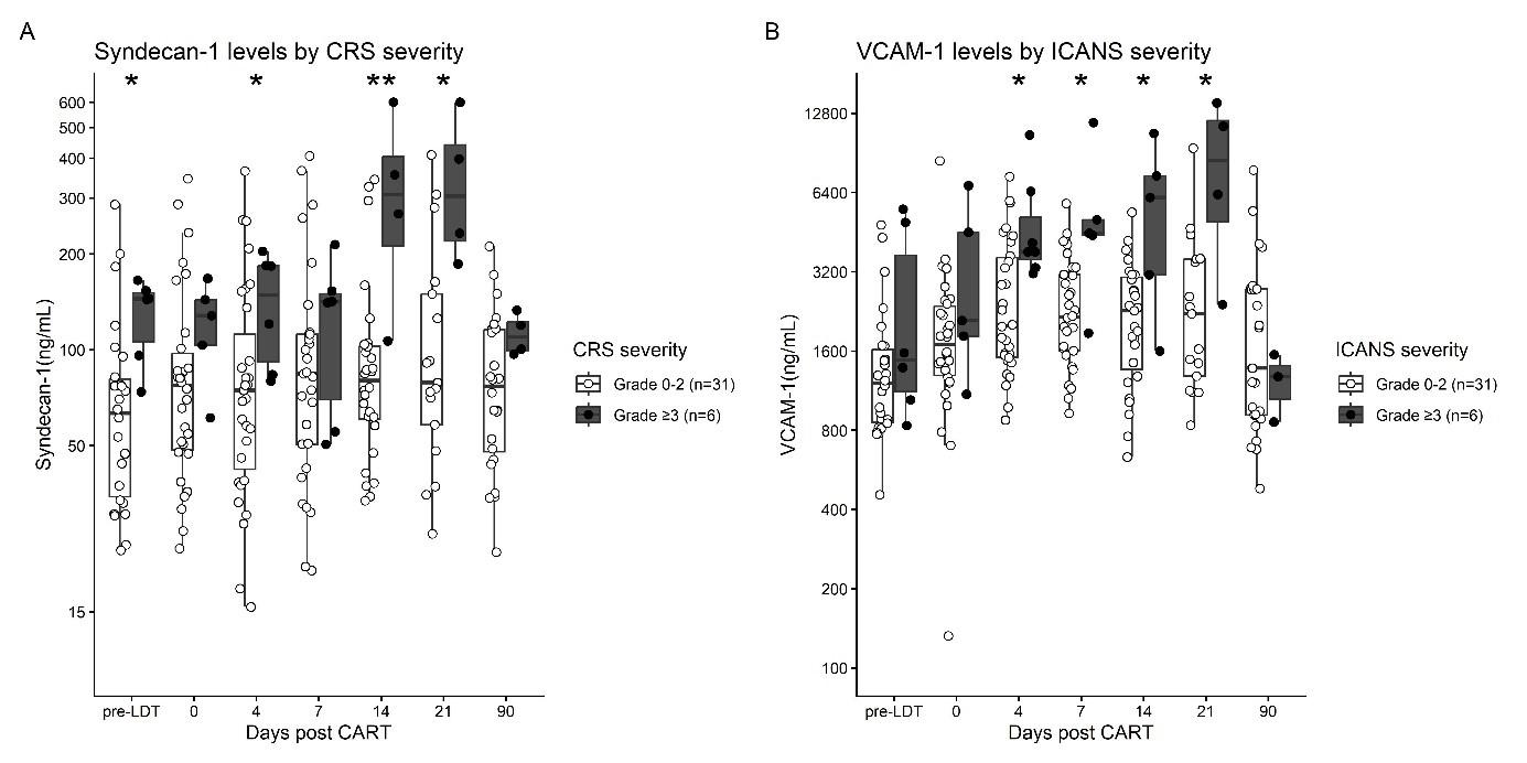
Following CAR-T infusion, nearly all endothelial markers temporarily decreased, except for VCAM-1, which increased, and syndecan-1, which remained stable
Patients developing CRS ≥3 (n=6) had significantly elevated syndecan-1 levels prior to LDT (Figure 1A) Furthermore, VCAM-1, vWF, thrombomodulin, PAI-1 and sVEGFR-1 levels rose during the progression of CRS and were highest in patients with severe disease (all p<0.05)
Patients developing ICANS ≥3 (n=6) had significantly higher levels of VCAM-1 (Figure 1B) and vWF on day +4 after CAR-T infusion, while angiopoietin-2, syndecan-1, thrombomodulin and sVEGFR-1 levels increased during the progression of ICANS (all p<0.05)
There were no significant associations between ICAM-1, P-selectin, E-selectin or PECAM-1 and these toxicities
Conclusion: Our findings support the emerging evidence of endothelial dysfunction in the pathogenesis of severe CRS and ICANS, and suggest syndecan-1, VCAM-1 and vWF as potential risk biomarkers, although confirmation in larger cohorts is needed.
Figure 1
Single-cell based multiomics profiling of tumour infiltrating lymphocyte in adoptive cell therapy reveals signatures of relevance for clinical response
Amalie Kai Bentzen* ,1,2,3, Grigorii Nos*,1, Kristoffer Haurum Johansen*,1 , Laura Stentoft Grand1 , Lasse Frank Voss1 , Edoardo Dionisio2 , Inigo Lacunza1 , Arianna Draghi2 , Janine Sophie Kemming1, Marcus Göransson1 , Marco Donia2, Inge Marie Svane2, Sine Reker Hadrup1
1 T-cells and cancer, Experimental and Translational Immunology Section, Department for Health Technology, Technical University of Denmark, 2800 Kongens Lyngby, Denmark
2 National Center for Cancer Immune Therapy (CCIT-DK), Department of Oncology, Copenhagen University Hospital, Herlev, Denmark
3 Department of Immunology and Microbiology, University of Copenhagen, Copenhagen, Denmark
Adoptive cell transfer (ACT) with tumour-infiltrating lymphocytes (TILs) can induce durable tumour regression, yet long-term clinical benefit remains inconsistent, and the determinants of anti-tumour T-cell persistence are poorly understood. To address this, we developed a multimodal platform, capturing pMHCspecificity and paired TCR sequences, transcriptomes and surface proteins in single cells, allowing us to link specificity, phenotype, and clonal dynamics. Across nine patients with diverse clinical outcome, we investigated a total of 67 longitudinal samples, including the infusion product, pre- and post-infusion PBMCs and pre-treatment tumour digest. From these patients we profiled T cells resognising neoepitopes, shared tumour associated antigens and viral epitopes. We often found several TCRs recognising the same tumour epitope and examples of TCRs with a flexible recognition profile, able to recognise different peptide targets. TCR specificity and functionality were experimentally validated by TCR CRISPR/Cas9 knock-in, confirming both the TCR-pMHC pairing and the direct tumour reactivity of both neoantigen and shared tumour antigenspecific TCRs. Across the longitudinal samples from the nine patients, we observed varying persistence of the tumour-specific T cell clones from the infusion product samples, with examples of clones detected up to 6 years after infusion. Infusion products and early post-infusion samples were consistently enriched for tumourspecific responses. However, in non-responders, we observed a rapid decline of tumour-reactive clonotypes. Additionally, these patients had fewer tumour-specific responses in blood prior to the TIL transfer. Based on the clonal dynamics and the phenotype of the T cell clones, we developed an “engraftment score” to compare the persistence of the anti-tumour T-cell clones and clinical outcomes. Interestingly, we observed that excessive expansion of T cell clones in the infusion product was linked to lower engraftment and expression of markers of exhaustion and terminal differentiation. These results highlight persistence of tumour-reactive clonotypes as a key determinant for ACT response and show potential for biomarker discovery.
Tracking antigen specific T cell responses in rapidly expanded TIL-ACT products of cervical cancer patients
Nithiyashri Jayashankar1,2, Mohammad Kadivar2 , Christina Friese1 , SiriA.
2Experimental and Translational Immunology, Department of Health Technology, Technical University of Denmark, Kongens Lyngby, Denmark
Email: nitjay@dtu dk
Persistent infection of human papillomavirus (HPV) causes the majority (>90%) cervical cancer (CC) cases. Tumor infiltrating lymphocyte based adoptive cell transfer (TIL-ACT) has previously shown an overall response rate of 44% in metastatic and recurrent CC. The manufacturing process of rapidly expanded TIL (REP TIL) product was further improved by the addition of monoclonal antibodies to the culture. However, there is still a lack of understanding with regards to theT-cell specificities in theTILproduct and its correlation with patient outcome, especially in CC. In this study both standard and improved REP TILs along with their precursor cultures (pre-REP) from five CC patients were screened for T cell clones against HPV, tumor associated antigens (TAA) and other common viral antigens (CEF), using DNAbarcoded peptide-major histocompatibility(pMHC) based multimer technology. Overall, 136 HPV responses were identified with 64 unique pMHC combinations, along with 23 TAA and 22 CEF responses. The improved REP process yielded a higher number of CD8 T-cell responsesthatweretraceableandpersistent frompre-REPtoREP,withan increasedexpression ofCD57,HLA-DR,and CD49dcomparedtostandardREP.Topresponses werevalidatedusing combinatorial tetramer staining. These findings reveal the antigenic landscape within CC TIL products, provide insight into the traceability of antigen-specific T-cell clones from culture initiation to infusion, and enable the discovery of novel TCRs for off-the-shelf therapies
Title: Nonviral CRISPR/Cas-engineered tumor-infiltrating lymphocytes with inducible cytokine release and checkpoint resistance for adoptive T cell therapy
1National Center for Cancer Immune Therapy (CCIT-DK), Department of Oncology, Copenhagen University Hospital, Herlev, Denmark
2Department of Biomedicine, Aarhus University, Aarhus, Denmark
3Section for Experimental and Translational Immunology, Department of Health Technology, Technical University of Denmark, Kongens Lyngby, Denmark
Adoptive cell therapy (ACT) with tumor infiltrating lymphocytes (TILs) has achieved durable responses in patients with metastatic melanoma. However, insufficient expansion and persistence of tumor-reactive T cells remainamajor causeofrelapse. Cytokines such as IL-7 can drive Tcells toward phenotypic subsetsassociated with enhanced persistence and antitumor activity, but toxicities are associated with systemic cytokine administration or constitutive expression of cytokines from TILs. Placing the cytokine transgene under control of a Nuclear Factor of Activated T cells (NFAT) inducible promoter restricts cytokine expression to antigendependent T cell activation, thereby minimizing such toxicities. CRISPR/Cas-mediated targeted integration into the PDCD1 gene locus further improves TILs by disrupting PD-1 to escape tumor-mediated immunosuppression. Here, we demonstrate efficient nonviral CRISPR/Cas-mediated targeted integration (>20%)ofaninducibleIL-7cytokinetransgeneandsimultaneousPD1knockout(>81%decreasedPD1surface expression) in patient-derived TILs. Edited TILs demonstrated NFAT-inducible IL-7 expression only in response to anti-CD3/CD28 stimulation (13.64-fold increase over unstimulated controls) and co-culture with autologous tumor cells (5.59-fold increase over unstimulated controls), thereby demonstrating stimulusdependent cytokine release. Furthermore, similar phenotypic and efficient cytotoxic profiles of edited and nonedited TILs were observed, confirming preserved antitumor function. This study establishes a clinically relevant, nonviral genome engineering strategy to generate TILs with inducible cytokine release and intrinsic checkpoint resistance. This approach is designed to enhance the persistence and antitumor activity, supporting translation into next-generation TIL therapies for patients with solid tumors.
Macrophage activation before lymphodepletion can predict CRS, ICANS, and late cytopenia after CD19 CAR-T cell therapy for hematological cancer
Monica Da-My Tran1, Marianne Ifversen1, Henning Grønbæk2, Holger J. Møller2, Sørine T. Neumann1 , Eva Kannik Haastrup1 , Inge Marie Svane3,4, Özcan Met3 , Søren L. Petersen1, Klaus Müller1,4, Katrine Kielsen1,4
1Rigshospitalet, Copenhagen University Hospital, Copenhagen, Denmark, 2Aarhus University Hospital, Aarhus, Denmark, 3Herlev Hospital, Herlev, Denmark, 4University of Copenhagen, Copenhagen, Denmark
Background: CAR-T cell therapy is challenged by immune-related toxicities, including cytokine release syndrome (CRS), immune effector cell-associated neurotoxicity (ICANS), and late cytopenia. Growing evidence suggests that macrophage activation plays a key role in the pathophysiology of these toxicities. We investigated whether circulating markers of macrophage activation (soluble mannose receptor, sMR and sCD163) could predict the severity of CRS, ICANS, and late cytopenia after CD19 CAR-T therapy.
Methods: We included 35 patients treated with CD19 CAR-T therapy for B-ALL (n=14) or B-NHL (n=21) at Rigshospitalet in Denmark from 2019-2025 (Table). Patients received tisagenlecleucel (n=13), axicabtagene ciloleucel (n=10), or an academic CAR-T product (DK-CLIC-1901, n=12). sMR and sCD163 levels were measured using in-house ELISA assays in plasma collected before lymphodepleting chemotherapy (pre-LD), on day of CAR-T infusion, daily from day +1 to +8, and on days +10, +12, +14, and +90 post-infusion. 100 healthy controls with a median age of 42 years (range: 19-69) were included for comparison.
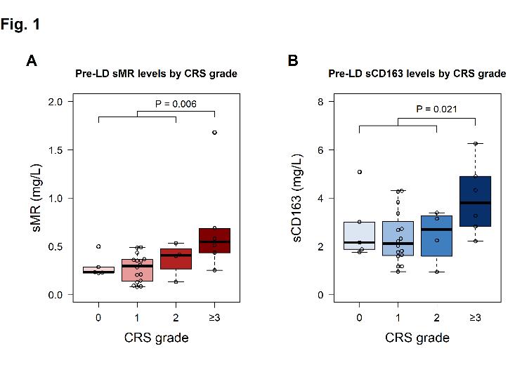
Results: Pre-LD levels of sMR and sCD163 were significantly elevated in patients compared with healthy controls (median0.35 vs. 0.22 mg/L, P<0.00054,and 2.32 vs. 1.58 mg/L, P<0.0001, respectively). After CART infusion, both markers rose and peaked at day +6 for sMR (0.50 mg/L) and day +12 for sCD163 (4.1 mg/L). Pre-LD levels of sMR and sCD163 were not associated with age, diagnosis, prior lines of therapy, bridging therapy, or same-day CRP, IL-6 or LDH levels (all P≥0.05).
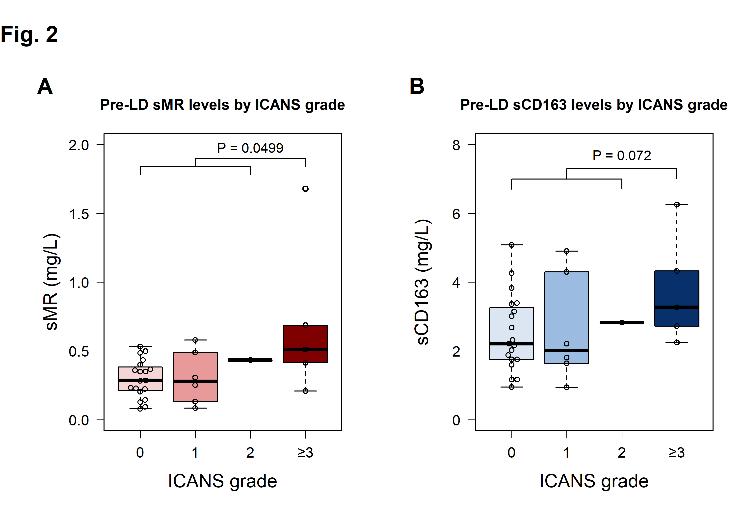
Patients developing grade ≥3 CRS (17%) and ICANS (14%) had higher levels of sMR and sCD163 before LD than those with grade 0-2 (Figure). In Cox analyses, the pre-LD levels of both markers were associated with increased risk of severe CRS (sMR: HR=2.7 per doubling, P=0.008; sCD163: HR=4.5 per doubling, P=0.034), and higher pre-LD sMR levels were associated with severe ICANS (HR=2.7 per doubling, P=0.027). Furthermore, high pre-LD LDH was associated with increased risk of severe CRS (HR=2.1 per doubling, P=0.011), while age, diagnosis, CAR-T product, and CAR-T cell dose were not associated with severe CRS or ICANS in our cohort.
Patients with grade ≥3 neutropenia (55% by ICAHT), thrombocytopenia (36%), and anemia (19%) beyond day +30 after CAR-T infusion had higher pre-LD sMR levels compared with grade 0-2 (P=0.006, P=0.002, and P=0.02, respectively), and higher pre-LD sCD163 levels were observed in patients with late grade ≥3 thrombocytopenia (P=0.04). In logistic regression analyses, higher pre-LD sMR was associated with increased risk of late grade ≥3 ICAHT (OR=3.6 per doubling, P=0.028), thrombocytopenia (OR=13.6 per doubling, P=0.016), and anemia (OR=8.2 per doubling, P=0.057).
Conclusions: Elevated levels of sMR and sCD163 before the start of lymphodepletion were associated with the development of severe CRS, ICANS, and late cytopenia in patients treated with CD19 CAR-T therapy, supporting the emerging evidence of a role of activated macrophages in the pathogenesis of CAR-T-related toxicities. These findings further suggest a clinical role of macrophage activation markers in guiding antiinflammatory prophylaxis in high-risk patients
Table:
Patient and treatment characteristics
Age at infusion (years), median (range)
Male sex, n (%)
Diagnosis, n (%)
B-ALL
B-NHL
No. of prior lines of therapy, median (range)
Prior HSCT, n (%)
Allogeneic HSCT
Autologous HSCT
Refractory to previous treatment line, n (%)
Pre-LD LDH (U/L), median (range)
Elevated pre-LD LDH, n (%)
Bridging therapy, n (%)
Chemotherapy-based regimens
IT therapy only
Radiotherapy ± steroids
Targeted therapy
Steroids only
None
CAR-T product, n (%)
Tisa-cel (Kymriah®)
Axi-cel (Yescarta®)
DK-CLIC-1901
CAR-T dose (cells x 106/kg), median (range)
(1.7-71.8)
(82.9%)
(40.0%)
(60.0%)
(20.0%)
(54.3%)
(0.23-4.8) Days from leukapheresis to infusion, median (range) 33 (13-58)
Figure:
Title: DAN-CART 1901: Preliminary results of safe and feasible administration of the fresh autologous anti-CD19 CAR-T product DK-CLIC-1901
Katrine Kielsen1,2, Özcan Met3, Eva Kannik Haastrup4, Klaus Müller1, Hilde Hylland1, Karsten Nysom1 , Stine E. Weis-Banke3, Lia Minculescu4, Niels Smedegaard Andersen5, Marietta Nygaard5, Brian Kornblit5 , Peter Brown5 , Inge Marie Svane3 , Søren L. Petersen5, Marianne Ifversen1
1 Department of Pediatrics and Adolescent Medicine, Copenhagen University Hospital – Rigshospitalet, Denmark
2 Department of Immunology and Microbiology, University of Copenhagen, Denmark,
3 National Center for Cancer Immune Therapy, Department of Oncology, Herlev Hospital, Denmark
4 Department of Clinical Immunology, Copenhagen University Hospital – Rigshospitalet, Denmark
5 Department of Hematology, Copenhagen University Hospital – Rigshospitalet, Denmark
Background: Treatment with chimericantigen receptor T-cells (CAR T) has demonstrated efficacyin patients with CD19-positive hematological malignancies and lower toxicity than other treatment options. We established an academic CAR T-cell program to broaden access for patients without other curative treatment options and to enable faster, more flexible local production.
We report preliminary results from the initial phase 1b/2 trial using the academic autologous 4 1BB anti-CD19 CAR-T product DK-CLIC-1901 (CTIS # 2024-515174-27-00). The primary objective was to evaluate the safety of DK-CLIC-1901. The secondary objectives were to assess feasibility, clinical efficacy, and immunological response after DK-CLIC-1901 infusion.
Methods: The main inclusion criteria were relapsed or refractory B-cell acute lymphoblastic leukemia (ALL) or non-Hodgkin lymphoma (NHL), age between 1-70 years, adequate performance status and organ function (lung, heart, liver, and kidney), life expectancy ≥ 12 weeks, and written informed consent.
Patients received a single infusion of fresh-to-fresh autologous, in vitro-expanded DK-CLIC-1901 CAR Tcells at a minimum dose of 1x10⁶ CAR T-cells/kg, with a dose reduction to 2x10⁷ CAR T-cells in total in case of high tumor burden. The DK-CLIC-1901 product was manufactured on the CliniMACS Prodigy platform (Miltenyi) using a lentiviral vector carrying a 4-1BB anti-CD19 CAR transgene [N Kekre et al, Front Immunol 2022].
Results: Thirteen patients passed the initial screening for participation. One patient was excluded before apheresis due to rapid progression of hematological malignancy; 12 patients underwent apheresis and received CAR-T infusion (Table).
Cytokine release syndrome (CRS) occurred in 9 patients (grade ≥3: n=3) and immune effector cell-associated neurotoxicity syndrome (ICANS) in 3 patients (grade ≥3: n=2), with a higher frequency in patients with high tumor burden (CRS grade ≥3: 67% vs. 11%; ICANS: 67% vs. 11%). One patient with a high tumor load died at day +26 from intracerebral hemorrhage following severe CRS/ICANS. Another patient developed HHV-6 encephalitis after CAR-T infusion with persistent neurological sequelae [NM Birk et al, Front Hematol 2025]. Late cytopenia was manageable, with grade ≥3 neutropenia (ICAHT), thrombocytopenia, and anemia occurring in 11%, 33%, and 0% of 9 evaluable patients at day 90 following CAR-T infusion.
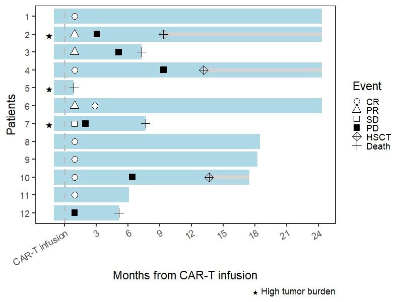
Median follow-up time was 12 months (26 days to 2.0 years). Nine of 11 evaluable patients (82%) responded to CAR-T infusion, of which 5 remain in complete remission more than 6 months from CAR-T infusion (Figure). Twopatientshadloss of B-cell aplasiawithinthefirst year after CAR-T infusion(day +83 and+125),
which was followed by a CD19-positive relapse in one patient. The estimated 1-year overall and progressionfree survival were 65% (95%CI: 42-100) and 39% (95%CI: 19-82), and the 1-year cumulative incidence of non-relapse mortality was 8%. Three of the 6 patients with relapse or progressive disease were confirmed CD19 positive, and three patients (25%) were subsequently rescued with allogeneic hematopoietic stem-cell transplantation.
Conclusion: This is the first trial of point-of-care manufactured CAR-T cells in Denmark and demonstrates that administering fresh CLIC-1901 product is safe, feasible and efficacious, especially in patients with low tumor burden.Our platform providesafeasibleandrapid manufacturingprocedureforfuture CAR-T products, which may also include targets outside commercial interests.
Table:
Clinical characteristics N=12
Age (years), median (range) 54 (16-67)
Male sex, n. (%) 11 (92%)
Diagnosis, n. (%)
B-cell ALL
B-cell NHL
Diffuse large B-cell lymphoma
Transformed follicular lymphoma
High-grade B-cell lymphoma
Burkitt lymphoma
High tumor load, n. (%)
ALL: >20% blasts in BM
(83%)
(25%)
(33%) 1 (8%) 1 (8%) 1 (8%)
Mantle cell lymphoma 2 (17%)
NHL: ≥10 cm tumor mass or BM involvement 0 (0%) 3 (25%)
LDH at the start of LDT (U/L), median (range) 229 (130-2200)
Prior lines of treatment, median (range) 3 (1-4)
CAR-T indication, n. (%)
Primary refractory
≥2 relapse
Relapse after autologous/allogeneic HSCT 5 (42%) 4 (33%) 3 (25%)
LDT with fludarabine (30 mg/m2 x4) and cyclophosphamide (500 mg/m2 x2), n. (%) 12 (100%)
Time from enrollment to apheresis (days), median (range) 6 (2-16)
DK-CLIC-1901 production time (days from apheresis to CAR-T infusion), median (range) 13 (13-19)
Cellular composition of DK-CLIC-1901 product, median (range)
CAR-T cell count (x106 CAR T-cells / kg bodyweight)
CAR-T cell count (x108 CAR T-cells in total)
CD4/CD8 T cell ratio
2 (0.23-3.1) 200 (20-200) 0.63 (0.29-3.69)
Figure:
GD2-targeting Tumor Infiltrating Lymphocytes for Pediatric Patients with CNS Tumors
Cecilie Ølvang Madsen1* , Thomas Morgan Hulen1 , Maria Ormhøj2 , René Mathiasen3, Marianne
Ifversen3 , Jane Skjøth-Rasmussen4 , Sine Reker Hadrup5 , Inge Marie Svane1 , Özcan Met1,5
1. National Center for Cancer Immune Therapy (CCIT-DK), Department of Oncology, Copenhagen University Hospital, Herlev, Denmark.
2. SDU Biotechnology, Department of Green Technology, University of Southern Denmark: Odense, Denmark
3. Department of Paediatrics and Adolescent Medicine, Rigshospitalet, Copenhagen University Hospital, Copenhagen, Denmark.
4. 4. Department of Clinical Medicine, University of Copenhagen, Copenhagen, Denmark
5. Section for Experimental and Translational Immunology, Department of Health Technology, Technical University of Denmark, Kongens Lyngby, Denmark * cecilie.oelvang.madsen@regionh.dk
Introduction: Central nervous system (CNS) tumors are among the most common pediatric cancers, but prognosis for several subtypes remains poor, particularly at relapse where curative options are lacking. Adoptivecell therapy withchimericantigenreceptor (CAR) T cellshasdemonstratedsuccess in hematological malignancies, while tumor-infiltrating lymphocyte (TIL) therapy has mediated durable responses in solid tumors. Combining these approaches may generate poly-specific TILs with improved tumor recognition and clearance
Method: TILs were expanded from tumor biopsies of pediatric patients with CNS tumors and transduced with a GD2-specific CAR using a lentiviral vector, followed by rapid expansion. TILs engineered with GD2-CAR weretested in vitro foractivation,effectorfunction,cytotoxicity,andreactivityagainstautologousGD2+ tumor cells when available.
Results: TILs could successfully be expanded from 6 out of 12 pediatric CNS tumor biopsies (50%), yielding a mean of 16.2×10⁶ CD3+ cells (range: 0.57×10⁶-34×10⁶), with up 8×10⁶ cells/fragment. All patient-derived TIL cultures were successfully transduced and expanded under clinically adaptable conditions TILs modified withGD2-CARshowedenhancedrecognitionandcytotoxicityagainst GD2+ tumorcells,includingautologous CNS tumor cell lines.
Conclusion: TILs engineered to express a GD2-targeting CAR demonstrated feasibility and enhanced antitumor activity in pediatric CNS tumor models. These findings provide proof-of-concept for combining TILtherapy with CAR engineering and support further preclinical development toward clinical translation for children with CNS tumors.
Impaired function of CLL-derived CD19 CAR T cells in a 3D tumor microenvironment reveals CXCR4 and IL-10 as potential targets
Janin Dingfelder1,2.3 , Michael Aigner1,2,3 , Jana Lindacher1,2,3, Pascal Lukas1, Katharina Zimmermann4, Axel Schambach4, Frederik Graw1, Andreas Mackensen1,2,3, Gloria Lutzny-Geier1,2,3
1 Department of Internal Medicine 5, Hematology and Oncology, University Hospital Erlangen, FriedrichAlexander University Erlangen-Nürnberg (FAU), Erlangen Germany
2 Bavarian Cancer Research Center (BZKF), Erlangen, Germany
3 Deutsches Zentrum für Immuntherapie (DZI), Friedrich-Alexander-Universität Erlangen-Nürnberg, Erlangen, Germany
4 Hannover Medical School (MHH), Institute of Experimental Hematology, Hannover, Germany
Introduction: Although targeted therapies have improved outcomes, CLL remains largely incurable, underscoring the need for novel therapeutic strategies. While CAR T-cell therapy has significantly advanced the treatment of acute lymphoblastic leukemia (ALL), its efficacy in relapsed/refractory CLL remains limited, suggesting disease-specific constraints on anti-tumor immunity. Increasing evidence indicates that malignant B cells shape the tumor microenvironment (TME) into protective niches, promoting immune dysfunction and therapy resistance. Understanding the mechanisms underlying reduced therapeutic responses, particularly the contribution of the TME totreatment failure,may reveal novel targets to enhance CAR T-cell function in CLL.
Methods: To dissect disease-specific differences in CAR T-cell functionality, we established a threedimensional (3D) scaffold-based co-culture system with primary CLL or ALL cells, autologous CAR T cells, and bone marrow-derived stromal cells (BMSCs). CAR T-cell function was assessed by flow cytometry for activation, cytotoxicity, proliferation, exhaustion, and differentiation in core and peripheral regions of the scaffold. Spatial distribution and target cell co-localization were analyzed by confocal immunofluorescence imaging. The soluble milieu was examined by multiplex cytokine assays and ELISA. Targeted interventions were tested in combination with CAR T cells in the CLL 3D model to evaluate functional improvements
Results: Comparative analyses of autologous CAR T cells in the 3D co-culture system revealed marked differences between CLL- and ALL-derived CAR T cells. Although exhibiting strong activation, CLL CAR T cells showed limited proliferation and reduced cytotoxicity, with decreased co-localization with malignant B cells and amore differentiated, exhausted phenotype.Functional impairmentsincytotoxicity and exhaustion were particularly pronounced in core regions, suggesting that TME-mediated factors exacerbate intrinsic defects. IL-10 secretion was elevated in CLL co-cultures, pointing to disease-specific immunosuppression. Furthermore, CXCR4+ CLL cells and CXCL12+ BMSCs were enriched in core regions, indicating spatially regulated stromal support via the CXCR4/CXCL12 axis. Targeting these TME-pathways partially improved CAR T-cell function, with both IL-10 and CXCR4 blockade enhancing cytotoxicity and reducing exhaustion, while IL-10 inhibition reduced CLL viability in a patient-dependent manner.
Conclusion: This study highlights key functional impairments of CLL-derived CAR T cells within the 3D model. These findings underscore the importance of elucidating mechanisms of CAR T-cell dysfunction and support the potential of TME-targeted combination strategies, such as IL-10 and CXCR4 blockade, to partially restore effector function. The autologous 3D co-culture system offers a translational platform to investigate CAR T-cell function and evaluate novel therapeutic approaches to improve CAR T-cell efficacy in CLL.
Generation and Validation of HER2-targeting CAR-TILs for Ovarian Cancer:
Investigating the potential as an adoptive cell therapy
Ashley Nicole Myers1*, Cecilie Ølvang Madsen1 , Thomas Morgan Hulen1, Marie Christine Wulff Westergaard1 , Inge Marie Svane1 , Özcan Met1,2
1. National Center for Cancer Immune Therapy (CCIT-DK), Department of Oncology, Copenhagen University Hospital, Herlev
2. Department of Health Technology, Technical University of Denmark, Lyngby * ashley.nicole.myers@regionh.dk
Introduction: Ovarian cancer (OC), the second most deadly gynecological cancer globally, remains challengingforimmunotherapyduetolowtumormutational burdenandlownumbersof tumor-reactive tumor-infiltrating lymphocytes (TILs). Current immunotherapy approaches have shown limited success. CAR-TILs, TILs engineered to express a chimeric antigen receptor (CAR), represent a novel OC immunotherapy and remain poorly explored. This study investigates whether engineering OCderived young TILs (yTILs) with a HER2-targeting CAR can improve adoptive cell therapy for OC.
Materials and Methods: To generate CAR-TILs, a low affinity HER2-targeting CAR was introduced to TILs via lentiviral transduction. Functional validation of CAR-TILs was performed using intracellular cytokine staining (ICS), flow cytometry, and 51Cr release cytotoxicity assays. Autologous patient-derived tumor cell lines (TCLs) and young TILs were used to investigate CAR-TIL potential and function.
Results: CAR-TILs were generated from three OC patients. Transduction efficiencies ranged from 113%. When co-cultured with autologous TCL, CD3+ CAR-TILs showed an increase in reactivity to 56%, compared to 1-3% reactivity from non-transduced (NTD) TILs, measured by CD107a, CD137, IFN-γ, and TNF co-expression Upon co-culture with HER2 target-bearing SKOV-3 cell line, CD3+ CAR-TILs were 4-20% reactive, compared to <1% reactivity in CD3+ NTD TILs. In short-term cytotoxicity assays, CAR-TILs produced 10-15% lysis of autologous TCL at an effector:target ratio of 90:1,comparedto1-5%lysisbyNTD.Overall, HER2-targetingCAR-TILsshowedincreasedreactivity and cytotoxicity towards high HER2-expressing and autologous TCLs compared to NTD controls. Increased HER2 expression was associated with stronger CAR-TIL responses.
Conclusions: HER2-targeting CAR-TIL production is feasible and may improve the limited baseline activity typical of OC-derived TILs. CAR-mediated effector functions appeared to be antigen densityand dose-dependent. Together, these results support further investigation of low-affinity HER2targeting CAR-TILs as a potentially safer and more effective OC immunotherapy.
T cell-targeted gene delivery for in vivo CAR-T cell therapy
Mar Guaza Lasheras1, Samuel A. Theuerkauf1, Luca J. Zinser1, Nandini Sheth1, Elham Adabi1, Johanna M. Gorol1,2, Fabian John1, Muhammed B. Demircan1,3,4, Lea R. Knapp1, Frederic Thalheimer1, Christian J. Buchholz1,5
1 Molecular Biotechnology and Gene Therapy, Paul-Ehrlich-Institut, 63225 Langen, Germany
2 Institute for Tumor Biology and Experimental Therapy, Georg-Speyer-Haus, 60596 Frankfurt, Germany
3 Goethe University Frankfurt, University Hospital, 60590 Frankfurt am Main, Germany
4 German Cancer Consortium (DKTK), Partner Site Frankfurt/Mainz, 60528 Frankfurt am Main, Germany
5 Frankfurt Cancer Institute, Goethe University, Frankfurt Germany
Chimeric antigen receptor (CAR)-T cell therapy has revolutionized the management of haematological malignancies, yet its application is limited by individualized, labor-intensive, and costly ex vivo manufacturing processes. In vivo genetic reprogramming of T cells offers a compelling alternative but relies on gene delivery systems that achieve high specificity, efficiency, and safety. In this study, we explored the use of rationally engineered, T cell–targeted adeno-associated virus (AAV) vectors as a modular platform for selective gene delivery and stable genome editing in human T lymphocytes, employing CAR-T cell generation as a translational model.
DARPin-retargeted AAVs (DART-AAVs) demonstrated highly selective binding and transduction of human CD4⁺ and CD8⁺ T cells within complex cellular environments. To support long-term CAR expression, AAVmediated delivery of donor template CAR constructs was combined with lipid nanoparticle (LNP)-mediated CRISPR/Cas9 editing, enabling targeted integration of the CAR transgene into the TRAC locus. This combined AAV–LNP strategy achieved cell-specific gene transfer, stable CAR expression, and sustained cytotoxic activity in vitro.
In addition, leveraging our expertise in viral vector retargeting, we developed RNA-LNPs directed toward CD8+ T lymphocytes through the incorporation of DARPins with high affinity for murine or human CD8. For surface display, DARPins were genetically fused to apolipoprotein E2 (ApoE2), produced in bacteria, and subsequently associated with RNA-LNPs. These engineered particles effectively distinguished between CD8+ and CD8- T cells in terms of both binding and reporter gene expression across murine and human systems. Importantly, when introduced into human donor blood or administered systemically in humanized mice, CD8LNPs labeled a significant proportion of CD8⁺ T cells, while showing minimal binding to CD4⁺ T cells. These findings indicate that ApoE2-DARPin constructs remain stably associated with RNA-LNPs even in serumcontaining conditions, highlighting this strategy as a novel platform technology based on biological engineering rather than chemical conjugation for precise targeting of RNA-LNPs to therapeutically relevant cell populations.
Overall, this study establishes DART-AAVs and RNA-LNPs as a promising platform for CAR-T cell engineering and provides mechanistic insights with broad relevance for cell type–specific and durable genetic modification beyond CAR-T cell therapy.
Adoptivecelltherapiescaninducedurabletumorregression,yetfunctionalheterogeneitywithininfusedTcell populationslimitsconsistentefficacy.Whilesingle-celltranscriptomicshasdefineddiverseTcellstates,direct links between these states and cytotoxic function remain unclear. Emerging evidence suggests that cellular state, rather than receptor specificity or abundance, is a primary determinant of T cell function1,2. We previouslydemonstratedthatXdrop®-basedmicrofluidicco-encapsulationenableshigh-throughputfunctional interrogation of CAR T cell–tumor interactions3. Here, we analyze Denmark’s first domestically produced CAR T product (DK-CLIC-1901) using Xdrop DE50 technology, integrating functional sorting with singlecelltranscriptomicsmappingcytotoxicactivityontotranscriptionalstateandclonalarchitecture.
Methods
CARTcellsfromasingleDK-CLIC-1901infusionproductwereenrichedusingCD19CARspecificmagnetic selection and co-encapsulated with CD19⁺ Daudi cells in Xdrop DE50 droplets as single-cell pairs. Droplets weresortedbyFACSaccordingtofunctionalsignalsindicatingwhethertheencapsulatedCARTcellexhibited a “killer” (caspase3/7⁺, granzyme B⁺) or “nonkiller” (caspase3/7–, granzyme B–) phenotype. Droplets were then broken, and the recovered cells processed for 10x Genomics single-cell RNA sequencing. Analyses includedunsupervised clustering, differential expression, geneontology, functional enrichment acrossstates, andassessmentofCARexpressionandTCRβ-definedclonotypes.
Results
Oftheco-encapsulatedCARTcells,11.7%wereclassifiedaskillerand13.2%asnonkiller,withtheremaining dropletsbeingnegativeorpositiveforonlyoneofthecytotoxicmarkers.Uponsinglecellsequencinganalysis, detectable CAR transcript expression comprised 77% of killer and 74% of nonkiller populations. Both populationsexhibitedsignificanttranscriptionalheterogeneity.Geneontologyanalysisrevealedthatbothkiller andnonkillerpopulationssegregateintoseveraldistinctbiologicalprograms.Nonkillerswerenotablyinstemlike/memory-likeandheat-shock/stress-associatedstates,indicatingthatlackofcytotoxicfunctionarisesfrom divergent cellular contexts. In contrast, killer cells were enriched in more functionally aligned effector programs,including proliferativeinterferon-primed,terminalcytotoxic andoxidative stress-associatedstates. TCR clonotype analysis revealed a broadly polyclonal repertoire with limited overlap between killer and nonkillerclonotypes.
Discussion
Thesefindingsdirectlylinkcytotoxicfunctiontotranscriptionalstateandsupportamodelinwhichfunctional capacity is primarilygoverned by cellular state rather thanreceptor abundance. The coexistence of killer and nonkiller cells within shared states highlights functional heterogeneity not captured by transcriptional clustering alone, consistent with emerging state-based models of T cell function. Ongoing analysis of additionalinfusionproductswillextendthesefindings.Together,thisworkprovidesaframeworkforresolving functionalheterogeneityinadoptivecelltherapyandidentifyingstatesassociatedwitheffectivetumorkilling.
References
1. Doan,A.E. et al. FOXO1isamasterregulatorofmemoryprogramminginCARTcells. Nature 629, (2024).
2. Prinzing,B. et al. Deleting DNMT3A in CAR T Cells Prevents Exhaustion and Enhances Antitumor Activity Sci. Transl. Med vol.13https://www.science.org(2021).
3. KielRasmussen,A.-C. et al. Analyzingfunctionalheterogeneityofeffectorcellsforenhanced adoptivecelltherapyapplications. Immuno-Oncology and Technology 100738(2024) doi:10.1016/j.iotech.2024.100738.
The FLIC facility at DTU provides comprehensive flow cytometry, cell sorting, and imaging services to support academic and industrial research. Established to deliver cutting-edge analytical capabilities, FLIC houses state-of-the-art instrumentation including spectral analyzers with imaging (BD FACSDiscover A8), spectralcellsorters(BDFACSDiscoverS8),high-parameterconventionalanalyzers(BDLSRFortessaSORP), and biosafety-equipped sorters (BD FACSAria Fusion). Additional platforms include confocal microscopy withAiryScan2super-resolutiondetection(ZeissLSM900),live-cellimaging(IncuCyteS3),andsingle-cell genomics systems (10x Chromium X, BD Rhapsody). FLIC operates as an open-access facility, providing hands-ontrainingandtechnicalsupporttoenableindependentuseroperationacrossallplatforms.Ourservices spanexperimentaldesignconsultation,multi-parameterpaneloptimization,rarecell sorting,high-throughput screening,anddataanalysissupport.ThefacilityisaccessibletoallDTUpersonnelandexternalpartnersfrom academia and industry. With expertise in spectral cytometry, high-parameter immunophenotyping, and advancedsortingtechniques,FLICserves as acentralresource for researchersrequiring advanced single-cell analysisandimagingsolutions.
Title: Adenoviral vectored full-length antigens for expansion of polyclonal CD4+ and CD8+ T cell responses in human dendritic cell and T cell co-culture.
Mikkel Dons Müller1,5 , Cristina Picon-Jara2,5 , Joana Daradoumis1,5 , Ditte Rahbæk Boilesen4,5 , Peter Johannes Holst3,5
1 Department for Immunology and Microbiology, University of Copenhagen
2Department of Biotechnology and Biomedicine, Technical University of Denmark
3Departement of Biomedical Sciences, University of Copenhagen
4Loma Therapeutics
5 HERVolution Therapeutics
Targeting large full-length antigens for polyclonal T cell expansions ex vivo is difficult using peptides but is attractive due to the possibility to expand broad responses in patients with different HLA alleles. We evaluated adenoviral vectored (AdV) full-length antigens in human monocyte derived dendritic cells as stimulators for human T cell expansions with viral antigens from human papillomavirus 16 (HPV16) and the human endogenous retrovirus K/HML-2 (HERV-K). Initially, both the AdV strategy and a strategy based on confirmed single peptides in HLA-A2 donors expanded T cell responses with a majority of responses toward HPV16 E1. Expanded cells were sorted for polyclonal rapid expansion using either peptides or AdV transduced DCs. Here the peptide-based strategy resulted in higher frequencies of specific T cells whereas the AdV strategy resulted in T cells that performed functionally better against cancer cells in a chromium release lysis assay. However, AdV vectors introduced substantial challenges for both HPV and HERV-K antigens including high background cytokine production (IFNγ, TNFα). Prime-boost strategies using heterologous AdV serotypes further exacerbated non-specific activation. This was partially mitigated by IFNγ-based magnetic enrichment followed by rapid expansion, where the final expanded cell exhibited low background and good tumor recognition. During repeated assays aiming at expanding HERV-K self-antigen specific responses we observed a considerable difference in the obtained T cell responses between assays and between donors and even within assays using the same donors with different expansion protocols. Overall, we were able to expand anti-viral and anti-HERV-K specific CD4+ and CD8+ T cell responses towards most donors with adequate quality of their stored antigen presenting cells. Further experiments are required to understand the sources of the pronounced variability, and to further increase the yield for possible therapeutic purposes, it seems possible to systematically expand functional, antigen specific CD4+ and CD8+ T cells, with relevant antigen recognition towards cancer cells, without prior consideration of donor HLA types.
Title: Harnessing Endogenous miRNAs for Targeted Activation of CRISPR/Cas Systems
Rasmus Møller1, Charlotte Risager Christensen1, Kristoffer Haurum Johansen1, Sine Reker Hadrup1
1Department of Health Technology, Technical University of Denmark
The therapeutic potential of CRISPR/Cas-based genome editing for treating genetic disorders is immense, yet its clinical translation remains hindered by challenges related to specificity, safety, and in vivo delivery. Current strategies to restrict CRISPR/Cas activity to target tissues are based on tissue-specific promoters or ex vivo manipulation, both of which can be either labor-intensive or suffer from lack of specificity and customizability. In this work, we present a novel approach to achieving tissue specificity through integration of endogenous microRNA (miRNA) biology with CRISPR-based technologies. Over 2,000 miRNAs have been annotated in the human genome; while many are broadly expressed, others are specific to particular cell types, tissues, or developmental stages. By making the correct processing of CRISPR RNA (crRNA) dependent on the presence of a specific miRNA, we confer cell-type specificity to CRISPR/Cas activity, ensuring that editing occurs only in the intended target cells. This system enables efficient genome editing while minimizing off-target effects and our approach represents a significant step toward safe, precise, and cell-specific genome editing for therapeutic applications.
Novel T-Cell Epitope Mapping and In-Depth Characterization of T-Cells in Respiratory Syncytial Virus Infection
1 Dept. of Health Technology, Section for Experimental and Translational Immunology, Technical University of Denmark, Lyngby, Denmark
2 European Commission – Horizion Europe call
3 Department of Experimental Medical Science, Adaptive immunity, Lund University, Lund, Sweden
4 Center for Infectious Medicine, Department of Medicine Huddinge, Karolinska Institutet, Stockholm, Sweden
5 Department of Virology and Microbiological Preparedness, Statens Serum Institut, Copenhagen, Denmark
Lower respiratory tract infections from seasonal and pandemic respiratory viruses remain a leading cause of morbidity and mortality worldwide Despite their global impact, therapeutic options remain limited and clinical management is predominantly supportive This highlights the need for novel immunological targets and biomarkers to improve prophylactic/treatment interventions, and predictive models for personalized care Our focus is to identify and characterize T-cells specific to Respiratory syncytial virus (RSV) antigens.
To achieve this, we employed a large-scale, high-throughput screening platform utilizing DNA-barcoded peptide–MHC (pMHC) multimer (MULT) libraries. We screened T-cell responses against a comprehensive RSV peptidome based on 2023–2024 Danish strains. The peptide library consisted of 1,791 unique peptides presented across 12 HLA alleles, yielding 2,642 distinct pMHC complexes.
Our initial screen was conducted in a cohort of 53 healthy individuals to define the baseline RSV-specific epitope landscape in a non-acute immunological setting. This enabled optimization of MULT libraries and insights into RSV-specific T-cell immunity in healthy individuals. Notably, we identified 278 unique pMHCs recognized by circulating T-cells, of which only 2 epitopes were previously reported in the Immune Epitope Database (IEDB), highlighting the substantial novelty and depth of the dataset. The screening results were validated using a combinatorial tetramer staining approach, using the ten most frequently recognized pMHCs in an HLA-matched subset of the donor cohort.
Then, we investigated 15 acutely RSV-infected patients with the established pMHC library, now in a single cell approach (10X platform) allowing us to identify intrinsic determinants in their gene expression, TCR clonality, and epitope-specificity, all at ones. This, to further characterize RSV-specific T-cells in a disease setting. In parallel, we sorted RSV-specific B cells using barcoded RSV-antigen-tetramers, to enable a multifaceted investigation of the adaptive immune system. Next, we will screen 16 lung-resident lymphocyte (LRL) samples from acutely RSV-infected infants with the same approach. The single cell data is being collected and analyzed, and preliminary results will be available for the conference.
Collectively, the initial healthy donor screen demonstrated the robustness of our epitope discovery platform and provided a novel reference map of RSV epitopes recognized by T-cells. The Single cell work provides an in-depth characterization on RSV specific T- and B-cells, where we can dissect the phenotypical and functional properties of antigen-specific T- and B-cell populations during active RSV infection. As part of the REACT consortium group, our T- and B-cell discoveries will be considered as one part out of many approaches across the REACT consortium group and collectively can be applied to improve on prophylactic/treatment interventions in the future.
Title: Bispecific immunotherapy of Multiple Myeloma: understanding the clonal diversity of treatment response
1Cancer Research Center of Toulouse, University of Toulouse, INSERM, CNRS, University of Toulouse III – Paul Sabatier
2 IUCT-Oncopole, Toulouse, France
3 Section for Experimental and Translational Immunology, Institute for Health Technology, Technical University of Denmark (DTU), Kongens Lyngby, Denmark
* presenting author
Bispecific antibodies that engage T cells represent a revolution in immunotherapy. By recognizing CD3 on one arm and a cancer-specific surface protein on the other, they bridge the recognition gap between T cells and poorly immunogenic cancer cells. Because hematologic malignancies often invade T cell-rich environments like the bone marrow, T cell-engaging bispecific antibodies (BsAbs) have proven effective against advanced blood cancers.
In multiple myeloma, three BsAbs are currently approved for multirefractory disease, including teclistamab, which targets CD3 and the plasma cell-specific protein BCMA. While more than two-thirds of patients present deep and durable responses, the median duration of response is around 14 months, indicating that both primary and acquired resistance occur1. Understanding these phenomena requires analysis of drug-tolerant cancer cells, antigen escape mechanisms, and the immune microenvironment. Accumulating evidence suggests that the baseline immune landscape could predict response, yet conflicting results have been reported on naive versus effector T cell populations2,3 and the role of T cell exhaustion4. Moreover, the impact of BsAb stimulation on T cell trafficking remains largely underexplored.
The ResisTec study addresses these questions through a design focused on the first months of teclistamab treatment in a real-world population. This single-arm, multicentric French study (NCT05945524) enrolled 100 multi-refractory myeloma patients treated with weekly teclistamab injections for three months, then bi-weekly. We collected bone marrow aspirates at baseline, 3 months, and recurrence; blood draws at baseline, monthly for 3 months, then at 6, 12, and 18 months. Single-cell RNA and TcR sequencing on purified CD45 immune cells revealed an enrichment of myeloid-derived populations in primary resistance patients, at the detriment of T cell and NK subsets. In blood, early changes at one month were more predictive of response than baseline frequencies notably, a failure of cytotoxic CD8 T cells to expand in non-responders alongside regulatory T cell expansion. Canonical T cell exhaustion was absent from circulating T cells over six months regardless of clinical response. Finally, T cell clones expanded in the bone marrow at baseline accumulated in the blood during treatment, particularly in responders.
Collectively, these results suggest that: 1) early immune changes in the circulation are important predictors of treatment response; 2) T cell responsiveness may not always be captured by baseline transcriptional phenotype, possibly implicating epigenetic mechanisms; and 3) treatment response may partly rely on enhanced T cell patrolling of disseminated lesions. Further work is needed to understand how these changes affect NK cells and to contextualize findings within the transcriptional and genomic landscape of malignant plasma cells.
1. Moreau, P. et al. Teclistamab in Relapsed or Refractory Multiple Myeloma. N. Engl. J. Med. 387, 495–505 (2022).
2. Verkleij, C. P. M. et al. T-Cell Characteristics Impact Response and Resistance to T-Cell-Redirecting Bispecific Antibodies in Multiple Myeloma. Clin. Cancer Res. Off. J. Am. Assoc. Cancer Res. 30, 3006–3022 (2024).
3. Cortes-Selva, D. et al. Correlation of immune fitness with response to teclistamab in relapsed/refractory multiple myeloma in the MajesTEC-1 study. Blood 144, 615–628 (2024).
4. Friedrich, M. J. et al. The pre-existing T cell landscape determines the response to bispecific T cell engagers in multiple myeloma patients. Cancer Cell 41, 711-725.e6 (2023).
CHARACTERIZING CD8⁺ KIR⁺ REGULATORY T CELL-MEDIATED IMMUNOSUPPRESSION IN CANCER
Sánchez-Sánchez M1, Draghi A2, Saini SK1
1Technical University of Denmark, Department of Health Technology; 2Herlev Hospital, Center for Cancer Immune Therapy (CCIT)
msasa@dtu.dk
Cancer immunotherapy relies on cytotoxic CD8⁺ T cells to eliminate tumor cells and can induce durable responses in some patients; however, many individuals either fail to respond or initially respond and later develop resistance. These outcomes highlight the role of immunosuppressive mechanisms within the tumor microenvironment. While CD4⁺ regulatory T cells have been extensively studied, CD8⁺ T cells expressing killer immunoglobulin-like receptors (KIRs) have recently emerged as a regulatory population capable of suppressing effector T cell responses, yet their role in cancer remains poorly understood. In my PhD project, I investigate CD8⁺ KIR⁺ regulatory T cells in solid tumors. Using peripheral blood and tumor-infiltrating lymphocytes from cancer patients, I analyze their frequency, phenotype, and KIR receptor expression using highdimensional flow cytometry. Antigen specificity is investigated using DNA-barcoded peptide–MHC multimers, while single-cell approaches integrate transcriptional and TCR clonotype information. Functional assays evaluate their suppressive effects on effector T cell responses. Through this work, I aim to better understand the contribution of CD8⁺ KIR⁺ regulatory T cells to immune suppression in solid tumors and identify potential strategies to enhance anti-tumor immunity and improve immunotherapy outcomes.
IDENTIFICATION AND CHARACTERIZATION OF SPLICEDERIVED T-CELL ANTIGENS IN BLOOD CANCERS
Ballocci T1, Saini SK1, Grønbæk K2, Supek F2
1 Technical University of Denmark, Department of Health Technology
2 University of Copenhagen, Biotech Research & Innovation Centre tobal@dtu.dk
Identification of tumor-specific antigens lays at the foundation of novel immunotherapy approaches, such as adoptive cell transfer and vaccine development. Although most research has studied somatic mutations as neoantigen sources, other cancer-specific phenomena, such as transcriptional aberrations, may have undervalued potential of generating targetable neoantigens. In this study, we are focused in understanding how somatic mutations in spliceosome genes can influence the transcriptional landscape of cancer cells and give rise to a pool of novel, splice-derived neoantigens that can be leveraged for T-cell based therapies
We will perform long-read RNA sequencing of splice-mutation bearing MDS and CLL patient samples to develop a computational pipeline for the generation of a cancer-specific, splicederived neoantigen library. By employing a DNA-barcode-labelled, peptide-MHC-I dextramer system, we will perform large-scale screenings of T cell reactivities against predicted peptides across multiple HLAs, in mutation-bearing patients and healthy donors to identify immunogenic targets. We will then validate these epitopes by performing a comprehensive characterization of epitope-specific CD8+ T cells, through the implementation of single-cell RNA sequencing, TCR sequencing and immunophenotyping assays to understand the actionability of splice-antigen reactive T cell clones.
Develop and validate DNA-barcode labelled MHCII multimers for detection of antigen-specific CD4 T cells displayed towards large libraries of epitopes
Yogesh Basavaraju1, Tripti Tamhane1, Signe Koggersbøl Skadborg1, Suzan Dijkstra3, Liying Lu2 , William W Kwok4, Lawrence Stern2, Georg M. Lauer3 ,Sine Reker Hadrup1
1 – Section of Experimental and Translational Immunology, Department of Health Technology, Technical University of Denmark, Lyngby, Denmark
2 – Department of Pathology, UMass Chan Medical School, Worcester MA, USA
3 – Gastrointestinal Unit, Massachusetts General Hospital and Harvard Medical School, Boston, MA, USA
4 – Beneroya Research Institute, Kwok lab, Seattle WA, USA
Methods for broad-scale detection of antigen-specific CD4 T cells are lacking, while such methods have demonstrated great value in exploring CD8 T cell response in health and disease. Here we use peptide-loaded major histocompatibility complex II (MHCII) proteins multimerized on a barcode- and fluorophore-labelled dextran backbone to provide a method for the detection of CD4 T cell responses to a large display of MHCII-associated peptides. We have established a protocol for MHCII production and peptide exchange suitable for generating large libraries of peptide-MHCII complexes. We validate the use of such pMHCII complexes in the form of barcode-labelled MHCII multimers to detect CD4 T cells. We demonstrate that antigen-specific T cell populations can be identified by amplifying the co-attached barcode by sorting the MHCII-multimer binding CD4 T cell population and that such signals correlate with the CD4 T cell population identified based on fluorescence-labeled MHC II multimers.
We demonstrate that individual CD4 T cell populations can be identified, with a peptide-MHC II panel consisting of 151 peptides derived from Flu, EBV, HCMV, and HCV loaded on DRB1*01:01 and DRB1*01:04. We could detect T cell populations at frequencies down to 0.001% of all CD4 T cells. Low-frequency responses were validated using peptide-driven expansion of antigen-specific CD4 T cells. This methodology was applied to identify MHC-II epitopes in HCV, recognized by CD4 T cells. Using PBMC samples from a patient cohort previously tested for CD4 functional reactivity to HCVderived 20mer peptides, we identified 15 minimal epitopes within such sequences, presented in DRB1*01:01 and DRB1*04:01, being recognized by CD4 T cells in these donors. This method would allow for in-depth analyses of the specificity of immune interactions driven by CD4 T cells, and provide a better understanding of the antigen-driven association between CD4 and CD8 T cell responses in infection and cancer. Large-scale screening would also facilitate the identification of neoepitopes targeting CD4 T cells providing more insight into immunotherapies and cancer vaccines.
In vivo Expansion of genetically engineered CAR T cells using antigen presenting polymer particles(T-Boost)
Keerthana Ramanathan*,1,Tao Zheng*,2 , Hólmfridur Rósa Halldórsdóttir3, Marcus Svensson-Frej3 , Erik Torgny Anders Ekengard3,Yi Sun2,†, Sine R. Hadrup1, †
1 T-cells and cancer, Experimental and Translational Immunology Section, Department for Health Technology, Technical University of Denmark, 2800 Kongens Lyngby, Denmark
2Nanomaterials and Nanobiosensors, Experimental and Translational Immunology Section, Department for Health Technology, Technical University of Denmark, 2800 Kongens Lyngby, Denmark
3Nano-X(BioStudio program), Bio Innovation Institute, Ole Maaløes Vej 3,2200Copenhagen N, Denmark
*These authors have contributed equally. †Corresponding author: Email: Sirha@dtu.dk
Abstract
The engineering of autologous T cells for the expression of chimeric antigen receptors (CAR) can induce profound clinical responses in haematological malignancies. However, the clinical production protocol for the engineered T cells is exhaustive and often leads to highly differentiated and exhausted effector T cells. To circumvent this, we have adapted an alternative approach in which we inject freshly CRISPR-engineered T cells followed by the injection of polymer nanoparticles (T-Boost) that present antigen and co-stimulation in vivo to support the expansion and survival of antigen-specific T cells.
Here we expand CRISPR/Cas9-engineered CAR-T cells with CD19 antigen for anti-CD19 CART cells, together with nanoparticle-bound IL2 and IL21. One stimulation with the T-Boost nanoparticles in vitro achieved more than 100-fold expansion of antigen-specific cells after 5-6 days of culture. The expanded CAR-T cells effectively killed antigen-presenting cancer cells in vitro.
Previous technologies were either non-biocompatible or inactivated by serum upon injection However, we have shown in vitro that pre-incubation with serum does not affect T-Boost activity. We also have preliminary data on in vivo maintenance of OT-1 T cells in pre-immunized BalbC mice, and we hypothesize that we can inject freshly engineered CAR T cells followed by T-Boost for stimulation, expansion, and maintenance of the engineered antigen-specific T cells. This approach will preserve T cell phenotypes and circumvent exhaustion due to ex vivo expansion.
Human Endogenous Retroviruses as novel targets for Cancer Immunotherapy
Andreas Kongsgaard1 , Konstantin Danilov1 , Marta Sánchez Sánchez1 , Kirsten Grønbæk2, Sunil Kumar Saini1
1 Technical University of Denmark, Department of Health Technology, Kgs Lyngby, Denmark
2 University of Copenhagen, Biotech Research & Innovation Centre, Copenhagen, Denmark
Myelodysplastic syndromes (MDS) are haematological malignancies with limited curative treatment options outside allogeneic stem cell transplantation. Because mutational neoantigens in MDS are often scarce and highly patient-specific, alternative tumour-associated antigen sources are needed. Human endogenous retroviruses (HERVs) are remnants of ancient retroviral infections that make up approximately 8% of the human genome and are normally epigenetically silenced but can become re-expressed in malignant cells and following hypomethylating agent treatment, making them attractive candidates for immunotherapy. Here, we investigate the immunogenicity and therapeutic potential of HERV-derived antigens in an MDS cohort treated with anti-PD-L1 and guadecitabine.
To identify candidate targets, we established an in-house HERV discovery pipeline based on Telescope and applied it to RNA-sequencing data from the MDS cohort. This identified 442 candidate HERVs, from which the most significantly upregulated HERVs were selected for downstream antigen prediction. Using patientspecific HLA profiles (n = 16), we predicted 2558 unique peptides corresponding to 3887 peptide-MHC complexes, which were screened in 17 patients at two timepoints, at screening andafter treatment, using DNAbarcoded peptide-MHC multimer technology. Ongoing analyses assess the frequency and activation state of HERV-reactive T cells to determine whether HERV-derived antigens elicit naturally occurring T-cell responses in MDS, whether these responses are enhanced by treatment, and whether shared immunogenic HERV targets exist across patients.
Title: CREATIC – Building a European Excellence Centre in Central Europe for ATMPs. From Capacity Building to Therapy Development to Patient Treatment
Nina Becker1, Stefanie Binder2, Jakob Wested3/4, Eduard Lenner5 et al.
1 Fraunhofer Institute for Cell Therapy and Immunology IZI, Perlickstr. 1, 04103 Leipzig, Germany
2 Faculty of Medicine, University of Leipzig, Philipp-Rosenthal-Str. 55, 04103 Leipzig, Germany
3 Centre for Advanced Studies in Biomedical Innovation Law (CeBIL), Karen Blixens Plads 16, DK-2300 Copenhagen S, Denmark
4 Faculty of Law, University of Copenhagen, Karen Blixens Plads 16, DK-2300 Copenhagen S, Denmark
Advanced Therapy Medicinal Products (ATMPs) offer major opportunities for patients with rare diseases and paediatric cancers, yet their widespread use is constrained by fragmented development pathways, regulatory complexity, and limited as well as costly access. CREATIC – a Horizon Europe Teaming for Excellence project- establishes a sustainable Central European Centre of Excellence that connects ATMP therapy development, GMP manufacturing, regulation, and fair access within an integrated ecosystem based on a strong partnership and leadership. Equally, it manifests a strong partnership between the widening-country coordinator Masaryk University (Czech Republic) and the partners Fraunhofer Institute for Cell Therapy and Immunology IZI (Germany), Leipzig University (Germany) and the University of Copenhagen (Denmark).
CREATIC focuses on highly personalised cell and gene therapies for patient groups underserved by conventional pharmaceutical models. In parallel, the project embeds intellectual property management, regulatory science, biomedical law, and health economics to support regulatory readiness, responsible innovation, and sustainable pricing and reimbursement.
By linking scientific excellence with manufacturing capability, and fair access, CREATIC delivers an end-toend ATMP ecosystem that accelerates translation to patients while strengthening European cohesion and competitiveness. In essence, the poster delivers:
• A snapshot of the CREATIC integrated ATMP ecosystem linking therapy development, GMP manufacturing, regulation, and fair access
• A demonstration for establishing a sustainable Centre of Excellence in Central Europe through strategic European partnerships and structured capacity transfer by looking at, CREATIC's scope, key challenges and impact
• An overview of existing project outputs and ecosystem, including webinars, ATMP newsletter, FAIR Medicine approach
Immune signatures associated with clinical response to the personalized neoantigen vaccine EVX-01 and pembrolizumab in advanced melanoma
M. A. Pavlidis1,2, M. Lausen1, R. Ohrt Andersen1, N. Viborg1, G. Long3, A. Khattak4, P. A. Ascierto5, C. Cimminiello6, T. Trolle1, N. Thuesen1, M. Chisamore7, B. O. Wolthers1, S. Kumar Saini2, S. Friis Thorsen1, B. Rønø1, D. Kleine-Kohlbrecher1
1Evaxion A/S, Hørsholm, Denmark
2Technical University of Denmark (DTU), Health Technology, Kongens Lyngby, Denmark
3Melanoma Institute Australia, The University of Sydney, and Mater and Royal North Shore Hospitals, Sydney, NSW, Australia
4Oncology, One Clinical Research, Hollywood Private Hospital & Edith Cowan University, Perth, WA, Australia
5Melanoma, Cancer Immunotherapy & Developmental Therapeutics, Istituto Nazionale Tumori- IRCCS- Fondazione Pascale, Napoli, Italy
6Divisione di Oncologia Medica del Melanoma, Sarcoma e Tumori Rari, IEO- Istituto Europeo di Oncologia IRCCS, Milan, Italy
7Merck & Co., Inc., Oncology Early Clinical Development, Rahway, NJ, United States
EVX-01, a personalized cancer vaccine (PCV) targeting neoantigens (neoAgs), is currently being evaluated in a phase 2 clinical trial with the Immune Checkpoint Inhibitor (ICI) pembrolizumab in stage III/IV unresectable melanoma patients with no prior therapy (NCT05309421) EVX-01 is a peptide-based PCV delivering up to 10 patient-specific neoAgs selected using Evaxion’s proprietary AI-Immunology™ platform At the two-year data cut-off, the objective response rate was 75% (12/16) in the overall cohort, with 54% (7/13) of the patients with stable disease or partial response after the 12-week pembrolizumab induction demonstrating additional tumour reduction following EVX-01 initiation Additionally, 100% (15/15) harbored EVX-01 neoAg-specific, long-lasting T-cell responses. However, pharmacodynamic biomarkers associated with clinical response to PCV-ICI combination therapy, including the combination of EVX-01 with pembrolizumab, remain poorly defined
To identify pharmacodynamic biomarkers of clinical response to EVX-01 and pembrolizumab, peripheral blood mononuclear cells (PBMCs) were collected from enrolled patients before, during and after study treatment and analyzed using immune assays. Strong and long-lasting immune responses specific to EVX-01 neoAgs were detected in all evaluable patients using IFNγ ELISpot Indepth characterization of EVX-01 neoAg-specific immune responses directly ex vivo using spectral flow cytometry revealed that EVX-01 vaccination induced both EVX-01 neoAg-specific CD4+ and CD8+ T cells in the majority of analysed patients, with CD4+ T cells predominating. EVX-01 neoAgreactive T cells harbored a functional and activated profile characterized by proinflammatory cytokine production, strong activation-induced marker and PD-1 expression indicative of antigen experience, and a predominant memory T-cell phenotype. In parallel, immune cell profiling using
a 25-color spectral flow cytometry panel revealed longitudinal, treatment-associated modulation of both myeloid and lymphoid compartments in peripheral blood.
Collectively, we report that EVX-01 induces strong, long-lasting IFNγ responses in all evaluable patients, driven predominantly by CD4+ T cells, and in a subset of patients, CD8+ T cells, with a proinflammatory, activated memory phenotype. Ongoing efforts to integrate the phenotypes of EVX-01 neoAg-specific T cells and treatment-induced peripheral immune changes with clinical outcomes will support the identification of biomarkers associated with clinical response.
Title: In-situ characterisation of T-cell reactivity
Erik Magnusson1 , Janine Kemming1 Hólmfridur Halldórsdóttir1 , Sebastian Sebbaha1 ,
Sine Reker Hadrup1
1 Technical University of Denmark, Health Tech
Immunotherapies have revolutionized cancer-therapies through leveraging the tumor-targeting capabilities of the adaptive immune system. Immune-checkpoint inhibitors (ICI) and chimeric-antigen receptor (CAR) Tcells have been at the forefront of these advancements with both trying to use CD8+-T-cells to eliminate cancer cells. ICIs blocks the immunosuppressive signals from the tumor, reinvigorating T-cell anti-tumor activity and CAR T-cells have customized receptors for targeting neo-antigens. ICIs show great efficacies in melanoma and CAR T-cells have been effective in leukemia but the overall response of immunotherapies for most other cancers have been limited. The tumor microenvironment (TME) has been proposed as a reason for the variability in treatment outcomes. Through a hypoxic environment with high pH, and tumor suppressive signals it lowers T-cell viability and efficacy. Tertiary lymphoid structures (TLS) have also been associated with improved patient outcomes.
Peptide bound Major Histocompability complex (pMHC) multimers target T-cell receptors allowing for recognition of their specificities. pMHCs have been used extensively in flow-cytometry and other single-cell applications but have seen limited use in spatial settings. Spatial investigation of tissues is important for understanding cellular interactions. This project aims to optimize staining methods for allowing for the recognition of specific T-cells in tissue sections utilizing immunofluorescence techniques. After achieving optimization, neo-antigen libraries will be screened for investigation of patient tumor samples. This will allow for study of tumor infiltration of neo-antigen specific T-cells and how they interact with the TME to give a deeper understanding on their impact on patient outcomes and formation of TLSs.
Cell avidity:thenext-genbindingassaytoadvancecell-basedandcell-engagingtherapeutic development
While conventional assays such as affinity, cytokine secretion, and cytotoxicity provide valuable data at a molecularlevel,thisinformationisinsufficienttofullycharacterizeandselectthebestcellulartherapies.There isstillalackofunderstandingaboutthebiophysicalcell-cellinteractionsthatdrivefunctionalprocesses.
Cellavidity,theintegratedstrengthofmultivalentinteractionsbetweenaneffectorcellanditstarget,canhelp elucidate the mechanism of action for therapeutic candidates. Our Cell Avidity platform challenges these interactingpairsusingcontactlessforceandquantifiesthestrengthofbindingbetweeneffectorandtargetcells to distinguish productive from unproductive cell binding in a physiological context. This biophysical metric providesauniqueviewintocellbindingcharacteristicstointerrogatebindingpotency,selectivity,sensitivity, andkinetics.
Assess the contribution of CD58:CD2 in CAR mediated cell avidity and how replacing that with syntheticPD-1:CD2interactionsreducesfratricidewhilemaintainingpotency.
Engineer CAR-T cells secreting a T-cell engaging molecule to overcome a challenging tumor microenvironmentinpancreaticadenocarcinoma.
Elucidate mechanism of action of tandem CAR-T targeting heterogenous solid tumors Phenotype tumor-primedNKcellsforcellbindingandfunction.
We developed a Cell Avidity platform that enables the characterization and screening of molecular binders andcellularproducts,includingantibodies,smallmolecules,andcelltherapies.CellAvidityprovidesessential informationrevealing potency, selectivity, sensitivity,and kinetics, offeringkeybiophysicalinsightsinto the mechanismofactionforimmune-basedtherapies.
[2] Montemurro A et al. NetTCR-2.0enables accurate prediction of TCR-peptide binding byusing paired TCRα and βsequencedata. Commun Biol.2021;4(1):1060.
Improving CAR-T Cell Persistence and Efficacy Through Epigenetic Reprogramming
Maria Letícia Rodrigues Carvalho1, Mariela Pires Cabral-Piccin1, Gabriela Sarti Kinker1,2, Raylane Adrielle Gonçalves Cambui1, Julianna Pereira1, Amanda Rondinelli1, Emmanuel Vinicius Oliveira Araujo1, Luiza Abdo3, Eduardo Mannarino3, Emmanuel Aragão3, Jordy Larco Lasso1, Maribel Limachi1, Oraci Leite1, Heloísa Monteiro5, Adriana Franco Paes Leme5, Marta Maria Moreira Lemos1, Marjorie Vieira Batista1, Jayr Schmidt Filho1, Cheryl H. Arrowsmith4, Vladmir Cláudio Cordeiro de Lima1, Martín Hernán Bonamino2, Tiago da Silva Medina1,2,6
1 International Research Center, A.C.Camargo Cancer Center, São Paulo, Brazil.
2 Department of Molecular Medicine, Faculty of Health Sciences, University of Southern Denmark (SDU), Odense, Denmark.
3 Cell and Gene Therapy Program - Research Coordination, Brazilian National Cancer Institute (INCA), Rio de Janeiro, Brazil.
4 Princess Margaret Cancer Centre, University Health Network, Toronto, Canada.
5 Laboratório Nacional de Biociências, LNBio- CNPEM, Campinas, Brazil
6 Danish Institute for Advanced Study (DIAS), University of Southern Denmark (SDU), Odense, Denmark.
Chimeric Antigen Receptor (CAR) T cell therapy has revolutionized the treatment of hematologic malignancies, leading to durable remissions in patients with B-cell acute lymphoblastic leukemia (B-ALL) and non-Hodgkin lymphoma (NHL). However, a substantial proportion of patients fail to achieve complete responses, and the efficacy of CAR-T cells against solid tumors remains limited. Key challenges include T cell exhaustion, expansion of regulatory CAR-T cells (CAR-Tregs), and the immunosuppressive tumor microenvironment. Although current strategies aim to generate less differentiated, stem-like CAR-T cells to improve persistence and reduce exhaustion, they do not fully prevent the emergence of dysfunctional or suppressive phenotypes. We hypothesized that epigenetic modulation could be a dual strategy by promoting a stem-like phenotype while limiting regulatory polarization in CAR-T cells. Here, we present an epigenetic therapy capable of enhancing stem-like features and reducing regulatory T cell frequency in healthy donor T cells, leading us to incorporate this epigenetic therapy during CAR-T cell manufacturing. In both anti-CD19 and anti-HER2 CAR-T cells, treatment with the epigenetic therapy enhanced and sustained a stem-like profile, reduced terminal differentiation and CAR-Treg development, increased IFNγ and TBET expression, and decreased inhibitory receptor markers. These effects persisted over time and under immunosuppressive conditions such as TGF-β exposure. Transcriptomics and proteomics analyses confirmed a shift toward persistence-associated programs while restraining regulatory gene signatures. Notably, treated CAR-T cells exhibited transcriptional profiles similar to those observed in B-ALL patients who respond to CAR-T therapy. Importantly, similar modulatory effects were observed in patient-derived CAR-T cells, where the epigenetic therapy also enhanced proliferation and antitumor activity in long-term killing and rechallenge assays against B-ALL and NHL cell lines. In vivo experiments using different tumor models (CD19⁺ Nalm-6 and HER2⁺ SKOV3) showed that epigenetically treated CAR-T cells achieved improved tumor control compared to control CART cells. Mice receiving treated CAR-T cells also demonstrated improved overall survival across models. Consistent with enhanced long-term persistence, epigenetic therapy led to increased accumulation of adoptively transferred CAR-T cells in the bone marrow, spleen, and blood of tumor-bearing mice three weeks after infusion of patient-derived CAR-T products. Together, our results support the use of an epigenetic therapy as a promising strategy to improve CAR-T cell potency, persistence, and resistance to suppression, with translational potential for both hematologic and solid tumors.
Overcoming Tumor Apoptosis Resistance by Enhancing Chimeric
Antigen Receptor T-cell Cytotoxicity by Delivery of Cathepsins through the Granzyme-perforin Pathway
M.R. Hansen 1 , K. Ramanathan 2 , H.R. Halldórsdóttir 2 , C. Rodriguez Pardo 2 , S. Reker
Hadrup 2 and M. Ormhøj 1
1University of Southern Denmark, Odense M, Denmark, 2Tecnical University of Denmark, Kgs. Lyngby, Denmark
Chimeric antigen receptor (CAR) T cell therapy has demonstrated remarkable success in treating B-cell malignancies, providing sustained clinical responses in patients with relapsed or refractory disease. However, tumor intrinsic resistance remains a significant barrier, with one contributing mechanism being the upregulation of anti-apoptotic proteins that interfere with programmed cell death. Proteins such as serine protease inhibitor B9 (SerpinB9), B-cell lymphoma 2 (BCL-2), and X-linked inhibitor of apoptosis protein (XIAP) disrupt distinct stages of the apoptotic cascade. They are highly associated with resistance to T-cell-mediated cytotoxicity.
To address these resistance mechanisms, we developed a strategy that enables targeted intracellular delivery of therapeutic proteins using the T cell’s native perforin-granzyme pathway. CAR T cells were engineered to co-express granzyme B fused to cathepsin D, a lysosomal protease with cytosolic activity. These engineered CAR T cells were evaluated for fusion protein localization, effects on T cell phenotype and activation, and their capacity to enhance cytotoxicity against wild-type and apoptosis-resistant tumor cells.
The fusion proteins localized efficiently to cytotoxic granules and were selectively delivered into tumor cells during CAR T cell engagement, without impairing granule function, T cell phenotype, or activation. Cathepsin D CAR T cells showed significantly enhanced cytotoxicity against wild-type NALM-6 cells and restored killing activity against apoptosis-resistant NALM6 cells overexpressing SerpinB9, BCL-2, or XIAP.
These findings establish a modular protein-delivery platform that enables CAR T cells to bypass intrinsic resistance mechanisms and execute potent cytotoxic responses. This approach represents a promising way to enhance the therapeutic efficacy of CAR T cell therapies.
Vaccine-induced PD-L1-specific T cells indirectly enhance antitumor immunity through myeloid cell modulation
1 National Center for Cancer Immune Therapy (DK-CCIT), Department of Oncology, Herlev Hospital, Herlev, Denmark
2IO Biotech ApS, Copenhagen, Denmark
3Department of Immunology and Microbiology, Faculty of Health and Medical Sciences, University of Copenhagen, Copenhagen, Denmark
Background
The immune modulatory vaccine (IMV) targeting IDO and PD-L1 is designed to enhance T cell responses against both cancer cells and immunosuppressive populations in the tumor microenvironment (TME). When combined with anti-PD-1 therapy, it showed clinical activity in patients with advanced metastatic melanoma in a phase I/II trial1. Here we investigate how vaccine-activated T cells modulate target-expressing cancer and immune cells and contribute to tumor elimination.
Methods
PD-L1-specific CD4+ T cell clones expanded from peripheral bloodmononuclear cells (PBMCs) of vaccinated patients from the phase I/II trial were co-cultured with autologous cancer cells and PBMC-derived CD14+ myeloidcells.TcellreactivitytowardcancerandCD14+ cellswasassessedusingIFN-γELISPOT.Phenotypic profiles of myeloid cells were analyzed with flow cytometry, and cancer cell killing was assessed using xCELLigence system, in the presence and absence of autologous tumor-infiltrating lymphocytes (TILs).
Results
Vaccine-induced PD-L1-specific CD4+ T cells showed reactivity against PD-L1+ myeloid cells co-cultured with PD-L1+ cancer cells in ELISPOT assays. Phenotypic analysis of myeloid cells revealed a shift toward an immunosuppressive profile induced by cancer cells, which was reversed by the addition of PD-L1-specific CD4+ T cells. In xCELLigence assays, targeting of CD14+ cells by PD-L1-specific CD4+ T cells promoted cancer cell killing and simultaneously enhanced the cytotoxic activity of tumor-reactive TILs.
Conclusions
Our data demonstrate that vaccine-activated PD-L1-specific T cells can directly target and reprogram PD-L1+ myeloid cells, leading to cancer cell elimination both through myeloid cell modulation and by improving TIL cytotoxicity. These results highlight the potential of IMVs to promote an immunopermissive TME by reprogramming immunosuppressive myeloid cells, supporting their combination with checkpoint therapies.
Reference
1. Kjeldsen, J. W. et al. A phase 1/2 trial of an immune-modulatory vaccine against IDO/PD-L1 in combination with nivolumab in metastatic melanoma. Nat Med 27, 2212–2223 (2021).
TR-TIL Platform: Antigen-agnostic pan-cancer identification of polyclonal tumor-reactive CD4⁺ and CD8⁺ TILs for TCR discovery
Authors: Arianna Draghi1# , Christopher Aled Chamberlain1# , Anne Weiss1 , Shawez Khan1, Katja Harbst2,3 , Edoardo Dionisio1 , Antonia Katharina Kurtz1 , Yago Garitaonaindia Diaz1 , Agnete Witness Præst Jensen1 , Andreas Sandnes1 , Mario Presti1 , Jakob Vasehus Schou4 , Göran Jonsson2,3, Inge Marie Svane1, Marco Donia1*
Affiliations: 1 National Center for Cancer Immune Therapy (CCIT-DK), Department of Oncology, Copenhagen University Hospital, Herlev, Denmark; 2 Division of Oncology and Pathology, Department of Clinical Sciences Lund, Faculty of Medicine, Lund University, Lund, Sweden; 3 Lund University Cancer Centre, Lund University, Lund, Sweden. 4 Department of Oncology, Herlev and Gentofte Hospital, Herlev, Denmark;
Correspondence addressed to: Marco Donia, National Center for Cancer Immune Therapy (CCIT-DK), Herlev Hospital, Borgmester Ib Juuls Vej 25C, 5th floor, 2730, Herlev, Denmark, phone: +4538681456 , +4538686688, fax: +4538683457, marco.donia@regionh.dk;
Background: Adoptive cell therapy (ACT) with tumor-infiltrating lymphocytes (TILs) has shown promising results in melanoma and other immunogenic solid tumors. For poorly immunogenic tumors, TCR T-cell ACT using genetically engineered T cells targeting tumor antigens may be more effective. Existing TCR-discovery approaches typically focus on MHC I–presented tumor antigens and thus depend on prior antigen knowledge. We introduce a high-performance, antigen-agnostic platform the TR-TIL Platform to identify polyclonal tumor-reactive CD4⁺ and CD8⁺ TILs for TCR discovery.
Methods: Wedeveloped an in vitro co-cultureassay recapitulating TIL–tumorinteractions to capture the transcriptomic landscape of tumor-reactive CD4⁺ and CD8⁺ TILs (TR-TILs). PreREP TILs from 10 patients across five tumor histologies were incubated with autologous or control tumor cells. Following tumor recognition, cells underwent single-cell RNA sequencing (scRNA-seq). We derived CD4⁺ and CD8⁺ transcriptional signatures (TR-TILsigs) by comparing autologous and control conditions. We then assessed TR-TIL sigs on an independent validation cohort of co-culture scRNAseq data.
Results: Recognition of autologous tumor antigens induced transcriptomic changes in TR-TILs, enabling the generation of TR-TILsigs that delineate distinct subclusters of CD4⁺ and CD8⁺ TR-TILs. To validate signature predictive accuracy, a total of 35 (to date) top-scoring TCRs from six patients (4 melanoma, 1 renal cell carcinoma, 1 colorectal cancer) were screened for tumor reactivity in coculture assays. Among CD8⁺ TCRs, 21/23 (91%) exhibited reactivity; among CD4⁺ TCRs, 10/12 (83%) exhibited reactivity. In melanoma, over 95% of selected top-scoring TCRs showed reactivity; in non-melanoma samples, 70% did so. The tumor-reactive TCRs were detectable in longitudinal peripheral blood samples from two long-term responders up to seven years after TIL-ACT.
Conclusions: We report a pan-tumor, antigen-agnostic TR-TIL Platform for discovery of CD4⁺ and CD8⁺ tumor-reactive TCRs. These TR-TIL signatures inform enrichment strategies and may enhance the therapeutic efficacy of adoptive TIL and TCR T-cell approaches.
Background:Pancreatic ductaladenocarcinoma(PDAC)ischaracterizedbyaprofoundlyimmunosuppressivetumormicroenvironment(TME),inwhichtransforminggrowthfactorbeta(TGFβ)plays a central role by promoting immune suppression, fibrosis, and, thus, resistance to immunotherapy. Immune-modulatoryvaccines(IMVs)targetingTGFβ-expressingimmunosuppressivecellsrepresent apromisingstrategytoreprogramtheTMEandenhanceT cell-mediatedanti-tumorimmunity.
Methods: In this study, we evaluated the immunogenicity and functional relevance of multiple TGFβ-derivedepitopesinperipheralbloodmononuclearcellsfromhealthydonorsandpatientswith PDAC.TcellresponseswereassessedbyIFNγELISPOTandintracellularcytokinestaining.CorrelationsbetweentheTGFβ-specificresponsesandclinicaloutcomeswereanalyzedinpatientstreated withcombinedimmunecheckpointinhibitorsandradiotherapy.FunctionalrecognitionofTGFβ-expressingtarget cellswas investigatedusingpeptide-pulseddendriticcells and TGFβ-expressingmalignant cells. Aside from peptide stimulation, a multi-epitope mRNA construct encoding several TGFβ-derivedepitopeswasdevelopedtoassesssimultaneousactivationofepitope-specificTcells.
Results: Robust T cell responses against multiple TGFβ-derived epitopes were detected in both healthy donors and PDAC patients. These responses were predominantly mediated by CD4⁺ T cells exhibiting pro-inflammatory and cytotoxic features, including granzyme B and TNFα expression. The TGFβ-specific T cells recognized target cells in a TGFβ-dependent manner, confirming their abilitytotargetrelevantimmunosuppressivecells.Importantly,higherbaselinefrequenciesofTGFβspecific T cells correlated with improved overall and progression-free survival following combined checkpoint blockade and radiotherapy. Notably, patients with responses to multiple TGFβ-derived epitopes demonstrated better clinical outcomes compared to those with baseline responses to only a single epitope or none. Lastly, a single mRNA construct encoding multiple TGFβ-derived epitopes enabled efficient activation of T cells with distinct specificities, supporting the feasibility of multiepitopevaccinationstrategies.
Conclusions:OurfindingsdemonstratethatTGFβ-specificTcellsarenaturallyoccurring,functionallyactive,andclinicallyrelevantinpatientswithPDAC.Moreover,thepresenceofTcellresponses targetingmultipleTGFβ-derivedepitopeswasassociatedwithimprovedclinicaloutcomes.Thissuggeststhat abroaderTGFβ-specificT cellrepertoiremay contribute tomore effectiveanti-tumorimmunity. These data provide a strong rationale for developing multi-epitope TGFβ-based IMVs, includingmRNAorpeptide-basedplatforms,asa strategy toreprogram theimmunosuppressiveTME andenhanceresponsestootherimmunotherapeuticinterventions.
MelTMG: A Tandem Minigene Platform Encoding Melanoma Minimal Epitopes for Detection of Tumor Antigen–Specific T Cell Reactivity in Melanoma
Benedetta Albieri1, Michael Douglas Crowther1 , Immaculada Creus-González2 , Alena Gros2 , Inge Marie Svane1, Özcan Met1,3
1 Danish National Center for Cancer Immune Therapy, Copenhagen University Hospital, Herlev, Denmark
2 Vall d'Hebron Institute of Oncology, Barcelona, Spain
3Section for Experimental and Translational Immunology, Technical University of Denmark, Kongens Lyngby, Denmark
Background: Robust functional assessment of tumor antigen-specific T cell responses remains a key limitation in the development and benchmarking of T cell-based immunotherapies Current approaches rely on autologous tumor material, which is not consistently available and limits standardization across patients and studies. This represents a major bottleneck for both translational research and clinical implementation. To address this, we developed a melanoma-specific tandem minigene (MelTMG) encoding shared tumorassociated antigen (TAA) epitopes as a standardized system for assessing T cell reactivity.
Methods: Feasibility was first established using a viral CEF-TMG construct encoding CMV, EBV, and influenzaminimalepitopes,demonstrating efficient antigenprocessingandpresentationintransfectedantigenpresenting cells (APCs) and robust recognition of minimal epitopes by autologous T cells. Epitope positioning did not affect T cell recognition, as demonstrated using MART-1- and gp100-specific T cells and HLAmatched K562 cells transfected with TMG constructs encoding MART-1- and gp100-epitopes in different positions. Based on this, two melanoma-specific constructs were generated: an HLA-A2–restricted MelTMG2 and a multi-HLA panMelTMG. Functional validation was performed using antigen-specific and bulk T cells. APCs included HLA-matched K562 cells and autologous immortalized B cells transfected with TMG mRNA T cell reactivity was assessed by ex vivo IFN-γ ELISpot.
Results: MelTMG constructs induced robust IFN-γ responses in both defined and bulk T cell populations. Antigen-specific responses were observed across experimental systems, including in 3 out of 6 bulk samples using K562-based assays and in a more physiological autologous B-cell setting in 1 out of 3 patient samples. Ongoing validation in expanded cohorts is expected to further establish reproducibility of MelTMG-mediated detection of tumor antigen–specific T cell responses.
Conclusions: MelTMG constructs encoding minimal epitopes represent a feasible and flexible tool for assessing T cell reactivity against shared melanoma antigens. The system supports antigen processing across multiple APCs, including K562 cells and autologous B cells, and enables detection of antigen-specific responses in bulk TIL populations MelTMG represents a promising standardized positive control for functional T cell assays, with ongoing validation to support broader application
Andreas Godsk Christiansen1, Thomas H. Lisle1, Shamaila Munir1, Emilie B. Jacobsen1, Josephine H. Ruders1 , Hannah J. Glöckner1, Simone K. Bendtsen1, Özcan Met1,2, Morten O. Holmström1,3, Mads H. Andersen1,3
Monocyte Chemoattractant Protein-1 (MCP-1/CCL2) is a chemokine expressed by tumor- and stromal cells in the tumor microenvironment (TME), where it recruits immunosuppressive myeloid cellsandtherebypromotesimmuneevasion.ElevatedMCP-1expressionassociateswithpoorclinical outcome across multiple malignancies. Other immunosuppressive molecules in the TME have been shownto harborimmunogenicepitopes and thereby act as attractive targets for immunemodulatory vaccines (IMVs). We therefore investigated whether MCP-1-derived epitopes are naturally recognizedbyTcells,andifMCP-1mayserveasatargetforimmunomodulatoryvaccination.
Peripheralbloodmononuclearcellsfromhealthydonorsandfrompatientswithcholangiocarcinoma and pancreatic cancer were stimulated in vitro with MCP-1-derived peptides. The resulting cell cultures were characterized by IFNγ ELISPOT and intracellular cytokine staining (ICS) upon restimulation with MCP-1-derived peptide. Recognition of endogenously processed MCP-1 was assessed against HLA-matched tumor cells and autologous monocytes. MCP-1 expression was quantified by flow cytometry and ELISA, and antigen dependency was probed through siRNAmediatedMCP-1knockdownandcytokine-drivenMCP-1induction.
Pro-inflammatory CD4+ and CD8+ T cell responses specific for MCP-1 were detected at high frequencies in both healthy donor and patient PBMC. MCP-1-specific T cell cultures recognized HLA-matched, MCP-1-expressing tumor cells. Silencing MCP-1 in these targets by siRNA significantly reduced T cell activation both in ELISPOT and ICS, confirming antigen-specific recognition.Conversely,TcellrecognitionofautologousCD14+ cellswasmarkedlyenhancedwhen targetcellswerestimulatedwithGM-CSFwhichenhancestheexpressionofMCP-1.
MCP-1 contains immunogenic epitopes, and T cells specific to these epitopes recognize target cells inanMCP-1-dependentmanner.Together,thesefindingsidentifyMCP-1asanimmunosuppressive TMEmoleculeharboringnaturallyrecognizedTcellepitopesinbothhealthyindividualsandcancer patients, with antigen dependency confirmed through complementary loss- and gain-of-expression approaches. This supports thefurther development ofMCP-1-derivedimmune modulatoryvaccines asastrategytoenhanceantitumorimmunity.
1National Center for Cancer Immune Therapy Denmark (CCIT-DK), Department of Oncology, Copenhagen University Hospital, 2730 Herlev, Denmark.
2Department of Health Technology, Technical University of Denmark, Lyngby, Denmark.
3Department of Immunology and Microbiology, University of Copenhagen, 2200 Copenhagen, Denmark.