
Test Bank for EKGs for the Nurse
Practitioner and Physician Assistant 4th
Edition by Knechtel
ISBN: 9780826155412

EKGs for the Nurse Practitioner and Physician Assistant
Fourth Edition
MAUREEN KNECHTEL, DMSc, PA-C
Copyright © 2026 Springer Publishing Company, LLC
All rights reserved.
This work is protected by U.S. copyright laws and is provided solely for the use of instructors in teaching their courses and as an aid for student learning. No part of this publication may be sold, reproduced, stored in a retrieval system, or transmitted in any form or by any means, electronic, mechanical, photocopying, recording, or otherwise, without the prior permission of Springer Publishing Company, LLC.
Springer Publishing Company, LLC www.springerpub.com
ISBN: 978-0-8261-4273-3
The author and the publisher of this Work have made every effort to use sources believed to be reliable to provide information that is accurate and compatible with the standards generally accepted at the time of publication. Because medical science is continually advancing, our knowledge base continues to expand. Therefore, as new information becomes available, changes in procedures become necessary. We recommend that the reader always consult current research and specific institutional policies before performing any clinical procedure or delivering any medication. The author and publisher shall not be liable for any special, consequential, or exemplary damages resulting, in whole or in part, from the readers’ use of, or reliance on, the information contained in this book. The publisher has no responsibility for the persistence or accuracy of URLs for external or third-party Internet websites referred to in this publication and does not guarantee that any content on such websites is, or will remain, accurate or appropriate.
MULTIPLE-CHOICE
1. The P wave on an EKG represents:
a. Atrial repolarization
b. Ventricular repolarization
*c. Atrial depolarization
d. Ventricular depolarization
Answer: C. Atrial depolarization
Atrial depolarization is represented by the P wave. Atrial repolarization is not readily represented on the standard EKG because it occurs at the same time as ventricular depolarization. Ventricular depolarization is represented by the QRS complex, and the electrical activity of the ventricles dominates the EKG. Ventricular repolarization corresponds to the T wave.
2. The PR interval corresponds to the time of:
a. Atrial depolarization
*b. Electrical conduction from the sinus node to the atrioventricular (AV) node
c. Conduction of the impulse down the bundle branches
d. Ventricular depolarization
e. Time of repolarization of the sinoatrial node
Answer: B. Electrical conduction from the sinus node to the AV node
The PR interval depicts the time of conduction spread from the atria to the AV node. Atrial depolarization is represented by the P wave. Conduction of impulses down the bundle branches is represented by the QRS complex. Atrial repolarization is not readily represented on the standard EKG.
3. A low atrial focus is most likely to cause which of the following findings on EKG?
a. Peaked T waves
*b. Inverted P waves
c. Biphasic P waves
d. Prolonged QT interval
Answer: B. Inverted P waves
Inverted P waves are associated with a low atrial focus. Peaked T waves may indicate hyperkalemia. This is due to the effect of high potassium on the cardiac action potential, namely that it lowers the
action potential and prevents repolarization. Biphasic P waves can be a normal finding in leads V1 to V2 and are not associated with hyperkalemia. A prolonged QT interval is more likely to be seen in hypokalemia because of the prolongation of ventricular repolarization.
4. The inferior leads on the EKG are leads:
a. I and aVL
*b. II, III, and aVF
c. I, aVL, and V5 to V6
d. aVR and aVL
e. V1 to V2
Answer: B. II, III, and aVF
The inferior leads are leads II, III, and aVF. Leads I, aVL, V5, and V6 are the lateral leads. Leads V1 to V4 are the anterior leads. The relationship of these lead groupings can be best visualized on the cardiac axis wheel.
5. The anterior leads on the EKG are:
a. Leads I and aVL
b. Leads II, III, and aVF
c. Leads V5 to V6
*d. Leads V1 to V4
Answer: D. Leads V1 to V4
Leads V1 to V4 are the anterior leads. The inferior leads are leads II, III, and aVF. Leads I, aVL, V5, and V6 are the lateral leads. The relationship of these lead groupings can be best visualized on the cardiac axis wheel.
6. The lateral leads on the EKG are:
a. Leads I, II, and III
b. Leads II, III, and aVF
*c. Leads I, aVL, V5, and V6
d. Leads V1, V2, and V3
e. Leads V4, V5, and V6
Answer: C. Leads I, aVL, V5, and V6
Leads I, aVL, V5, and V6 are the lateral leads. The inferior leads are leads II, III, and aVF. Leads V1 to V4 are the anterior leads. The relationship of these lead groupings can be best visualized on the cardiac axis wheel.
7. On an EKG, one large block corresponds to how many seconds?
a. 0.04
b. 0.1
*c. 0.2
d. 0.25
e. 0.5
Answer: C. 0.2
One large block on the EKG corresponds to 0.2 seconds, or 200 milliseconds. The other answer options do not correspond to a measurement of a large box on EKG.
8. Hyperkalemia is most likely to cause which of the following findings on EKG?
*a. Peaked T waves
b. Inverted P waves
c. Biphasic P waves
d. Prolonged QT interval
Answer: A. Peaked T waves
Peaked T waves indicate hyperkalemia. This is due to the effect of high potassium on the cardiac action potential, namely that it lowers the action potential and prevents repolarization. Inverted P waves are associated with a low atrial focus. Biphasic P waves can be a normal finding in leads V1 to V2 and are not associated with hyperkalemia. A prolonged QT interval is more likely to be seen in hypokalemia caused by the prolongation of ventricular repolarization.
9. Normally, the QT interval is:
*a. Half the duration of the RR interval
b. Double the duration of the RR interval
c. The same duration as the PR interval
d. Double the duration of the PR interval
Answer: A. Half the duration of the RR interval
At normal rates, the QT interval is approximately half the length of the RR interval, but this is a rough guideline and not a fixed rule. The ratio of the QT interval to the RR interval is helpful for assessing QT prolongation or QT shortening relative to heart rate. If the QT interval is disproportionately long or short relative to the RR interval, this could suggest electrical abnormalities, such as long QT syndrome or other arrhythmic disorders.
10. Which of the following most accurately describes preexcitation on the baseline resting EKG?
a. Long PR interval and wide QRS
*b. Short PR interval and notching in upstroke of the QRS
c. Short PR interval and normal-appearing QRS
d. Long PR interval and normal-appearing QRS
Answer: B. Short PR interval and notching in upstroke of the QRS
Explanation: Preexcitation describes an electrical impulse from the atria arriving early to the ventricles, resulting in a shortened PR interval. The ventricles are then activated outside of the AV node, resulting in a wide QRS with a slurring in the upstroke.
11. A ventricular escape focus will fire at a rate of:
a. 60 to 100 beats per minute (bpm)
b. 40 to 60 bpm
*c. 20 to 40 bpm
d. 100 to 120 bpm
Answer: C. 20 to 40 bpm
The ventricles can initiate a pacemaker impulse if all other areas of automaticity above it fail. The ventricles will beat at a fixed rate of 20 to 40 bpm. Normal sinus node activity occurs at 60 to 100 bpm, an AV nodal focus initiates at 40 to 60 bpm, and there is no escape focus that beats at 100 to 120 bpm.
12. The left bundle is composed of which type of fascicles?
a. Right and left
*b. Anterior and posterior
c. Lateral and inferior
d. East and west
Answer: B. Anterior and posterior
The left bundle branch is comprised of an anterior and posterior fascicle. The anterior fascicle is located anteriorly in the left ventricle and toward the left side. The posterior fascicle is located posteriorly and toward the right side of the left ventricle.
13. Which of the following types of heart block is most likely to be associated with structural heart disease?
a. First-degree atrioventricular (AV) block
b. Right bundle branch block
*c. Left bundle branch block
d. Mobitz type I second-degree AV block
Answer: C. Left bundle branch block
Left bundle branch block is more likely to be associated with structural heart disease, in part because of the size of the left ventricle as well as the left bundle branches. In general, it would take significant disruption in this area to result in a block or delay of impulses through it. First-degree AV block alone is usually asymptomatic and can be a result of an aging conduction system. Right bundle branch block is often a benign finding, unless it is new in the setting of cardiac ischemia. Mobitz type I second-degree AV block alone is often benign as long as the patient is not having symptoms associated with it and may be reversible.
14. A 70-year-old woman presents to the emergency department with near syncope. She has a history of sick sinus syndrome and has a dual chamber cardiac pacemaker in place. Please identify the most likely cause of the findings on this telemetry strip.

a. Undersensing resulting in overpacing
*b. Oversensing resulting in underpacing
c. This represents normal device function
d. Full pacemaker battery depletion
Answer: B. Oversensing resulting in underpacing
This tracing reveals a period of normal atrioventricular sequential pacing before the disappearance of pacing activity. The disappearance of pacing activity suggests underpacing caused by oversensing. Overpacing would result in pacing activity in the presence of intrinsic beats, and battery depletion would result in no pacing activity at all.
15. Which of the following conditions is typically a benign finding, requiring no intervention?
a. Atrial fibrillation
*b. Sinus arrhythmia
c. Second-degree atrioventricular (AV) block type II
d. Complete heart block
Answer: B. Sinus arrhythmia
Sinus arrhythmia is usually a normal variant typically seen in healthy, young patients and requires no intervention. Atrial fibrillation can result in stroke if not recognized and treated. Second-degree AV block type II and complete heart block represent an indication for cardiac pacing, if not immediately reversible.
16. Choose the EKG findings that are most specific to pulmonary embolism.
a. ST segment elevation in leads V1 to V3
b. ST segment depression in leads V1 to V3
*c. S wave in lead I, Q wave in lead III, and T wave inversion in lead III
d. Sinus bradycardia with diffuse Q waves
Answer: C. S wave in lead I, Q wave in lead III, and T wave inversion in lead III
A significant pulmonary embolus can cause right heart strain, resulting in an S wave in lead I, a Q wave in lead III, and T wave inversion in lead III. The other answer options are not consistent with expected EKG findings in the presence of right heart strain.
17. The first step in diagnosing a bundle branch block is to identify:
a. RsR in leads V1 to V2
b. Deep Q waves in leads V1 to V2
*c. A wide QRS
d. Atrioventricular (AV) dissociation
Answer: C. A wide QRS
For there to be a bundle branch block, there must be conduction delay in the ventricles, which is represented by a widening of the QRS complex. An RsR complex in leads V1 to V2 in the setting of a normal QRS duration is not consistent with right bundle branch block. Deep Q waves in leads V1 to V2 and AV dissociation are not diagnostic criteria for right bundle branch block.
18. Which of the following EKG findings is the most compelling indication for permanent pacing, if it persists once precipitating factors are removed?
a. Asymptomatic sinus bradycardia
b. Left bundle branch block
c. Second-degree atrioventricular (AV) block type I
*d. Second-degree AV block type II
Answer: D. Second-degree AV block type II
Of the options, only second-degree type II is a stand-alone indication for permanent pacing.
19. Hypokalemia is most likely to cause which of the following EKG changes?
a. Bradycardia and heart block
*b. QT prolongation
c. Narrowing of the QRS
d. ST segment elevation
e. Peaked T waves
Answer: B. QT prolongation
Hypokalemia causes a delay in ventricular repolarization, resulting in QT prolongation.
Bradycardia, heart block, and peaked T waves are more likely to be seen in hyperkalemia. Narrowing of the QRS complex is not consistent with hypokalemia. ST segment elevation is consistent with an injury pattern.
SHORT-ANSWER QUESTIONS
20. What is the correct interpretation of this rhythm strip?
Answer: Atrial flutter
There are sawtooth P waves present in between R waves, consistent with atrial flutter.
21. Using the 300 method, determine the approximate heart rate, in beats per minute (bpm), of this rhythm strip:

Answer: ~75 bpm
Starting from an R wave that falls along a thick dark line and counting over by large boxes to the next R wave, we can estimate the heart rate to be 75 bpm because one large box represents 0.2 seconds, or 300 bpm.
22. Identify the EKG abnormality in this rhythm strip:
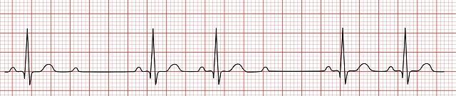
Answer: Second-degree atrioventricular (AV) block type II
There is a fixed PR interval present throughout the tracing with an unpredictable dropped QRS complex, consistent with second-degree type II AV block.
23. Identify the culprit artery causing the EKG findings in this tracing:
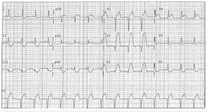
Answer: Left anterior descending artery
There is ST segment elevation of greater than 2 mm in leads V1 to V4 with reciprocal ST segment depression in leads II, III, and aVF, consistent with anterior wall injury. The anterior wall is supplied by the left anterior descending artery.
24. State the correct interpretation of this tracing:

Answer: Atrial fibrillation
The QRS complexes are irregularly irregular, and there is no identifiable P wave in between them, consistent with atrial fibrillation.
25. State the correct interpretation of this 10-second rhythm strip:
Answer: Sinus tachycardia
Counting every QRS complex in this strip and multiplying by 6 gives a rate of 120 beats per minute. There is a normal-appearing P wave before every QRS complex, consistent with sinus tachycardia.
26. Interpret this 12-lead EKG in its entirety:
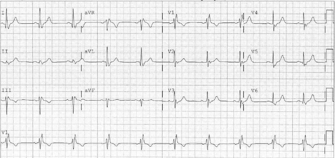
Answer: Normal sinus rhythm with right bundle branch block
The QRS duration is greater than 120 ms. There is an RSR pattern in lead V1 and an RS in lead V6, consistent with right bundle branch block. The heart rate is 60 beats per minute, and there is a normal-appearing P wave before each QRS.
27. Interpret this rhythm strip:
Answer: Sinus bradycardia with premature atrial contractions
The underlying rhythm is sinus bradycardia, as seen in beats one and two of the rhythm strip. Beats three and seven arrive early, lead with a P wave, and are associated with a resetting of the sinus rate.
28. Interpret this 12-lead EKG in its entirety: