
9 minute read
an alpaca Peter Briston Multicentric lymphoma in two firstcalved heifers Peter Briston


Peter Briston, Cambridge Vets
Multicentric lymphoma in two first-calved heifers
Advertisement
Abstract
Case 1 - Electronic diagnostics combined with physical palpation and FNA cytology gave a definitive and terminal diagnosis of multicentric lymphoma.
Case 2 - Clinical diagnosis from palpation was confirmed as lymphoma by postmortem and histopathology.
Case 1:
History
It was a dark and stormy night, and my phone shrieked like a harpy. Well, it was a mild Sunday evening and the cell phone chirruped.
A client of mine with an autumn calving dairy herd had installed the Cow Manager system. Every cow has a “SenzTag” ear tag which measure various biological parameters, and if thresholds are crossed the farmer automatically receives notification on an app, with the data on a chart. She had been informed by the system that one of her first calvers was not ruminating (see fig 1). Having drafted her out to examine her, they noticed a “lump in the vagina” in addition to a raised rectal temperature.
However, we were at “Covid Defcon 4”, so being a dutiful member of society, I attempted a teleconsult. Distance-guided aspiration with a new needle did not yield the fluid I was expecting from a presumed cyst, but I assured my client this was probably an incidental finding, and decided we should treat the pyrexia with antibiotics and an anti-inflammatory. I was to examine the animal myself if there was no improvement.
There was no improvement. In fact, she became increasingly “touchy and nervous, and kicked out if people or another cow touched her”. Her appetite fell off and she would occasionally stumble.
Examination
So two days later I examined her. She looked a well-conditioned, healthy cow, but there was a tennis ball size lump at the mucocutaneous junction of the vulva. It was indeed solid. As I palpated her per vaginum, more and more

tumours became apparent, ranging in size from a marble to a melon. They felt to be located intramural vaginal. Doing a rectal exam confirmed multiple lumps in the reproductive tract.
At this point I suspected cancer which had metastasized to multiple locations. The owner did not want to spend any money on her as she was now a cull, and refused the suggestion of biopsy and histopathology to confirm diagnosis and prognosis. I was able to do Fine Needle Aspirates (FNA) and made smears on three slides.
Differential Diagnoses
Ð Lymphoma Ð Squamous cell carcinoma Ð Leimyoma / leiomyosarcoma Ð Fibroma / fibrosarcoma
Outcome
The next day she had deteriorated so much that the owner promptly called petfood. Fortunately, the company was very willing to set aside the beast’s viscera for me to examine and photograph. Unfortunately, the viscera actually got chucked into a 1 tonne offal bin and were unable to be examined.
However, the slides yielded some useful cytology.
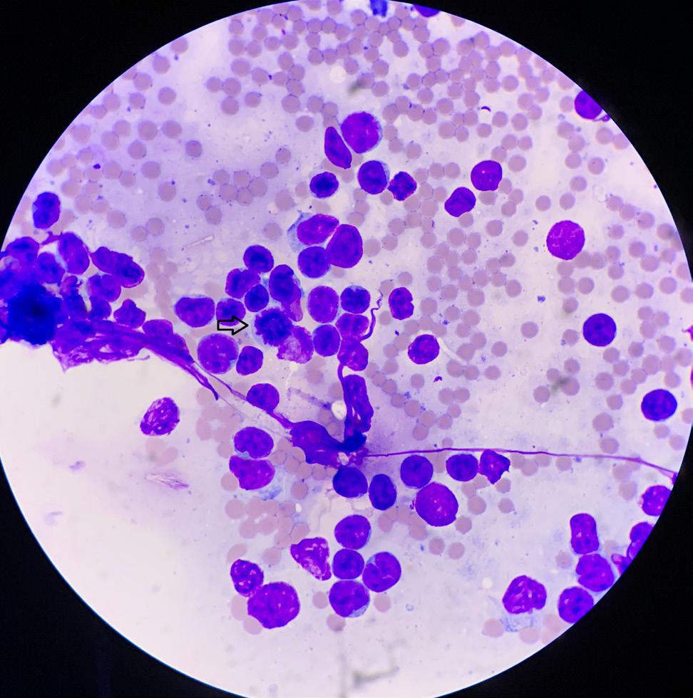
Cytology
The slides contain large numbers of a monotypic population of round cells admixed with moderate background haemorrhage. Many ruptured cells (common with neoplastic lymphocytes) and free chromatin is evident in places. The cells are medium to large (approximately 1-3 x the size of a neutrophil) and have scant basophilic cytoplasm. Nuclei are slightly cleaved and have dense chromatin, often containing 1-2 large pale nucleoli. Anisocytosis is mild to moderate, and there are scattered mitotic figures throughout.
Diagnosis:
The predominance of these large round cells (lymphoblasts) from a mass is consistent with lymphoma.
Case 2:
History
A month later I was called out to a different farm to see a Rising 3 year old cross-bred dairy cow, who was walking at the back of the mob and not eating much.
Clinical exam
Normal on appearance and most parameters, but with very reduced rumination. Rectal examination revealed a large, solid mass in the wall of the vagina and a lump within the intestinal tract.
Blood samples were taken but with a provisional diagnosis of lymphoma and a poor prognosis, she was sent to pet food the following week.
continued
continued
Serology EBL negative
Haematology / CBC
No abnormality
Post-mortem
In addition to the large mass in the wall of the vagina, multiple lymph nodes throughout the intestinal tract were enlarged, as well as a section of the wall of the small intestine. Samples were taken and fixed in formalin.
Histopathology
The sections from the lymph node and vagina are similar and consists of a neoplasm with no distinct margins. The neoplasm consists of neoplastic lymphocytes forming large sheets with minimal stroma. The lymphocytes have scant to moderate light eosinophilic cytoplasm and round to oval nuclei measuring 2-3 red blood cells in diameter with multiple prominent nucleoli. There are 5-10 mitotic figures per high power field. In the intestinal section, the same neoplastic lymphocyte population infiltrates the submucosa, muscularis and serosa.
Definitive Diagnosis
Lymphoma
Discussion
The first case was interesting for several reasons:
Covid and tele-diagnosis – it may be that Covid-19 will be seen as a black swan event in terms of setting a precedent for distance diagnosis being


more accepted. When you are dealing with clients who have a lot of experience and knowledge about animal health and husbandry, you can glean a lot of information and work through multiple ruleouts over the phone. Admittedly, it didn’t help this time, and a physical exam by a veterinarian was necessary. Which is also a valid point when discussing cases with clients.
Electronic health aids – the system used on this farm has been invaluable in the early detection of problems. Electronic ear tags measure rumination, eating, activity and temperature, and alerts are sent to the farmer by an app. We have been able previously to pick up cases of LDA and a flurry of ketosis in a 2-stage model: the tags alert the farmer to a health issue who can then draft out and check the cows. But as the signs are often vague, a veterinary exam is required to elucidate the exact diagnosis and offer a treatment / prevention plan. I suspect the prevalence of this sort of technology will only increase, and it is a great opportunity for improved veterinary outcomes.
FNA / cytology – farm medicine is still a very clinical, hands-on practice, and it was pleasing to be able to do FNA and get a diagnosis from cytology. Donald (2011) points out the utility of aspiration and smears from skin masses or enlarged lymph nodes to differentiate inflammation from neoplasia. I

benefitted from a recent webinar (Jenkins, 2020) and implemented several of the tips:
Ð Ð
Ð Ð Ð Ð Size of needle (19 - 22g) and syringe (5ml) Multiple advances of needle in multiple directions Release vacuum before withdrawal of needle Gentle expulsion and smear Make multiple slides Do not refrigerate
The second case was interesting because, like waiting for a bus, I had only seen 1 case of adult lymphoma in 2 decades, then I diagnose 2 within a month. Previously I have seen one similar case in a bull (diagnosed by exploratory laparotomy), and one case of thymic lymphoma in a young calf.
This case enabled me to do the extended work-up, with blood tests, post-mortem and histopathology. The local Pet Food company are very helpful, and by being willing to take some time to follow up individual cows and examine them in the abattoir my own education is improved. I find a box of chocolates help grease the wheels of industry here.
Lymphoma
Lymphosarcoma or bovine leukosis is one of the more common tumours of cattle (Parkinson et al, 2019), and can be divided into Enzootic Bovine Leukosis (EBL), or Sporadic Bovine Leukosis, which is seen in younger animals and has an unknown aetiology. The latter type is further categorized into:
Ð Juvenile Form – seen in calves less than six months, with a general enlargement of lymph nodes. Signs include weight loss, rumen tympany, anaemia, ataxia, fever, diarrhoea. CBC reveals a leukaemia and non-regenerative anaemia (Thompson J. et al, 2013) Ð
Ð Thymic Form – seen between six and 24 months as a large firm mass in the ventral caudal neck area, resulting in dyspahagia, jugular engorgement and bloat (Milnes, 2015). Atypical lymphocytes are seen in the blood.
Cutaneous Form – seen at 1-3 years as grey-white raised hairless circular plaques over skin, which eventually infiltrates organs. CBC has anaemia and neoplastic lymphocytosis.
These cases would be a multicentric adolescent form of the Juvenile presentation, but in heifers.
EBL is caused by Bovine Leukosis Virus (BLV), which is predominantly spread via infected blood, so iatrogenic transmission from herd vaccination is possible, as is transplacental transmission. Once infected, the animal is a carrier for life, but only a small percentage of BLV positive cows develop lymphosarcoma. Signs are generally seen between 5-8 years (Thompson KG, 1993); chronic disease, weight loss, weakness, inappetence, and other signs depending on organ involvement.
ELISA tests for EBL can be run on blood and milk. LIC runs the national control scheme, and bulk milk tests were down to 99.98% herd EBL negative status by 2008 (Voges, 2012), meaning NZ is essentially EBL free.
All forms of lymphoma are fatal, with no treatment.
REFERENCES:
Donald J. (2011). Cytology as a diagnostic tool in cattle. VetScript, Volume 24, Issue 1, p 26
Jenkins, K. (2020). Cytology Tips on how to maximise a diagnostic sample with a focus on FNA technique, “Gribbles Veterinary Pathology & Lincoln Institute Online Training Events” Apr 22, 2020, https://lincolninstitute.wistia.com/ medias/jhmfognf0i
Milnes, E. (2015). Thymic lymphoma in a dairy heifer. VetScript, Volume 28, Issue 5, pp 26-27
Parkinson TJ, Vermunt JJ, Malmo J, Laven R. (2019). A comprehensive textbook – diseases of cattle in Australasia, 2nd ed, Massey University Press
Thompson J, Vaatstra B, Norris L. (2013). Case report - lymphoma and leukaemia in a six-month old calf. VetScript, Volume 26, Issue 3, pp 36-38
Thompson KG, Johnstone AC and Hilbink F. (1993). Enzootic bovine leukosis in New Zealand – A case report and update. New Zealand Veterinary Journal 41(4): 190–194.
Voges, H. (2012). Reports from industry surveillance and disease control programmes: New Zealand dairy enzootic bovine leukosis (EBL) control scheme. Surveillance, 39(3, article 41).
Acknowledgements
Bill & Michelle of Burgess Farm
Duncan Wait of Grail Farm
Dr. Sarah Lee of Cambridge Vets (cytology) and
Dr. Genevieve D’Amours of SVS Laboratories (histopathology) for doing the actual skilled diagnostics.
AC PetFoods


