SWT/KWS Mt Kenya Veterinary Unit Report April 2026
SWT/KWS MT KENYA MOBILE VETERINARY UNIT
APRIL 2026
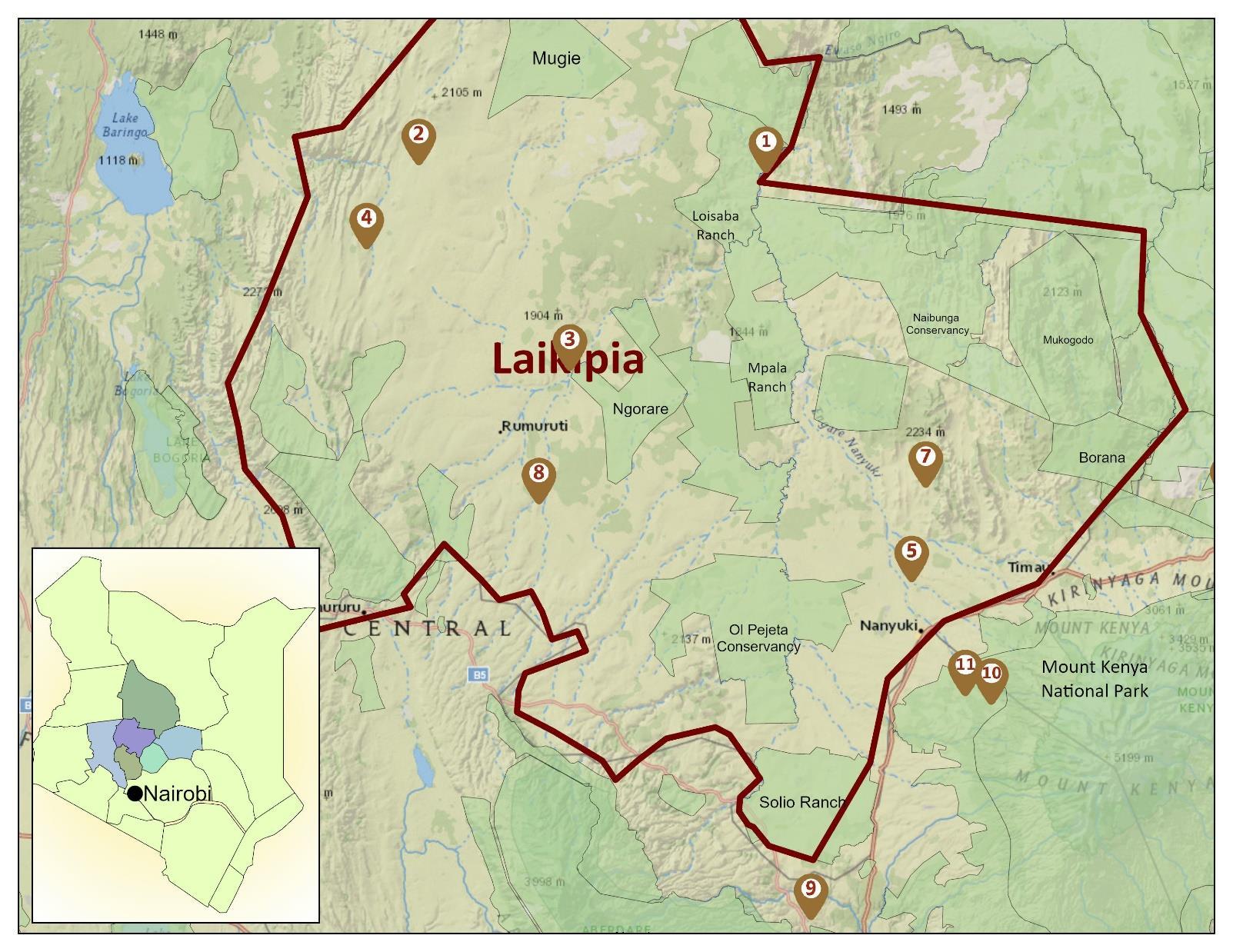
11 Cases in April 2026
April Report by Dr. Jeremiah Poghorn
1 Poaching Case 3 Elephant Cases
Mount Kenya Mobile Veterinary Unit had a series of activities during the April 2026 reporting period including clinical interventions, translocations, collarings and disease surveillance. Interventions include treatment of a wounded lion and wire snare removal from an adult elephant. Two wild dogs and two lionesses were collared for monitoring purposes. Disease surveillance activities included elephant, a giraffe calf and a zebra necropsy.
Acknowledgement
Sincere appreciation to the Sheldrick Wildlife Trust for logistical support that enabled prompt response to cases. We are grateful for the collaboration and coordination extended from the various conservancies we have worked with during this period, this partnership has been instrumental in ensuring timely and effective veterinary interventions when required.
Case Details
14-Apr-26
15-Apr-26
16-Apr-26
23-Apr-26
25-Apr-26
patient was observed to have nonweight bearing left hind limb lameness
elephant was observed with a wire snare attached to the left hind limb
carcass was fresh with death estimated within 24 hours
28-Apr-26
April 2026
Wild Dog Collaring Loisaba Conservancy
Wild dog collaring was indicated to monitor pack movement patterns and home range to gain a better understanding of the newly sighted pack of this endangered species. Data collected from use of the digital collars was to serve as a research tool and inform conservation efforts.
Immobilisation and collaring
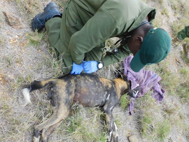
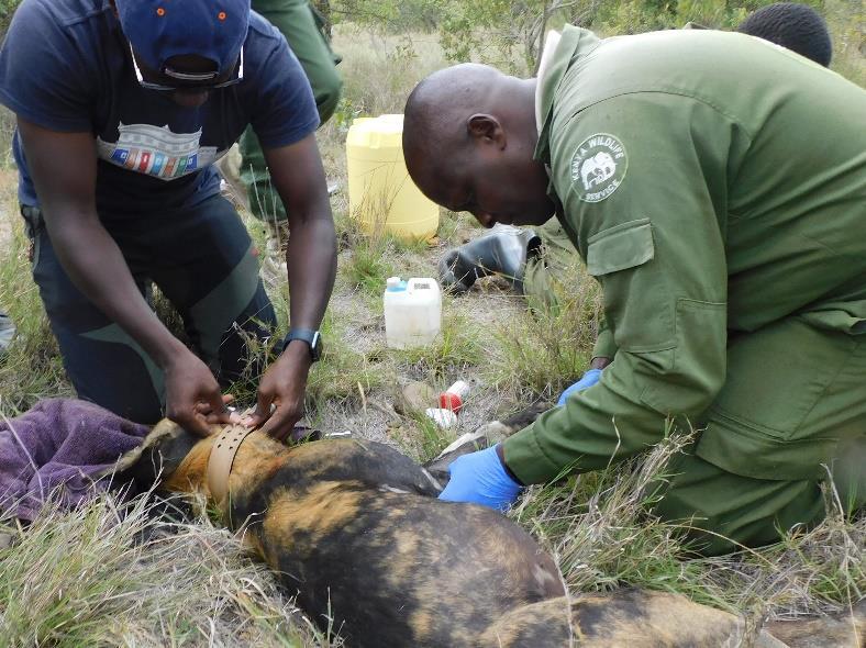
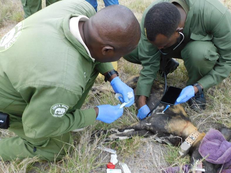
One adult male and one adult female were targeted for collaring. They were darted with a combination of Medetomidine and Ketamine with assistance from drone technology to facilitate pack visualisation.
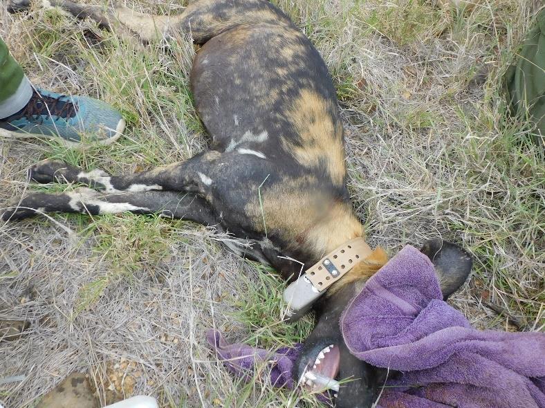
The animals were collared with a digital GPD neck collar and body measurements were taken and recorded. Blood samples, ear tissue samples and tick samples were also collected. Prophylactic antibiotic cover treatment was done by intramuscular administration of 750mg Amoxicillin trihydrate. Anti-parasitic treatment was done using topical fipronil spray (Frontline) applied along the backline to control extreme ectoparasites. The subjects received an excellent prognosis with rapid recovery from anaesthesia expected since they were in good health.
The wild dog collaring activity was successfully completed. Smooth coordination between the Loisaba Conservancy team, the Wild Dog Research team and the KWS veterinary team is commendable and was key.
Case # 2&4 – 14
and 17th April 2026
Lion Collaring
Mukutan Conservancy
Lion collaring was indicated to monitor individual and pride movement patterns and home range to proactively prevent and manage human wildlife conflict by deterring livestock predation as well as to serve as a research tool from the lion location data collected.
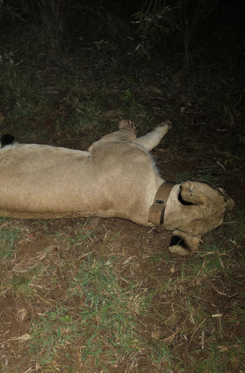
Immobilisation and collaring
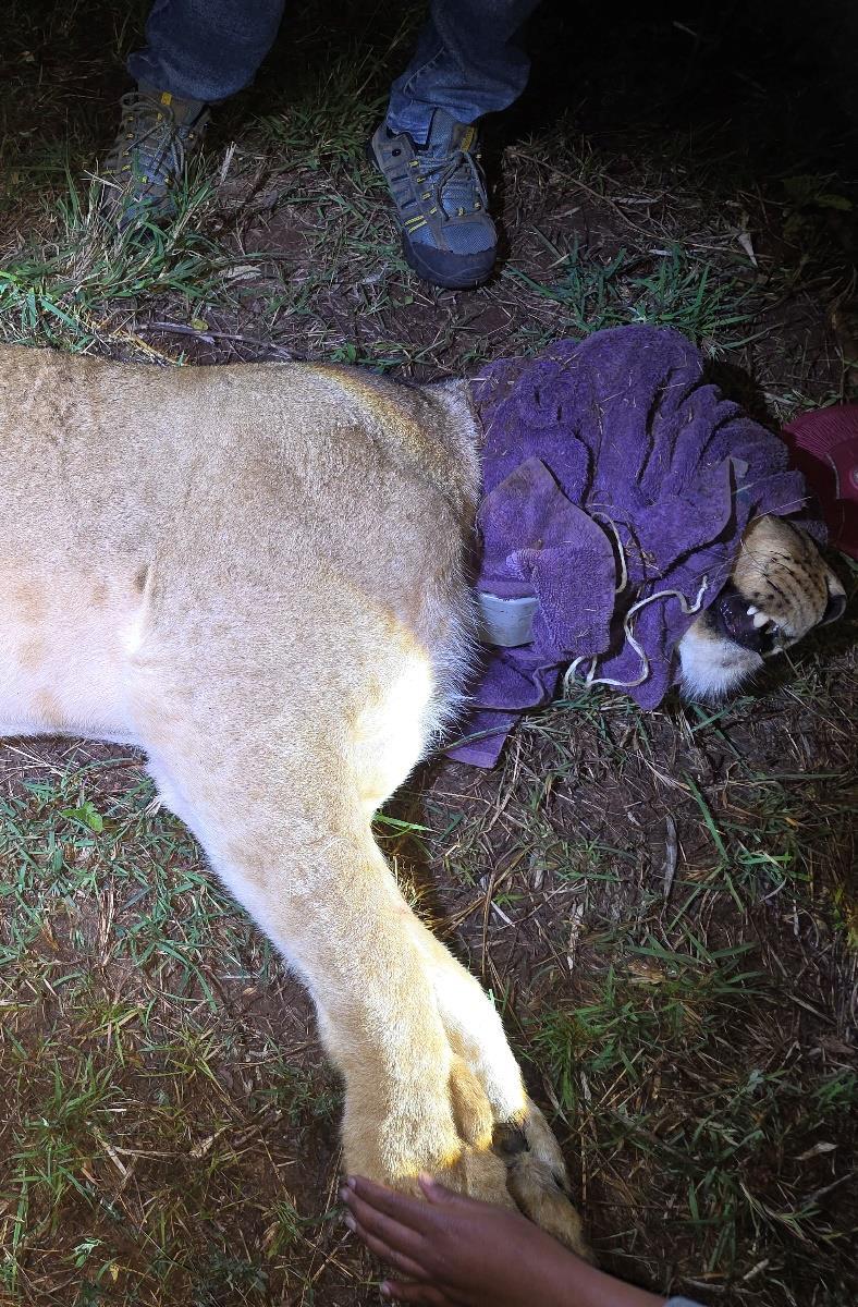
An adult and subadult lioness were selected for collaring and darted using a drug combination of 6mg Medetomidine and 300mg Ketamine on 2 different days A top up of 200mg Ketamine was administered by intramuscular injection.
Digital collaring was done by GPS neck collar placement. Body measurements of the lioness’ were taken and recorded. Blood samples were collected in EDTA tubes, and a serum sample was collected in a clot activator tube. Prophylactic antibiotic cover treatment was done by intramuscular administration of 3000mg Amoxicillin trihydrate. Anti-parasitic treatment was done using topical fipronil spray (Frontline) applied along the backline to control extreme ectoparasites.
The lioness’ received an excellent prognosis with rapid recovery from anaesthesia expected as they were in good health. The lion collaring activity at Mukutan Conservancy was successfully completed. Two lionesses from different prides were collared in line with the set target.
.
Lion Natural Causes
Ol Maisor Farm
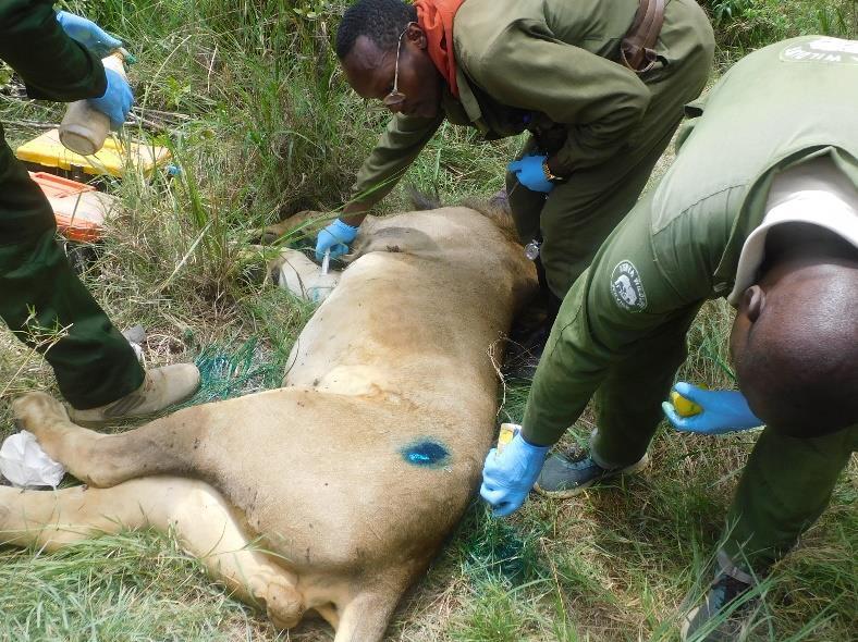
A report was received from the Lion Landscapes Monitoring Team of a collared lion that was observed with lameness and was stationary for extended periods on the monitoring systems.
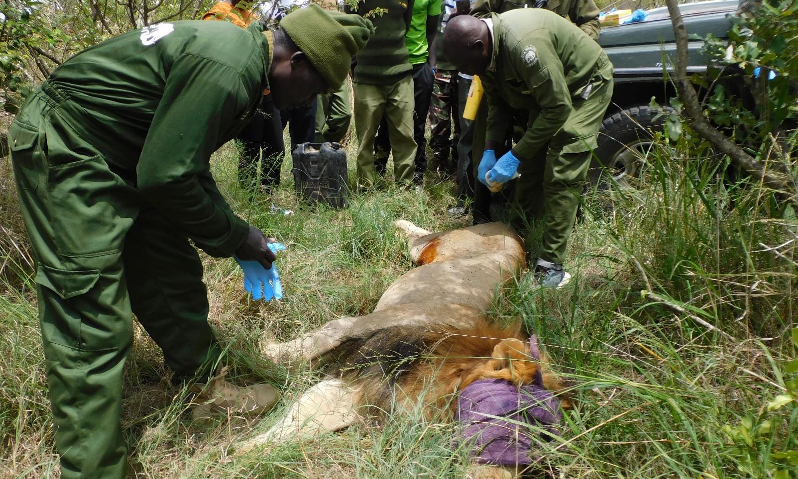
Immobilisation examination and treatment
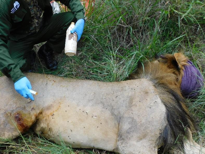
The subject was darted with 300mg Ketamine and 8mg Medetomidine. The anesthetised subject was found in a thicket, blindfolded and moved to a more open place for subsequent treatment. A top up of 250mg Ketamine and 2mg Medetomidine was given by intramuscular hand injection as the anaesthesia was light.
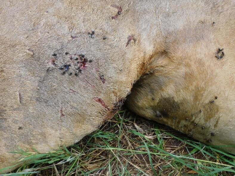
Multiple exudative penetrating wounds approximately 1cm in diameter were noted on the right stifle area, left pelvic area, along with the left and right antebrachium. These wounds were accompanied by extensive subcutaneous undermining. The wounds were suspected to be bite and claw injuries sustained in a fight. The wounds were lavaged with Iodine, infused with Cloxacillin ointment and packed with green clay to minimize secondary contamination. The lion was also administered 80mg Dexamethasone, 6,000mg Amoxicillin, 3,000mg Butaphosphan and 1.5mg Cyanocobalamin. Frontline was applied to control extreme ectoparasites.
Prognosis
Prognosis is guarded due to the extensive nature of the injuries. The lion will be monitored by the GPS collar.
Case
Elephant Postmortem Salama, Laikipia County
A report was received of an elephant carcass that was spotted next to a river in Salama Ward. The tusks were removed as it was late for the veterinary team to arrive.
Postmortem examination
A carcass of an adult male elephant was found in right lateral recumbency facing a Southern direction.The carcass which was in good body condition was distended with multiple vesicles on the ventral thoracic region which oozed froth. It was determined that the carcass was in early autolysis and death was staged to have occurred within the previous 72 hours. Hepatomegaly was observed with rounded liver edges and extensive pseudo melanosis. Severe splenomegaly was noted accompanied by multifocal ecchymotic serosal haemorrhages. Good gut fill was observed in the stomach with ingesta present along the alimentary tract, pointing to a possible acute death. Generalised pseudo melanosis was noted in the liver, spleen and intestines. Liver tissue, spleen tissue, stomach contents and intestinal tissue samples were collected.
Cause of death
Septicaemia was determined to be the preliminary cause of death. Differential diagnoses include bacterial septicaemia with Clostridia spp.
Case 6 – 21st April 2026
White rhino Natural Causes
Lewa Conservancy
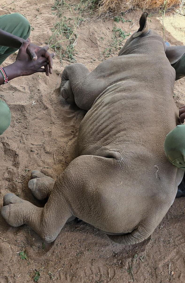
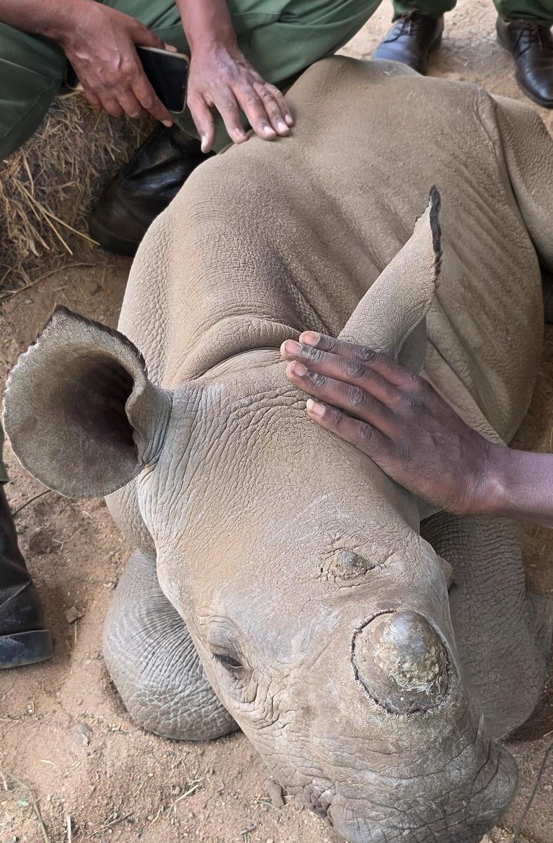
A report was received from Lewa Conservancy about an orphaned rhino calf with left hind limb lameness requiring a review.
Examination and treatment
The rhino calf had been rescued three months prior and placed in an orphanage at the Conservancy. Prior radiographic imaging was unremarkable. The patient was observed to have non-weight bearing left hind limb lameness, dragging the affected leg while walking. There was asymmetry of gluteal muscle masses which was attributed to disuse atrophy with the left side appearing reduced compared to the right side. An orthopaedic examination elicited a pain response during hock and hip joint palpation. Based on radiographic imaging data and clinical examination findings, this case was determined to be a coxofemoral subluxation. 1 week analgesia therapy was prescribed using Phenylbutazone to control pain while healing progressed. Restricted exercise was prescribed for one month with running to be avoided.
Prognosis
The rhino calf received a fair prognosis with the completeness of healing highly dependent on the endogenous factors and nursing care
Elephant Natural Causes
Enasoit Conservancy
A report was received from the Enasoit Conservancy of an adult male elephant that was observed by field staff to be inappetant with a gait suggestive of lameness
Immobilisation, examination and treatment
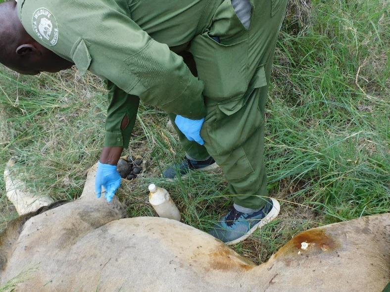
The patient was found standing stationary in an abnormal posture with the hind limbs extended backwards while leaning on the fore limbs associated with abdominal pain. The elephant was reluctant to move on auditory stimulation; a normal gait was observed when the elephant finally moved. Colic was deduced due to the behaviours observed and the tentative diagnoses suspected included abdominal obstruction and poisoning since the elephant was frequently trespassing onto neighbouring farms to raid on crops. The elephant was darted from the vehicle with 20mg Etorphine On rectal exploration, it was observed that there were no faecal contents in the rectum and colon consistent with complete gastrointestinal obstruction. 40lt of water was used as a rectal enema to facilitate unblocking the obstruction. 80mg Dexamethasone, 4000mg Butaphospan, 2mg Cyanocobalamin and 30,000mg of Amoxicillin were also administered.
Prognosis
The patient had a guarded prognosis due to suspected complete obstruction of the gastrointestinal tract. Sadly, despite treatment the patient collapsed and died after anaesthetic reversal. An autopsy revealed septic shock secondary to a colonic perforation which led to leakage of ingesta with subsequent fatal peritonitis.
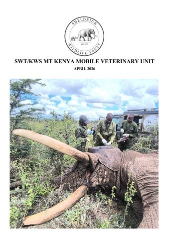
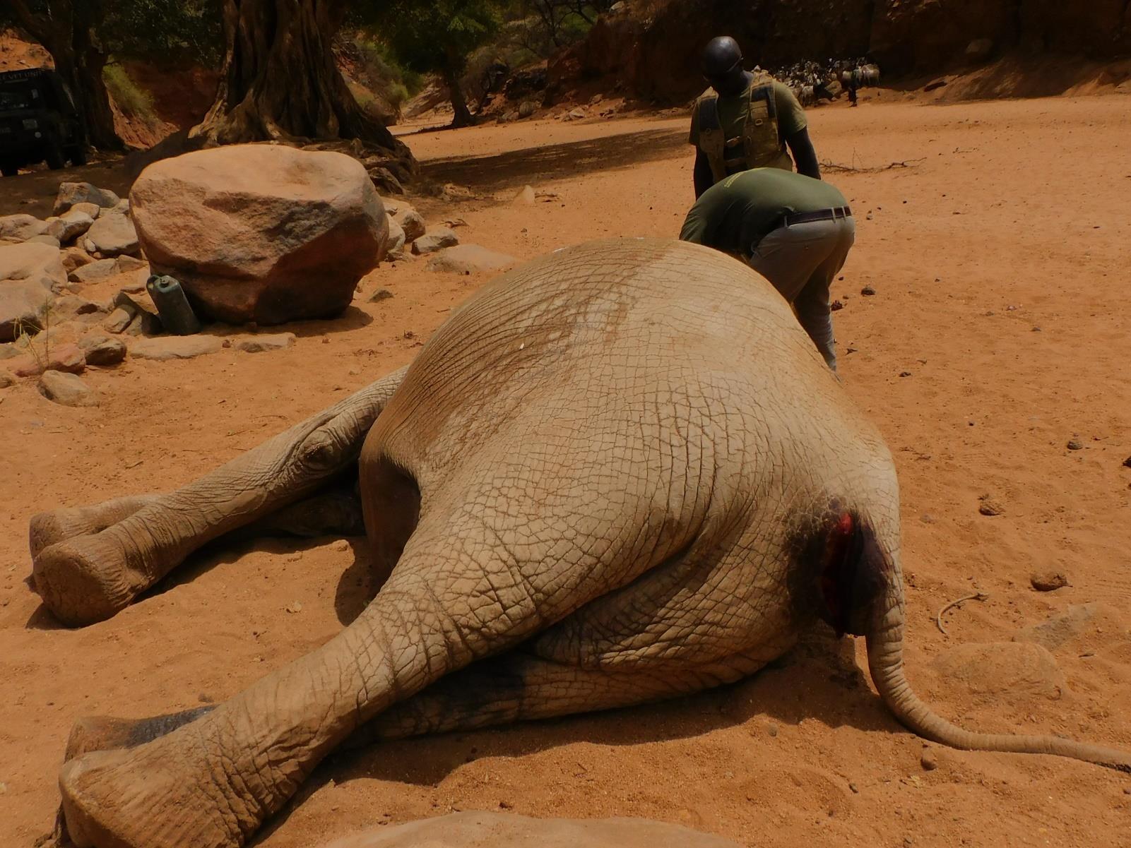
Elephant Snare
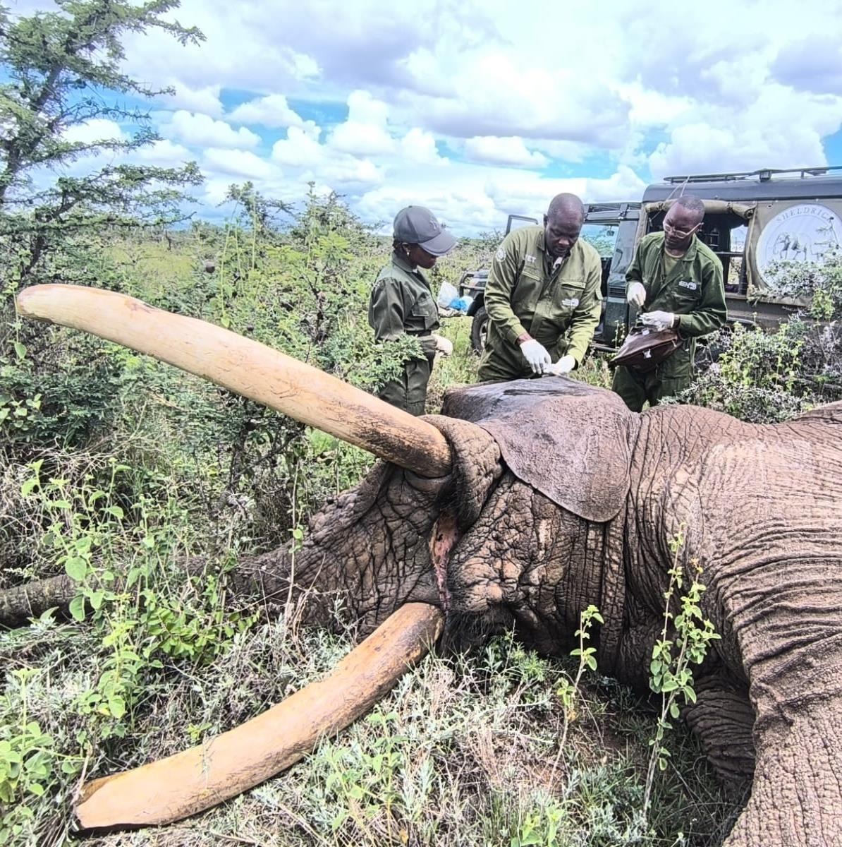
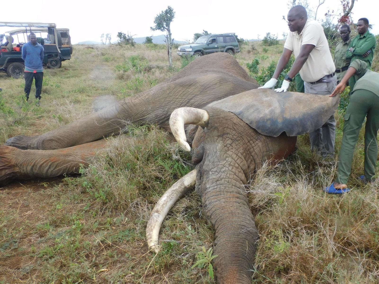
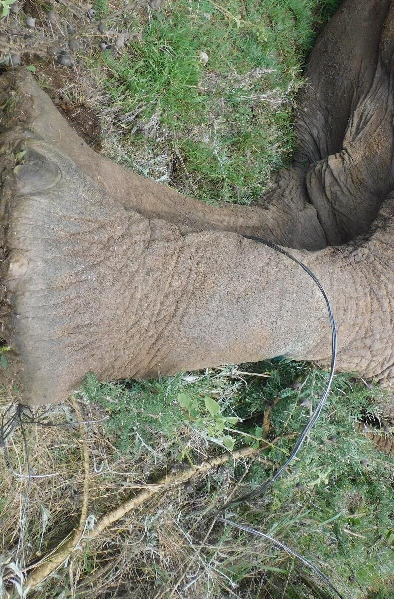
A report was received from Kifuko Ranch of an adult male elephant that was observed to have a wire snare lodged around the left hind limb. A ranger team tracked the animal and maintained distance surveillance.
Immobilisation, examination and treatment
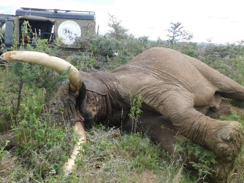
The adult male elephant herd was observed with a wire snare attached to the left hind limb while moving at a normal gait. The elephant was free-darted from the vehicle with 18mg Etorphine hydrochloride and fell into right lateral recumbence.
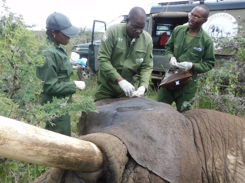
The wire snare lodged superficially at the proximal tibio-fibular region of the left hind limb was cut and removed using a wire cutter. Topical Oxytetracycline aerosol wound spray was then applied to superficial skin lesions caused by snare friction. 2000mg Flunixin meglumine was administered intravenously via an ear vein as an anti-inflammatory agent.
Prognosis
The patient received an excellent prognosis since the wire snare was found early with injury limited to superficial skin grazing. Prompt recovery was expected with rapid return to functionality.
Kifuko Ranch
Case 9 – 26th April 2026
Giraffe Postmortem Abedares Country Club
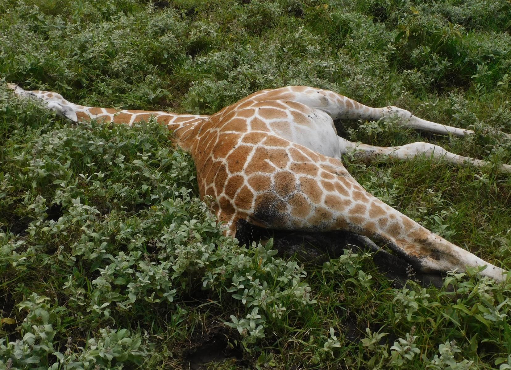
The giraffe calf was reported to have diarrhoea on Saturday 25th by the patrol officer. He died on Sunday.
Postmortem examination
The carcass was fresh with death estimated to have occurred within the previous 24 hours. The calf had matting on the perineum with dark brownish diarrhoea. Trachea had brownish fluid with food material in the proximal part and the distal 1/3 had froth. Tracheal mucosa was dark reddened due to congestion. Lungs were congested and frothy. Pericardial blood vessels were prominent. Liver was enlarged and had a cooked liver appearance. Intestines were gas filled and empty with scanty mucoid intestinal contents. Intestinal mucosa and serosa were severely congested. Mesenteric blood vessels were congested. Mesenteric lymph nodes were enlarged
Possible diagnosis were Tracheitis, pulmonary oedema, Pneumonia, acute mucoid enteritis or Hepatitis.
Cause of death
Postmortem findings of inflamed intestinal mucosa with mucoid intestinal contents were consistent with acute enteritis suspected to be of pathogenic origin. Immediate cause of death is attributed to respiratory failure due to aspiration pneumonia caused by the food material within the airway.
Zebra
Human-Wildlife Conflict
Mt Kenya Forest
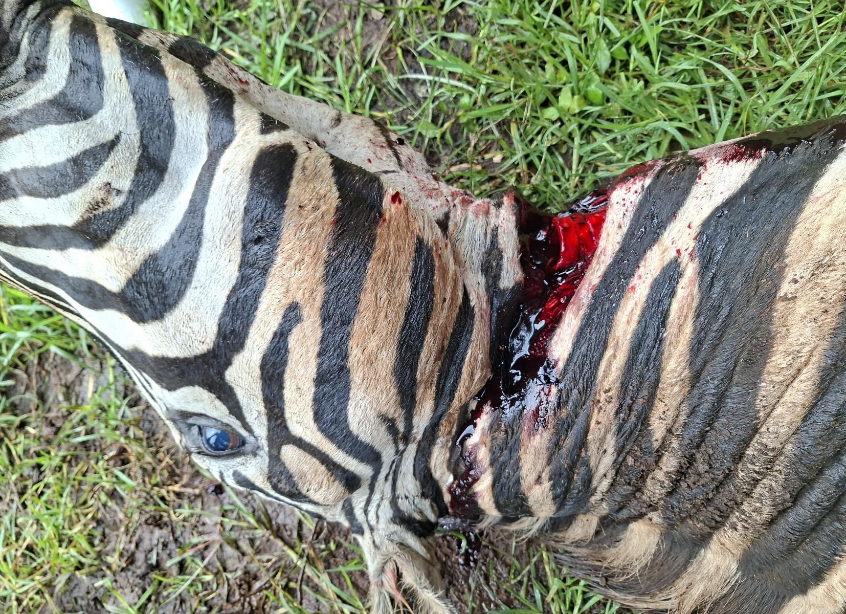
The zebra was reported dead after attempting to break through a barbed wire fence to join a female and calf on the other side of the fence in the army compound. After several attempts of hitting the fence, the zebra was exhausted and was sighted at about 8:00 in the morning.
Postmortem examination
The barbed fence was kinked inside the perimeter wall where the animal lay. The fresh carcass, with death estimated to have occurred within the previous 12 hours, was found in right lateral recumbency and with the head tilted to the left. The throat was slit and there was clotted and dried blood matting the neck region.
Externally, there were no signs of struggle. Scanty blood was seen in the internal organs including the heart, spleen and major blood vessels. There was an antemortem clot on the heart. Internal organs were unremarkable.
Cause of death
The immediate cause of death was hypovolemic shock caused by excessive bleeding from the slit on the throat which severed jugular and carotid blood vessels.
Case
Bongo Relocation
Mt Kenya Wildlife Conservancy
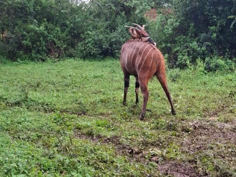
Five female mountain bongos were translocated from Boma Kubwa to the McKeand breeding boma. The five females named Limuru, Oburu, Wema, Herper and Namunyak were to be introduced to four male mountain bongos which had been imported from Europe for the purpose of gene conservation.
Relocation
The mountain bongos were chemically immobilized by hand darting using a dan inject rifle and 1.5ml darts loaded with Etorphine and Azaperone, amounts depending on size and weight. All bongos were given 15ml Oxytetracycline, 10ml Phenylbutazone, 10ml Multivitamins IM, Ivermectin 3ml, Fenbendazole 5ml and tick control spot on 10ml. Namunyak had lameness and examination revealed that her right forelimb had a fracture on the lateral claw. Wound management was done by cleaning the hoof with water, Hydrogen peroxide and Iodine then covered using clay. The vital parameters including respiratory rate and rectal temperature were recorded, and the subjects were supplemented with oxygen during the immobilization. Faecal material, whole blood and tick samples were collected.
The mountain bongos were carried using an animal stretcher and loaded on a land cruiser. They were transported to the release site and occasionally monitored during transit. The anaesthesia was reversed at the release site. The mountain bongos exhibited smooth and rapid recovery within less than one minute.