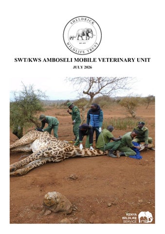
SWT/KWS AMBOSELI MOBILE VETERINARY UNIT JULY 2026
Introduction
19 Cases in July 2026
5 Poaching Cases
15 Elephant Cases
June Report by Dr. Kariuki Edward The Amboseli Ecosystem experienced a dry and cold period in July 2026. 19 cases were attended to in the Amboseli Ecosystem with elephants being significantly affected by multiple mysterious deaths. Laboratory examinations revealed cyanide and bacterial pathogens suggesting the elephants were likely poisoned due to crop raiding. Two giraffe and two zebras were treated for snare/arrow injuries. Acknowledgement We thank Sheldrick Wildlife Trust (SWT) for material support and Mobile Unit funding. KWS Staff, ATE and BLF rangers for the field support and the monitoring of distressed wildlife are highly acknowledged and appreciated.
SWT/KWS Amboseli Mobile Vet Unit Treatment Locations July 2026
Case Details Date
Species
Area Found
1-Jul-26
Giraffe
Kuku Group Ranch
1-Jul-26
Elephant
2-Jul-26
Reason for Intervention
Outcome
Snared
A tightly constricting braided wire snare firmly embedded in the neck
Successfully Treated
Amboseli NP
Natural Causes
A fresh puncture wound with traumatic penetration by an elephant tusk
Died
Elephant
Amboseli NP
Postmortem
Lesions are highly suggestive of toxaemia, and septicaemia
Died
4-Jul-26
Elephant
Amboseli NP
HWC
Cardiopulmonary failure associated with severe systemic circulatory disturbance
HWC Death
5-Jul-26
Elephant
Amboseli NP
HWC
Pathological condition characterised by severe multisystemic disease
HWC Death
5-Jul-26
Elephant
Amboseli NP
HWC
Prolonged recumbency, inability to rise, paddling movements while recumbent
HWC Death
7-Jul-26
Elephant
Kimana Sanctuary
HWC
Acute cardiopulmonary failure associated with severe multisystemic disturbance
HWC Death
7-Jul-26
Elephant
Kuku Group Ranch
HWC
Prolonged lateral recumbency, inability to stand, paddling movements and circling
HWC Death
8-Jul-26
Elephant
Kimana Sanctuary
HWC
To rise, paddling movements and circling while recumbent
HWC Death
9-Jul-26
Elephant
Amboseli NP
HWC
Cause of death was undetermined but likely same ailment as other elephants
HWC Death
9-Jul-26
Elephant
Kimana Sanctuary
HWC
Severe cardiopulmonary compromise and multisystemic failure
HWC Death
13-Jul-26
Elephant
Kimana Sanctuary
HWC
Observed in prolonged left lateral recumbency
HWC Death
17-Jul-26
Giraffe
Kimana Sanctuary
Snared
A braided wire snare tightly positioned around the cranial cervical region
Successfully Treated
18-Jul-26
Elephant
Kimana Sanctuary
HWC
Similar conditions to other mysterious deaths in the area
HWC Death
18-Jul-26
Elephant
Amboseli NP
HWC
She continuously paddled her limbs but was unable to lift herself from the ground
HWC Death
19-Jul-26
Elephant
Amboseli NP
HWC
The elephant remained unable to rise and showed no signs of clinical recovery
HWC Death
19-Jul-26
Elephant
Kimana Sanctuary
Spear
Three deep penetrating wounds to the right side of the neck
Successfully Treated
25-Jul-26
Zebra
Olasira Conservancy
Arrow
A penetrating puncture arrow wound situated on the right lateral cervical region
Successfully Treated
25-Jul-26
Zebra
Olasira Conservancy
Arrow
A penetrating arrow puncture wound on the right aspect of the cervical region
Arrow
Case 1 – 1st July 2026 Giraffe
Snared
Kuku Group Ranch
Big Life Foundation Rangers reported an adult male giraffe with a wire snare tightly constricting the cervical region, with a long trailing section of wire extending behind the animal. Immobilisation, examination and treatment The giraffe was approached cautiously using a vehicle and darted with 15mg Etorphine combined with 60mg Azaperone. Progressive sedation characterised by generalised ataxia and reduced coordination occurred within 10 minutes. Once adequate immobilisation was achieved, trained personnel guided the giraffe to the ground and the anaesthetic reversed whilst the giraffe was manually restrained for treatment. Although the snare was firmly embedded within the skin folds of the neck, there was no evidence of cutaneous laceration, skin perforation, subcutaneous emphysema, haemorrhage, tracheal compression, or major vascular injury. The snare was carefully severed using wire cutters and removed completely from the neck. Although no overt skin wounds were identified, prolonged constriction by the snare was considered sufficient to induce localised soft tissue contusion, vascular compression, inflammation, and pain. Consequently, prophylactic medical therapy with Amoxicillin and Flunixin was administered. Prognosis The giraffe recovered uneventfully, rose to standing without assistance. Prognosis is good.
.
Case 2 – 1st July 2026 Elephant
Postmortem
Amboseli National Park
A male adult female elephant was reported dead by a local herdsman within the Kimana Gate area of Amboseli National Park. The Vet team found the carcass in left lateral recumbency and was subjected to systematic gross pathological examination. Postmortem examination Assessment of the surroundings revealed disturbance of the vegetation surrounding the carcass. The trampling of vegetation, displaced soil and multiple areas of ground disruption were consistent with premortem struggling and prolonged attempts by the animal to regain a standing position. These environmental findings suggested that the elephant had experienced an extended period of terminal recumbency accompanied by physiological distress prior to death. Extensive postmortem scavenging had resulted in substantial loss of the internal organs, preventing comprehensive pathological assessment and determination of the primary cause of death. Cause of death In the absence of evidence indicative of human interference or predatory attack and considering the social behaviour of elephants, the wound was considered most consistent with an accidental tusk-inflicted injury.
Case 3 – 2nd July 2026 Elephant
Human – Wildlife Conflict
Amboseli National Park
The Vet Unit received an urgent field report concerning an adult female elephant and her 9-month-old calf that were found in lateral recumbency within the Empaash region. Upon arrival at the scene, the veterinary team established that the adult female was critically recumbent, whereas the juvenile calf had already died. Postmortem examination The carcass was found in lateral recumbency with rigor mortis fully established, indicating that death had most likely occurred within the preceding 12–24 hours. The calf was in good body condition, with no significant traumatic injuries, external haemorrhages or skin lesions. The mucous membranes were markedly pale. Similar pallor was evident throughout the skeletal musculature, particularly within the forelimbs, indicating widespread tissue hypoperfusion and ischemia. Within the abdominal cavity, the liver was enlarged, congested and diffusely inflamed. The spleen appeared pale and contracted. There was severe pulmonary congestion characterized by excessive intravascular blood accumulation within the lung. The most significant pathological findings were observed in the heart. Cause of death The immediate cause of death was determined to be acute cardiovascular collapse accompanied by systemic shock possibly due to poisoning.
. Cause of death In the absence of evidence indicative of human interference or predatory attack and considering the social behaviour of elephants, the wound was considered most consistent with an accidental tusk-inflicted injury.
Case 4 – 2nd July 2026 Elephant
Human – Wildlife Conflict
Amboseli National Park
An adult female elephant named Isobar was reported with prolonged lateral recumbency with inability to rise. Supportive therapy, including intravenous dextrose, normal saline, lactated Ringer's solution, and atropine sulphate, was administered but the elephant succumbed. Postmortem examination There were ticks, a fresh wound on the vulva, bloated abdomen, mucopurulent discharge on the medial aspect, conjunctiva of the left eye and bilateral flexion of the forelimbs. Internal examination revealed circular area of oedema and congestion in the subcutis of the left lumbar region, focal area of congestion on the left dorsal abdominal wall, trauma wound on the left ribcage, severe congestion on the mucosal surface of the pharynx and congested left lung. There were petechial haemorrhages on the pericardium of the ventricles and foul-smelling whitish mucoid substance with white precipitate in the oesophagus as well as congestion of the omentum, enlarged liver, extremely watery digesta in the colon, focalised ulceration of the colon. Cause of death The pathological picture was consistent with acute cardiopulmonary resulting in multisystemic congestion and terminal respiratory insufficiency possibly due to poisoning.
Case 5 – 5th July 2026 Elephant
Human – Wildlife Conflict
Amboseli National Park
Noonkotiak was reported with prolonged lateral recumbency, inability to stand, paddling movements and circling while recumbent. Owing to the grave prognosis and poor welfare outlook, humane euthanasia was performed followed by a comprehensive necropsy. Postmortem examination The carcass was in good body condition with tick infestation and a fresh traumatic wound involving the perineal region extending into the rectum. There was bilateral flexion of the forelimbs. There was generalised enlargement of all the lymph nodes, oedema and congestion of bronchial lymph nodes; multifocal petechial and ecchymotic haemorrhages on the diaphragmatic and visceral surface of the spleen. Localised haemorrhages on the subscapularis muscles. Cyanotic pharynx, focal whitish abscess on the left lung, oedematous and congested lungs. Mild increase in peritoneal fluid, blood, enlarged liver with a cooked appearance, irregular yellowish patches in the liver parenchyma on cross-section, yellowish coloured ingesta in the stomach and duodenum, focal congestion in the mucosal ridges of the colon. Cause of death Severe multisystemic disease with cardiopulmonary compromise, systemic vascular injury and chronic inflammatory changes possibly due to poisoning.
Case 6 – 5th July 2026 Elephant
Human – Wildlife Conflict
Amboseli National Park
Observations indicated prolonged lateral recumbency, inability to rise, paddling movements and circling while recumbent. Before a complete veterinary intervention could be undertaken, the elephant succumbed. Postmortem examination At the time of examination, the carcass had undergone extensive predation. Nearly all soft tissues had been removed, leaving only remnants of the skin, ribs and skull available for inspection. The advanced degree of carcass destruction precluded systematic examination of major organ systems including the cardiovascular, respiratory, digestive, urinary and nervous systems. Significant internal pathological lesions could not be evaluated. The findings correspond with the field necropsy observations recorded for this case. Cause of death The cause was inconclusive due to advanced post-mortem predation that obscured the primary disease process. Absence of internal organs prevented definitive pathological diagnosis but due to its location and proximity to other elephant deaths it is a suspected HWC/poisoning case.
Case 7 – 7th July 2026 Elephant
Human – Wildlife Conflict
Amboseli National Park
The elephant was reported after developing prolonged lateral recumbency characterised by inability to rise, paddling movements and circling while recumbent. Supportive therapy, including intravenous Dextrose, normal saline, Dexamethasone, lactated Ringer's solution, and Atropine sulphate, was administered but the elephant succumbed and a postmortem performed. Postmortem examination Examination revealed tick infestation, blood oozing from the trunk and bilateral flexion of the forelimbs with rigor mortis. There were multifocal haemorrhages on the subcutis of the left abdominal wall, oedema and congestion of the prescapular and mediastinal lymph nodes, and multifocal haemorrhages on the spleen. The left kidney had a cooked appearance. The left lung was congested and there was an increase in pericardial fluid with a reddish tinge; multifocal areas of petechial haemorrhages on the pericardium; increased coronary fat; streaks of pale and reddish areas on the myocardium. The liver was enlarged and congested liver. A tiny piece of watermelon rind was found in the stomach. Cause of death Fatal acute cardiopulmonary failure associated with severe multisystemic circulatory disturbance and generalised vascular congestion possibly due to poisoning.
Case 8 – 7th July 2026 Elephant
Human – Wildlife Conflict
Kuku Group Ranch
The elephant exhibited prolonged lateral recumbency, inability to stand, paddling movements and circling while recumbent. Supportive treatment consisting of intravenous Dextrose, normal saline, lactated Ringer's solution and Atropine sulphate but owing to the poor clinical response, grave prognosis and compromised welfare, humane euthanasia was performed. Postmortem examination The elephant was in good body condition with generalised tick infestation, bilateral flexion of the forelimbs and fecal material lodged within and partially protruding from the anus. Internally, there was oedema and congestion of the prescapular and precrural lymph nodes. Left kidney had a cooked appearance and there was congestion and frothing affecting the apical and cardiac lobes of the left lung as well as slight increase in pericardial fluid with a reddish tinge, multifocal ecchymosis haemorrhages in the aorta. The liver was congested and enlarged. There were large quantities of tomatoes were in the stomach ingesta. Cause of death Severe cardiopulmonary compromise with generalised circulatory failure, pulmonary dysfunction and multisystemic vascular congestion possibly due to poisoning through crop raiding.
Case 9 – 8th July 2026 Elephant
Human – Wildlife Conflict
Kuku Group Ranch
HIP was reported after approximately six days of progressive illness characterised by prolonged lateral recumbency, inability to rise, paddling movements and circling while recumbent. The elephant was given intravenous Dextrose, normal saline, Lactated Ringer's solution and Atropine sulphate but despite intensive intervention, the prognosis remained grave and humane euthanasia and a postmortem was performed. Postmortem examination There was a heavy tick infestation together with multiple decubital sores over prominent bony protuberances, consistent with prolonged recumbency. There were oedema and congestion of the prescapular lymph nodes, areas of petechiation and congestion on the spleen. The bladder was full and there was congestion and frothing in the lung parenchyma mostly affecting the right lung. There was also increased pericardial fluid with a reddish tinge, and focal haemorrhages on the coronary fat. The liver was enlarged and yellowish in colour with blackish spots on the bile ducts. The faecal matter was found in the descending rectum with impaction of the rectum and ulceration of the colonic mucosa. Cause of death Severe cardiopulmonary compromise with generalised circulatory failure, pulmonary dysfunction and multisystemic vascular congestion possibly due to poisoning.
Case 10 – 9th July 2026 Elephant
Human – Wildlife Conflict
Amboseli National Park
Personnel from the Kenya Wildlife Service (KWS) conducting routine wildlife surveillance patrols within the Tortilis sector of Amboseli National Park reported the presence of an elephant carcass. Postmortem examination The remains of the adult male elephant were in an advanced stage of decomposition. The carcass exhibited severe postmortem alteration consistent with prolonged environmental exposure under field conditions. Extensive scavenging had resulted in substantial destruction of the carcass, thereby compromising pathological evaluation. The abdominal cavity was open, and the visceral organs had either been consumed or displaced from their normal anatomical locations. Musculature surrounding the axial skeleton exhibited advanced autolysis and decomposition. No recognisable traumatic lesions, penetrating wounds, fractures or other gross pathological abnormalities could be identified. The advanced stage of decomposition, together with complete removal of the major visceral organs, rendered evaluation of organ morphology impossible. Cause of death Exact cause of death could not be determined but due to location this elephant is likely to be another poisoning victim.
Case 11 – 9th July 2026 Elephant
Human – Wildlife Conflict
Amboseli National Park
Clinical examination revealed prolonged lateral recumbency with inability to rise. Supportive therapy consisting of intravenous Dextrose was instituted but the animal failed to respond. Owing to the grave prognosis and poor welfare outlook the elephant was euthanised. Postmortem examination Examination revealed generalised tick infestation and bilateral flexion of the forelimbs. Internally there was multifocal haemorrhages in the subcutis of the right abdominal body wall, oedema of the prescapular lymph nodes and multifocal ecchymosis haemorrhages on the visceral surface of the spleen. There was froth in the trachea and the diaphragmatic lobe, congestion in the entire right lung as well as focal petechial haemorrhages on the pericardium, coronary fat, and the left ventricle. Blood vessels in the omentum were congested, with congestion and enlargement of the liver, ulceration and erosion of the gastric mucosa. Cause of death Severe cardiopulmonary compromise with generalised circulatory failure and multisystemic vascular injury resulting in terminal respiratory insufficiency most likely due to poisoning.
Case 12 – 13th July 2026 Elephant
Human – Wildlife Conflict
Amboseli National Park
The Big Life Foundation reported a male elephant calf, that remained in recumbency despite repeated attempts by the family herd to get it to stand. The Amboseli Veterinary Unit responded promptly to the scene to undertake a comprehensive clinical assessment, institute supportive therapy and evaluate the prognosis. Examination and treatment The calf had a good body condition score but had multiple superficial cutaneous abrasions and contusions distributed over the body consistent with tusk-inflicted trauma sustained during repeated rescue attempts by members of the family herd. Supportive therapy consisted of intravenous administration of Atropine to provide cardiovascular support. This was followed by intravenous isotonic fluid therapy using Normal Saline in combination with 5% Dextrose and an additional 30ml of 50% glucose was administered intravenously to correct potential hypoglycaemia. Following medical stabilisation, ropes and a vehicle were used to assist the calf into a standing position. Despite repeated efforts, the animal failed to regain weight-bearing ability. The prolonged recumbency, absence of clinical response to intensive supportive therapy and inability to stand indicated a grave prognosis with poor likelihood of recovery and the animal was euthanised. Cause of death The postmortem revealed similar symptoms as the other elephant deaths attributed to poisoning.
Case 13 – 17th July 2026 Giraffe
Snared
Kimani Sanctuary
The Amboseli Mobile Veterinary Unit (AMVU) responded to a report from members of the local community regarding an adult male giraffe observed roaming within Elangata Oor Kaputiei with a wire poaching snare tightly encircling the cervical region. Immobilisation, examination and treatment The giraffe was approached cautiously using a vehicle, and darting with 15mg Etorphine combined with 60mg Azaperone. The animal became sedated in 10 minutes, when it was assisted to the ground with ropes and the anaesthesia reversed to prevent complications frequently seen in giraffes. The giraffe was then restrained manually for treatment. Inspection revealed a braided wire snare tightly positioned around the cranial cervical region. The snare had not penetrated the skin or underlying musculature. There was no evidence of cutaneous ulceration, subcutaneous tissue damage, vascular compromise, tracheal compression, haemorrhage, or secondary infection. The wire snare was carefully severed using heavy-duty wire cutters. No treatment was necessary owing to the absence of tissue penetration or associated inflammatory lesions. Prognosis The giraffe regained full balance, stood unassisted, and moved away with normal posture. Prognosis is good.
.
Case 14 – 18th July 2026 Elephant
Human – Wildlife Conflict
Amboseli National Park
Personnel from the Big Life Foundation reported the carcass of an adult female elephant discovered within the Lemong’o area. Postmortem examination External examination revealed that the carcass had undergone severe postmortem predation by carnivores and scavenging birds. Most of the soft tissues surrounding the head had been consumed, leaving the skull exposed. Similar extensive tissue loss was observed over the ventral thorax and abdomen, where the body cavity had been opened, exposing the rib cage and internal structures. Further scavenger damage was evident along the dorsal thoracic region and around the perineal and anal areas, where large amounts of soft tissue had been removed. Based on the degree of decomposition, insect colonisation, and scavenger activity, the elephant was estimated to have died more than 12 hours before the veterinary team’s arrival. Cause of death No definitive gross pathological lesions could be identified to establish the primary cause of death however, due to the location this is possibly another poisoning victim.
Case 15 & 16 – 18th July 2026 Elephant
Human – Wildlife Conflict
Amboseli National Park
A local herdsman reported an adult female elephant that had been found recumbent within the Discovery area of Amboseli. Although alert and responsive, she was unable to stand despite repeated attempts. She continuously paddled her limbs and struggled to regain her footing but was unable to lift herself. Examination and treatment The elephant was in fair body condition with no external wounds, fractures, snare injuries or other traumatic lesions that could readily explain the prolonged recumbency. Recognising that prolonged recumbency can rapidly lead to muscle damage and circulatory compromise, the veterinary team attempted to assist the elephant to her feet using heavy-duty ropes attached to a vehicle. Despite several carefully coordinated lifting attempts, the elephant was unable to achieve a standing position. During examination the elephant urinated normally and displayed normal thermoregulatory behaviour. Supportive medical therapy was instituted in an effort to improve her physiological status but despite supportive therapy and repeated mechanical attempts to assist her, no meaningful improvement was observed, and she remained persistently recumbent Cause of death The prognosis was grave and the animal was euthanised when it deteriorated further. Possibly another poisoning case.
Case 17 – 19th July 2026 Elephant
Spear
Kimana Sanctuary
Big Life Foundation reported an adult male elephant observed with severe injuries in the Osero House area.
Immobilisation, examination and treatment The elephant was chemically immobilised using 20mg Etorphine delivered remotely by darting from a safe distance as the drug took effect. Examination revealed one large wound together with two additional deep penetrating wounds situated on the right side of the neck. The wounds contained extensive necrotic tissue, thick purulent discharge and maggots, indicating a chronically infected lesion. The surrounding tissues were inflamed and emitted a foul odour consistent with advanced bacterial infection. The infection had extended cranially and involved the right eye. These findings were suggestive of severe inflammatory changes. All necrotic tissue and maggots were removed before the wounds were flushed with Hydrogen peroxide. The wound cavities were then irrigated with Iodine solution to reduce bacterial contamination. Ten oxytetracycline pessaries were inserted deep into the wounds to provide prolonged local antimicrobial activity, after which Oxytetracycline wound spray was applied over the affected tissues. To control the extensive bacterial infection, the elephant received Amoxicillin and Dexamethasone was also administered to reduce inflammation and minimise tissue swelling. Prognosis Prognosis was fair, although continued monitoring was recommended because of the severity of the lesions.
.
Case 18 – 25th July 2026 Zebra
Arrow
Olasira Conservancy
Big Rangers reported a juvenile male zebra with an arrow embedded within the neck. By the time the veterinary team was mobilised, the projectile had become dislodged leaving a residual puncture wound.
Immobilisation, examination and treatment The zebra was approached using a motorcycle to minimize disturbance and was remotely immobilised with an anaesthetic combination of Etorphine hydrochloride (2mg) and Azaperone (30mg). Induction was smooth, with satisfactory immobilization achieved approximately 15 minutes after dart administration. The penetrating puncture wound was to the right lateral cervical region, consistent with the reported arrow injury. The surrounding hair coat was heavily contaminated with dried purulent exudate, while the wound itself actively discharged purulent material, confirming the presence of a localised suppurative infection. No evidence of major vascular injury or neurological impairment associated with the cervical structures was observed. Hydrogen peroxide was used to remove necrotic tissue and accumulated purulent debris from the wound cavity. Following debridement, the lesion was thoroughly irrigated with water and Iodine then sprayed with Oxytetracycline. The zebra was also given Amoxicillin (10ml) and Dexamethasone (8ml) was administered to mitigate infection, inflammation and improve patient comfort during the healing process. Prognosis The prognosis was considered good, owing to the localised nature of the lesion.
Case 19 – 25th July 2026 Zebra
Arrow
Olasira Conservancy
Big Rangers from Olasira Conservancy reported an adult male zebra that had sustained an arrow injury to the cervical region. The animal had initially been observed with the arrow protruding from the neck. On subsequent monitoring the following day, the arrow shaft had detached, raising concern that the metallic arrowhead remained deeply embedded within the cervical musculature. Immobilisation, examination and treatment Chemical immobilization was successfully achieved using 5mg Etorphine combined with 70mg Azaperone administered remotely via a dart syringe. Physical examination identified a penetrating puncture wound on the right lateral aspect of the cervical region. The surrounding skin and coat were contaminated with dried purulent exudate and coagulated blood. Exploration of the wound tract confirmed the presence of a retained metallic arrowhead deeply embedded within the cervical musculature. Following aseptic preparation of the affected area, the wound tract was carefully explored using surgical forceps, enabling successful extraction of the retained arrowhead without complication. Removal of the foreign body exposed a deep penetrating wound extending into the cervical musculature. The wound cavity underwent debridement and lavage with Hydrogen peroxide to remove necrotic tissue and accumulated debris. This was followed by rinsing with clean water and irrigation with povidone-Iodine solution to establish effective antisepsis throughout the wound tract. Topical Oxytetracycline aerosol spray was then applied to provide localized antimicrobial therapy while minimizing the risk of fly strike and myiasis. Systemic therapy consisted of 20ml Amoxicillin administered by deep intramuscular injection to control secondary bacterial infection and 10ml Dexamethasone administered intramuscularly to reduce inflammation and tissue oedema associated with the penetrating cervical injury. Prognosis The overall prognosis for complete recovery was good. The successful removal of the retained arrowhead, combined with thorough wound management was expected to facilitate uncomplicated wound healing.