ISSN 2744-6824
JULY 2025
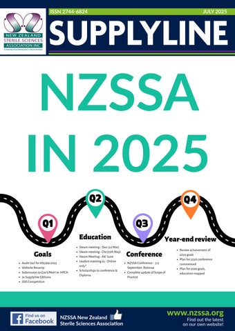
NZSSA IN 2025
NZSSA IN 2025 Q2
Q4
Q3
Q1 Education Goals Audit tool for AS5369:2023 Website Revamp Submission to Gov’t/MoH re: HPCA 3x Supplyline Editions SSD Competition
Find us on Facebook Badge
Steam meeting - Dun (22 Mar) Steam meeting - Chc(25th May) Steam Meeting - Akl June Leaders meeting x2 - Online only? Scholarships to conference & Diploma
Year-end review Conference NZSSA Conference - 3-5 September, Rotorua Complete update of Scope of Practice
Review achievement of 2025 goals Plan for 2026 conference commenced Plan for 2026 goals, education mapped
CMYK / .ai
NZSSA New Zealand the journal of NEW ZEALAND STERILE SCIENCES ASSOCIATION INC Sterile Sciences Association
www.nzssa.org
S U PonPFind L Y out L I NtheE latest 1 our own website!
Business Card Directory A QUICK AND HANDY DIRECTORY OF OUR STERILE SERVICES SUPPLIERS
Lon Hua Account Manager Infection Control & Life Science
Customer Service Phone: 0800 257 352 PO Box 22-519, Otahuhu 1640, Auckland www.whiteley.co.nz
Annette Moffatt
New Zealand
Getinge Australia Pty Ltd Level 2, Building B Millenium Centre 600 Great South Road Ellerslie Auckland New Zealand 1051
Mobile +64 21 438 827 Toll free 0800 143 864
Account Manager 021 587 031 annette.moffatt@opritech.co.nz Customer Care 0800 32 40 32
lon.hua@getinge.com www.getinge.com/anz • NZBN: 9429032644500
Neil Maclennan Customer Service
opritech.co.nz
Follow us:
Neil Maclennan
Medical Products Manager
Medical Products Manager
M: +64 21 499 101 E: nmaclennan @ crknz.co.nz
P: +64 9 276 3271 M: +64 21 499 101 E: nmaclennan @ crknz.co.nz
3 Hotunui Drive Mount Wellington, Auckland P: +64 9 276 3271 F: +64 9 276 9645 www.crkennedy.co.nz
3 Hotunui Drive Mount Wellington, Auckland P: +64 9 276 3271 F: +64 9 276 9645 www.crkennedy.co.nz
Phone: 0800 257 352 PO Box 22-519, Otahuhu 1640, Auckland P: +64 9 276 3271 www.whiteley.co.nz
Aidan Kersley Director
JAMES HENDERSON 027 543 2266 | jhenderson@nzvalidation.co.nz 615 Peacockes Road, RD 2, Tamahere, Hamilton nzvalidation.co.nz
m 021 0221 4984 | p 0800 224 224 e aidan@sterilesolutions.co.nz e office@sterilesolutions.co.nz
Daria Nikolaeva
Steve Tarring
National Sales Manager
Managing Director
40 Naples Street, PO Box 64 Martinborough, 5741. New Zealand
40 Naples Street, PO Box 64 Martinborough, 5741. New Zealand
T: 0800.224.426 M: 027.385.9966 E: daria.nikolaeva@micronz.co.nz
T: 0800.224.426 M: 021.952.082 E: steve.tarring@micronz.co.nz
Rebecca Heney
Richard Steingold
SALES MANAGER - NEW ZEALAND HEALTHCARE DIVISION ANZ rebecca.heney@ecolab.com T 0800 425 529 M +64 21 335 925 W healthcare-nz.ecolab.com
BUSINESS DEVELOPMENT MANAGER HEALTHCARE DIVISION ANZ richard.steingold@ecolab.com
261 Annex Road, Middleton, Christchurch 8024
T 0800 425 529 M +64 21 0816 2290 W healthcare-nz.ecolab.com
7A Pacific Rise, Mt Wellington, Auckland 1060
INTERMED MEDICAL LIMITED
Geneviève Boyer BHSc (Nursing) RCN
Product Specialist
71 Apollo Drive, Albany, Auckland 0632 T 0800 333 444 W www.intermed.co.nz E genevieve.boyer@intermed.co.nz
Craigs Design & Print
021 719 712
Editor’s Message July 2025 Kiaora, Welcome to the July 2025 edition of Supplyline. Half way through the year and winter is well and truly here. The months have just flown by this year. In this edition there are updates on this year’s conference and information around the review of the NZSSA Constitution and Rules. Our Constitution and Rules need to be updated so that we can apply to reregister with New Zealand Companies. This all has to be done and in place by April 2026. The organising for this year’s conference is well underway. Venue has been booked in Rotorua, the speakers are booked and the program is already available for people to view. Well done to the conference committee, they have done an amazing amount of work to have the program all sorted. Donna and I would really like to see more information from you, our members, to be published in Supplyline. So send us
President’s Message Kiaora I hope you’re keeping warm and dry during these colder winter months. MEETING WITH CATHY CUMMINGS FROM DAA Shelagh Thomas attended a meeting with Cathy Cummings from DAA on 11 June 2025. The purpose of the meeting was to explore how the NZSSA and DAA could collaborate more closely moving forward. They had an in-depth discussion about the Standard, with a particular focus on Quality Management Systems and the use of Auditing or Gap Analysis Tools. Following a further conversation with Victoria, she has agreed to establish a reference group to explore the auditing requirements in Sterile Services in more detail. NZSSA representation on this group will include myself and Shelagh, and we will attend the upcoming meeting. PROFESSIONAL LEADERS MEETING Aileen attended the Professional Leaders meeting with Jacqui Lunday (National Chief Allied Health Scientific & Technical) yesterday. Interesting meeting. Some of the Chiefs Allied Health throughout NZ have been assigned specific portfolios. Some are related to Health NZ’s targets. the journal of NEW ZEALAND STERILE SCIENCES ASSOCIATION INC
a photo of your team or your unit or both. Or let us know of any upgrades, or changes that have happened in your unit. We would be happy to include your information in Supplyline. Ngā mihi Aileen Derby Editor NZSSA Supplyline
Another edition of Supply line means another opportunity for the NZSSA team to reach out to all of you through this publication, ensuring you are well updated on the happenings in our profession and Sterile Sciences community. In this edition, you will also find announcements about the exciting contest we are currently running. We would love to see all your applications! I’m sure your hospital will be proud! See you at the Conference. Donna Dador National Sterile Service Adviser Southern Cross Healthcare
Hannah McCarrison, Chief Allied Health, Scientific and Technical Officer has been assigned a portfolio that relates to the different groups that make up Allied Health, Scientific and Technical. She is interested in hearing from any groups to look at what support we need, how we work together with other groups and would like to see our Terms of Reference. Jacqui is going to send out information from yesterday’s meeting which I’ll forward onto you when I receive it. CONFERENCE – ROTORUA, SEPTEMBER 2025 Jenny, John, Charanjeet, Britta, and I have been working hard to organise the upcoming conference, and everything is progressing well. Our keynote speaker, Hank Balch, has already generated significant interest. So far, attendee numbers are up compared to last year, and the calibre of speakers is strong, with a range of engaging and relevant topics. Just a quick reminder that early bird registrations close on 31 July 2025 – don’t miss out! We held our first leaders’ meeting on 30 June 2025. Our guest speaker was Ross Hanson from HR, who covered a range of topics, including sick leave management and strategies for dealing with difficult staff. Next Leaders meeting is October 17th, 2025 See you all at the NZSSA conference Martin Bird President NZSSA
S U P P LY L I N E
3
Join us for the 49th Annual Conference of the New Zealand Sterile Sciences Association. This year our theme is entitled: 'Growing a Professional Profession' This year’s theme highlights the ongoing development, innovation, and collaboration needed to elevate industry standards and strengthen our community. The trade exhibition will be showcasing their product range and consumables and offer great learning opportunities. Rotorua offers a fantastic setting for networking with colleagues and gaining insights from industry experts. Scan the QR Code and Register Now! Earlybird rate finishes on Thursday 10 July 2025
For any conference queries, please contact: Britta van Uden, Event Manager, Britta@mtanz.org.nz
4
S U P P LY L I N E
SUPPLYLINE – JULY 2025
the journal of NEW ZEALAND STERILE SCIENCES ASSOCIATION INC
S U P P LY L I N E
5
Scan to Download the Power of Plasma Whitepaper
Join us
O H
H O
O
O
O
H
H
Plasma Ignites and Dissociates the H2O2
H
H
H
H
H O H O
O
O
O
Recombines as water (H2O) and oxygen (O2)
O
O
Research has demonstrated that HPGP systems offer a higher Margin of Safety compared to VHP,particularly in the presence of challenging contaminants such as salt and blood plasma.
Hydrogen Peroxide (H2O2) Enters Chamber
H
O
H
Higher Cycle Concentrations of HP: Plasma allows for higher cycle concentrations of HP, increasing the Margin of Safety compared to VHP platforms. Rutala’s study showed a 76.3% sterilization failure rate for VHP versus only a 1.9% failure rate for HPGP under conditions of salt and serum residual. This represents an over four-fold advantage in Margin of Safety under conditions where cleaning is not complete prior to sterilization.
Reduced HP Vapor Release: Plasma breaks down HP into benign components (H2O and O2), preventing a burst of HP vapor when opening the sterilization chamber door and subsequent release into the environment.
Quick Breakdown of Residual HP: Plasma technology enables the rapid breakdown of residual HP, whereas VHP without plasma technology has a gradual release.
Benefits of Plasma Technology
for Effective Low-Temperature Sterilization
The Power of Plasma Website
A brief review of ISO 15883-5: 2021 and cleaning validation acceptance criteria. Author: Dr Trevor Glasbey | Chief Scientist at Whiteley Cleaning validation in healthcare settings plays a critical role in ensuring the safety and effectiveness of medical devices processed through washer-disinfectors. As clinical practices evolve and regulatory expectations heighten, the standards governing automated cleaning processes must also adapt. ISO 15883 is the international benchmark for washer-disinfector performance, providing guidance on cleaning, disinfection, and validation requirements. This article focuses on the 2021 revision of ISO 15883-5, which introduces significant changes in the approach to test soils and establishes clear, quantitative acceptance criteria for cleaning efficacy. The ISO 15883 series of standards relating to washer disinfectors currently have 7 parts. Of these, most have revised versions published since 2021. The two exceptions are the Part 6 document which is expected to publish very shortly, and the Part 4 document, published in 2018, for which work has recently commenced on its revision. The titular Part 5 document, ISO 15883-5: 2021 – “Washer-disinfectors – Part 5: Performance requirements and test method criteria for demonstrating cleaning efficacy” has undergone the most significant revision, the main features of which are presented here. This document replaces the former technical specification, ISO/TS 15883-5:2005 – Washer-disinfectors – Part 5: “Test soils and methods for demonstrating cleaning
efficacy” which contained a compendium of various national soils to be used for demonstrating cleaning efficacy across the range of device types. Rather than publishing a list of test soils, the new standard leaves the choice of test soil and soiling method to the user, however these choices must be justified and documented. The development of new test soils is also permissible. 4.2.1 The rationale for the choice of test soil and soiling method shall be justified and documented. Test soil formulations may be chosen or developed based on a review of the literature and demonstration of its relevance based on the use of the devices in clinical practice, 4.2.2 The test soil must also conform to the performance criteria specified in Annex B (of the new standard). CLEANING EFFICACY Of note here are the acceptance criteria for quantifiable tests may be specified in terms of both an alert level and an action level. Action levels are the maximum criteria for acceptable cleaning efficacy during testing, but the desired criteria are given as the alert levels. If values are detected between the alert and action levels they shall be investigated and considered to pass cleaning requirements if justified. The alert and action level for the suggested analytes are given in the table below
Acceptance criteria Analyte
Alert level
Action level
Protein
≥ 3 µg/cm2
≥ 6 µg/cm2
Total organic carbon (TOC)
≥ 6 µg/cm2
≥ 12 µg/cm2
Carbohydrate
≥ 0.9 µg/cm2
≥ 1.8 µg/cm2
Haemoglobin
≥ 1 µg/cm2
≥ 2.2 µg/cm2
ATP
≥ 10 femtomoles ATP/cm2
≥ 22 femtomoles ATP/cm2
Endotoxin
≥ 2.2 EU/cm2
≥ 20 EU/cm2
In conclusion, the revision of the part 5 document now provides quantitative acceptance criteria for cleaning efficacy. The use of visual assessment is still permissible but should be reserved for non-invasive devices, such as those that do not penetrate inside the body, either through a body orifice or through the surface of the body, might only require visual examination.
6
S U P P LY L I N E
Some non-invasive devices can represent higher levels of risk, eg. infant formula bottles or contact tonometers, perhaps requiring a more quantitative approach. Reference is made to ISO 14971:2007 for an approach to risk assessment to support justifications. For further details on how these updates may impact your cleaning validation processes, please contact your Whiteley representative on 0800 257 352 or email whiteley@whiteley.co.nz
SUPPLYLINE – JULY 2025
Retirement Acknowledgement – Auckland CSSD KATHY MOKOTUPU – Sterilising Technician
JANET MACDONALD – Role(s) Title – Quality Lead
Kathy Mokotupu began her journey at Te Toka Tumai in February 1974, dedicating an extraordinary 51 years of service in SSS - truly a remarkable length of service. During her time in SSS, Kathy held the role of a Shift Coordinator and Sterilising Technician. Kathy worked in various areas within our service but respiratory was her favourite and she was a steadfast advocate for this area. Kathy was a caring and supportive team member, always quick to share her food, ensuring everyone got their breaks, and would offer help when others were busy. She looked out for her teammates, speaking up when needed and sharing her knowledge—especially in respiratory, offering valuable tips and tricks to improve processing efficiency.
Janet MacDonald joined Te Toka Tumai in Oct 2007, almost 18 years of outstanding service. Janet started as a Sterilising Technician before stepping into the vital role of Quality Lead, where she played a key part in maintaining our Quality Management System (QMS) and upholding our ISO accreditation. Even as she prepares to retire at the end of May, Janet continues to support the team in working towards another successful certification later this year. She is passionate about quality and tries her absolute best to improve departmental practices and processes. She is always willing to help, especially by supporting technicians with their studies, assignments, and understanding of QMS.
MINNIE HAIMAU – Role(s) Title – Area Lead Minnie Haimau started at Te Toka Tumai in April 1986, giving an incredible 39 years of service. Minnie held various positions with SSS over years, she served as a Sterilising Technician, Shift Coordinator, and as an Area Lead, overseeing operations in the decontamination area. Minnie was known for many wonderful things one of which was her commitment to bringing people together. She would often organise shared lunches, Christmas celebrations and sometimes putting on a feast for the entire team at her own expense. She played an active role with collections for the employee support centre, offering immense help to staff during COVID by providing meals - often delivering them herself and would bring in a car boot full of bread for staff to enjoy. This generous gesture speaks volumes about the kind and thoughtful person she is, and she will be deeply missed for the motherly figure she was to many.
We want to take this moment to express our heartfelt thanks and gratitude to Kathy, Minnie, and Janet, for their unwavering commitment, hard work, and the countless contributions made over the many years of service at Te Toka Tumai. Their leadership, friendship, and presence will be deeply missed, and we wish them all the best with their well-deserved retirement.
Janet MacDonald, Quality Lead, Auckland Sterile Sciences
the journal of NEW ZEALAND STERILE SCIENCES ASSOCIATION INC
S U P P LY L I N E
7
pH
8
S U P P LY L I N E
SUPPLYLINE – JULY 2025
Heliyon 10 (2024) e37281
Contents lists available at ScienceDirect
Heliyon journal homepage: www.cell.com/heliyon
Research article
Microbiological evaluation of ultraviolet C light-emitting diodes for disinfection of medical instruments Hannah Siwe a, b , Annelies Aerssens c , Mieke V. Flour b , Silke Ternest a, c , Leen Van Simaey d , Duncan Verstraeten b , Alain F. Kalmar e , Isabel LerouxRoels c, f, 1, * , Philip Meuleman a, 1, ** , Piet Cools d, 1, *** a
Laboratory of Liver Infectious Diseases, Department of Diagnostic Sciences, Faculty of Medicine and Health Sciences, Ghent University, Corneel Heymanslaan 10, entrance 38, Medical Research Building 2, 9000, Ghent, Belgium Research and Development, eLEDricity, Hundelgemsesteenweg 446A, 9820, Merelbeke, Belgium c Department of Infection Control, Ghent University Hospital, Corneel Heymanslaan 10, entrance 12, Clinical Building 12E, 9000, Ghent, Belgium d Laboratory Bacteriology Research, Department of Diagnostic Sciences, Faculty of Medicine and Health Sciences, Ghent University, Corneel Heymanslaan 10, entrance 38, Medical Research Building 2, 9000, Ghent, Belgium e Department of Electronics and Information Systems, IBiTech, Ghent University, Technologiepark-Zwijnaarde 126, 9052, Ghent, Belgium f Department of Diagnostic Sciences, Faculty of Medicine and Health Sciences, Ghent University, Corneel Heymanslaan 10, entrance 38, Medical Research Building 2, 9000, Ghent, Belgium b
A R T I C L E I N F O
A B S T R A C T
Keywords: UVC LED Sustainability Staphylococcus aureus Reprocessing
Background: Despite the many guidelines for reprocessing of medical instruments, challenges persist such as microbial resistance to biocides, corrosive effects on materials, and timeconsuming reprocessing procedures. Ultraviolet (UV) C light-emitting diode (LED) chambers might provide a solution but the integration in healthcare is still in its infancy. Here, we evaluated the efficacy of a novel ZAPARAY™ UVC LED chamber as a time and energy-efficient alternative for reprocessing of medical instruments for which current disinfection protocols exhibit limitations. Methods: We verified the disinfection efficacy of the UVC LED chamber on a Petri dish and contaminated several medical devices with Staphylococcus aureus ATCC 25923. The bacterial reduction was assessed after 5 min of UVC LED exposure. Additionally, we investigated the impact of rinsing before UVC exposure. Results: We demonstrated a bacterial reduction of 9 log10 on a Petri dish. Non-rinsed dental tools exhibited varied reduction levels ranging from a 3.23 log10 to a 6.25 log10 reduction. Rinsing alone yielded an average reduction of 2.7 log10 and additional UVC exposure further reduced the bacterial load by an average of 3.65 log10. We showed an average 4.90 log10 reduction on thermistors, 2 log10 or less on orthodontic pliers, and no reduction on handpieces. Conclusions: This study demonstrates that UVC LED chambers may be used as a standardized substitute for specific (manual) disinfection procedures of certain medical devices, offering a
* Corresponding author. ** Corresponding author. *** Corresponding author. E-mail addresses: Isabel.LerouxRoels@UZGent.be (I. Leroux-Roels), Philip.Meuleman@UGent.be (P. Meuleman), Piet.Cools@UGent.be (P. Cools). 1 Shared senior authors with equal contribution. https://doi.org/10.1016/j.heliyon.2024.e37281 Received 6 May 2024; Received in revised form 28 August 2024; Accepted 30 August 2024 Available online 2 September 2024 2405-8440/© 2024 The Authors. Published by Elsevier Ltd. This is an open access article under the CC BY-NC-ND license (http://creativecommons.org/licenses/by-nc-nd/4.0/).
the journal of NEW ZEALAND STERILE SCIENCES ASSOCIATION INC
S U P P LY L I N E
9
Heliyon 10 (2024) e37281
H. Siwe et al.
time-efficient and more sustainable alternative. However, its use should be preceded by efficacy testing for each specific type of instrument.
1. Introduction Correct reprocessing of reusable medical devices is an essential cornerstone of a good infection prevention and control program in healthcare facilities [1]. Reprocessing entails cleaning, disinfection and/or sterilization procedures to ensure the safe reuse of medical instruments [2]. This can be obtained through (the combination of) a variety of physical and chemical methods that include the use of detergents, oxidizing agents, aldehydes, radiation, and/or heat [3,4]. Spaulding’s classification is a commonly used system to assess which procedure is required for adequate reprocessing [4]. This classification defines non-critical (in contact with intact skin), semi-critical (in contact with non-intact skin or mucosae), or critical instruments (in contact with blood or sterile tissues), with the latter posing the highest risk for infection [3]. Despite the many reprocessing guidelines and protocols, device-associated outbreaks have been increasingly reported [5–7] and several challenges and limitations remain. Critical devices (e.g. surgical instruments) and some semi-critical devices, such as heat-sensitive flexible endoscopes, undergo a largely automated process of sterilization or high-level disinfection (HLD) [3] but these procedures are often time-consuming. Many non- and semi-critical medical instruments still undergo (semi-) manual reprocessing, but these manual procedures are not standardized and therefore highly error-prone, particularly when it concerns devices with a complex design [8–11]. The use of chemical disinfectants is associated with a high environmental burden [12,13], and the risk of inducing or increasing microbial resistance to biocides and cross-resistance to antibiotics [14,15]. The recommended dwell time of disinfectants may also be compromised under time pressure [16]. Furthermore, corrosive effects on instruments have been reported as a result of heat sterilization or chemical disinfection [17,18]. Disinfection by ultraviolet (UV) C generated by mercury discharge lamps is well established in the context of environmental disinfection [19–21] and might provide an answer to some of these challenges. UVC inactivates microorganisms either by direct absorption or indirectly via reactive oxygen species, targeting their DNA, RNA, proteins, and lipids [22–24]. However, there are more energy-efficient alternatives available including UVC light-emitting diodes (LEDs). These LEDs offer various other advantages such as being more compact and eco-friendly as they are free of toxic mercury [25,26]. Nevertheless, UVC LEDs are at present not widely used which could be as a result of their lower irradiance efficiency as compared to mercury discharge lamps [26]. However, UVC LEDs remain highly effective in deactivating pathogens and the technology is continuously evolving, as forecasted by Haitz’s law [27]. Ongoing research aims to further enhance their efficiency [28], indicating a promising future for UVC LEDs in various applications. The implementation of UVC chambers for instrument disinfection in healthcare is still in an early stage due to several limitations that need to be considered such as the low quality of available research, the lack of standardization for UVC chambers, the low penetration depth of UVC, and the necessity for optimal exposure of the instrument without shadow formation [29]. UVC mercury chambers have been researched and shown to be effective for disinfection of electronic devices such as tablets [30,31] used in hospitals, and to some extent on medical devices including intravaginal probes [32–34], flexible fiberoptic laryngoscopes [35] and silicon dental impressions [36]. UVC LED has been investigated for stethoscope disinfection, revealing its potential for effective decontamination of these medical devices [37]. To the best of our knowledge, the efficacy and broad applicability of UVC LED chambers for instrument disinfection have not been thoroughly investigated, highlighting the need for further research to ensure their correct and optimal implementation. We aimed to evaluate the efficacy of a novel UVC LED chamber for disinfection of non- or semi-critical instruments, particularly those instruments for which current disinfection protocols exhibit limitations. In this study, we focused on how the shape and technical characteristics of the medical instruments influence the disinfection efficacy, using Staphylococcus aureus as a clinically relevant model pathogen. 2. Methods 2.1. Identification and selection of instruments As an initial market survey, open interviews were held with potential end-users in health care (e.g. head nurses, dentists …), which led to the selection of dental tools (hammer shaped needle, sawtooth needle, and chisel needle) as proof of concept for microbiological testing (Table 1). At 14 departments of the Ghent University Hospital, semi-structured interviews, and questionnaires (Supplementary Figure A1) were used to identify those instruments for which there is a high need for an alternative disinfection strategy. In total, 29 identified instruments were given a high, medium, or low score for six parameters (i.e. correct implementation of current protocol, impact of current protocol on lifespan of instrument, processing time of current protocol, use frequency, cost of instrument and risk of a healthcare-associated infection (HAI)) (Supplementary Tables A1 and A2) and plotted in two heatmaps displaying instrument clusters sharing similar characteristics (Supplementary Figure A2). Based on feasibility and accessibility, a subset of instruments were selected for microbiological testing, more specifically, a nasal/oral thermistor (sensor) (REF: P1222, Pro-Tech, Downers Grove, Illinois, USA), a dental handpiece (REF: SYNEA WA-99 LT, W&H Dentalwerk, Bürmoos, Austria), a round ended orthodontic plier (REF: 4151-640, Masel Orthodontics, Carlsbad, California, USA), and a sharp ended orthodontic plier (REF: 678-106, Hu-Friedy, Chicago, Illinois, USA) (Table 1).
2
10
S U P P LY L I N E
SUPPLYLINE – JULY 2025
Heliyon 10 (2024) e37281
H. Siwe et al.
2.2. UVC LED device The UVC LED disinfection device evaluated in this study was the ZAPARAY™ RAY-ONE prototype 0/102 (eLEDricity, Merelbeke, Belgium) and further designated in this manuscript as “UVC LED chamber”. The device consists of an enclosed chamber with a drawer made of a quartz bottom in which objects can be placed with a maximum size of 200 mm × 300 mm x 70 mm (width x depth x height). The chamber and drawer are lined with UVC reflective material. This reflective material, along with LEDs mounted at the top and bottom of the chamber that emit UVC radiation at 275 nm, ensure homogenous UVC exposure with an irradiance of approximately 0,395 mW/cm2. 2.3. Contamination Staphylococcus aureus (ATCC 25923) was aerobically cultured at 37 ◦ C for 24 h on tryptic soy agar with 5 % sheep blood (TSA-5% SB) (BD, Franklin Lakes, New Jersey, USA). For each experiment, an inoculum suspension was freshly prepared by resuspending S. aureus colonies in 0.9 % sterile NaCl solution (saline). The optical density (OD) at 600 nm was measured using the Denovix DS-11 spectrophotometer (Denovix, Wilmington, USA) with the OD ranging between 1,0 and 1,7 to obtain a concentration of approximately 109 colony forming unit (CFU)/mL. As initial verification and reference, a 10 μL droplet of the inoculum suspension was pipetted onto the center of a sterile 60 × 15 mm polystyrene Petri dish and then immediately exposed to UVC. Dental tools, orthodontic pliers and handpieces were steam sterilized before the start of each experiment and thermistors were decontaminated with 70 % ethanol. Dental tools were contaminated by applying the inoculum through partial submersion in 7 mL of inoculum suspension in a 15 mL tube. Orthodontic pliers were partially submerged in 80 mL inoculum suspension in a 100 mL multipurpose beaker. Handpieces were contaminated through partial submersion of the instrument in 10 mL of inoculum suspension in a 50 mL tube. All instruments were incubated for 10 min at room temperature. Afterward, a tapping motion was applied to the instruments to remove the excess liquid by tapping the instrument four times against the border of its container, used during contamination. Thermistors were contaminated around the three sensor tips by rubbing a cotton swab that had been soaked with inoculum in a circular motion around the tips. To investigate whether rinsing before UVC exposure would improve the outcome of the disinfection procedure; dental tools, handpieces, and orthodontic pliers, were rinsed for 3 s under a jet of water as a simplified cleaning step. Pliers were held in an open orientation during rinsing for water to flow through the hinge. Afterward, instruments were dapped on tissue paper to remove excess liquid. All instruments were exposed to UVC immediately after the contamination procedure without allowing the instruments to dry up. 2.4. UVC disinfection Petri dishes and medical devices were positioned directly in the center of the drawer, except for dental tools which were positioned in a fixture for stability. Orthodontic pliers were placed in an open orientation. Petri dishes and medical devices were subjected to a 5min disinfection cycle which equated to a UVC dose of approximately 118 mJ/cm2. Contaminated Petri dishes and instruments that did not undergo UVC treatment were handled in parallel and served as an untreated control. 2.5. Assessment of log reduction factors 2.5.1. Bacterial recovery The 10 μL droplet in the Petri dish was collected by locally rinsing the area of the droplet with 1 mL of saline which was then added to 9 mL saline and resuspended. Afterward, it was locally rinsed for a second time with 1 mL originating from the already collected volume to apply additional friction for the collection of residual material on the surface of the Petri dish. Bacteria on dental tools were recovered by partial submersion in 7 mL saline in a 15 mL tube. Handpieces were placed in a 50 mL tube containing 10 mL saline. All tubes with objects inside were vortexed for 20 s. Orthodontic pliers were placed in an open orientation inside a 100 mL multipurpose beaker and rinsed five times with the same 10 mL saline. Thermistors were positioned in a fixture above a 60 × 15 mm polystyrene sterile Petri dish. Subsequently, the three sensor tips were each rinsed with 1 mL saline. Finally, all rinse fluids were collected in a 15 mL tube and vortexed. 2.5.2. Bacterial enumeration The bacterial load was quantified by performing a tenfold serial dilution of the collected suspension. A droplet of 5 μL was pipetted onto TSA-5%SB for each dilution, and afterward, plates were slanted vertically at a nearly 90◦ angle to create a droplet line. Subsequently, plates were incubated at 37 ◦ C for 18–24 h and colonies were manually counted. The dilution with the highest countable value within the range of 0–100 CFU was used to calculate the bacterial concentration of the original suspension. The log reduction was calculated as followed: log10 reduction factor = log10 (CFU/mL untreated) – log10 (CFU/mL treated)
3
the journal of NEW ZEALAND STERILE SCIENCES ASSOCIATION INC
S U P P LY L I N E
11
Table 1 Medical devices. dental tools sawtooth
chisel
orthodon
round-en
H. Siwe et al.
hammer-shaped
handpiece
H. Siwe et al.
4 handpiece chisel
orthodontic pliers round-ended
orthodontic pliers round-ended
oral/nasal thermistor sharp-ended
oral/nasal thermistor sharp-ended
Heliyon 10 (2024) e37281
S U P P LY L I N E
SUPPLYLINE – JULY 2025
Heliy
12
Heliyon 10 (2024) e37281
H. Siwe et al.
2.6. Confirmation of the absence of bacterial growth after UVC exposure To confirm the lack of viable bacteria in the absence of growth with the droplet line technique, a 10 μL droplet was spotted on a Petri dish and treated as previously described. The droplet was then collected by locally rinsing the area with 90 μL saline. This total volume of 100 μL was plated onto TSA-5%SB according to the spread plate technique. Similarly, thermistors and dental tools were prepared and handled as previously described and after bacterial recovery, the instruments were directly imprinted onto TSA-5%SB. 2.7. Statistical analysis Statistical analysis was performed using GraphPad Prism version 8.0.1. Bar charts in all figures were depicted as mean ± standard deviation with dots representing individual data points. Data was verified for normal distribution using the Shapiro Wilk test. A paired t-test was used when the data was normally distributed, otherwise, the Wilcoxon matched-pairs signed rank test was used. Statistical significance was expressed as * (p < 0.05), ** (p < 0.01), *** (p < 0.001), **** (p < 0.0001), or ns (non-significant). 3. Results 3.1. Verification of the UVC LED chamber as a disinfection device Before we initiated the evaluation of the device to disinfect medical instruments, we wanted to assess its usefulness in a disinfection experiment using Petri dishes as a simple surface. Collected suspensions from the untreated control (n = 14) had an average bacterial concentration of 1.02 × 10⁹ CFU/mL (Fig. 1). Exposure of the droplet to a 5-min disinfection cycle resulted in a complete reduction below detectable levels across all experiments (Fig. 1), with an average log10 reduction factor of 9. The absence of growth was confirmed when the treated droplet was spread-plated (Supplementary Figure A3). 3.2. Evaluation of the efficacy of the UVC LED chamber for disinfection of non-rinsed medical instruments To evaluate the efficacy of the device for disinfection of medical instruments, various identified instruments were artificially contaminated with S. aureus and exposed to UVC. The bacterial load of the collected suspensions of the different dental tools and pliers was highly comparable for the untreated conditions, resulting in an average concentration of 106 CFU/mL (Fig. 2A). UVC exposure yielded significantly lower bacterial concentrations for all three dental tools, with a complete reduction for the chisel needle (Fig. 2A). Remarkably, for the sawtooth needle results varied from partial to complete reduction across different experiments. The average log10 reduction factor was 3.23, 5.18 and 6.25 for the hammer-shaped needle, sawtooth needle, and chisel needle, respectively (Fig. 2B). Pliers showed a significant, but lower reduction of the bacterial load after UVC exposure with a log10 reduction factor of 2 or less (Fig. 2B). UVC exposure of handpieces did not result in any bacterial reduction whereas thermistors showed the absence of growth after exposure, resulting in an average log10 reduction factor of 4.90 (Fig. 2A and B). Additionally, the agar imprinting method confirmed the absence of bacterial growth on the UVC treated thermistors (Supplementary Fig. A4). However, a few colonies were observed for the treated chisel needle as compared to the dense growth of S. aureus for the untreated control (Supplementary Fig. A4).
Fig. 1. Bacterial load of a contaminated Petri dish without UVC exposure (untreated) and after 5 min of UVC exposure (treated). Log-transformed colony forming units (CFU)/mL of S. aureus under control and treatment conditions. Dots represent individual data points. Bar charts are shown as mean ± standard deviation (n = 14 treated and 14 untreated) (14 independent experiments), *** (p < 0.001) (Wilcoxon matched-pairs signed rank test). 5
the journal of NEW ZEALAND STERILE SCIENCES ASSOCIATION INC
S U P P LY L I N E
13
Heliyon 10 (2024) e37281
H. Siwe et al.
Fig. 2. Bacterial load and log reduction factors of non-rinsed medical instruments. Dots represent individual data points. Bar charts are shown as mean ± standard deviation (SD). (A) Log-transformed colony forming units (CFU)/mL of S. aureus under control and treatment conditions collected from dental tools (n = 6) (6 independent experiments), round and sharp ended orthodontic pliers (n = 3) (3 independent experiments), handpieces (n = 4) (2 independent experiments), and thermistor (n = 4) (2 independent experiments). * (p < 0.05), **p < 0.01), *** (p < 0.001), **** (p < 0.0001), ns (non-significant) (paired t-test except for data on sharp ended plier and sawtooth, Wilcoxon matched-pairs signed rank test) (B) Log10 reduction factors for the different medical instruments.
6
14
S U P P LY L I N E
SUPPLYLINE – JULY 2025
Heliyon 10 (2024) e37281
H. Siwe et al.
3.3. Evaluation of the efficacy of the UVC LED chamber for disinfection of rinsed medical instruments To evaluate whether rinsing improves the overall disinfection procedure for those instruments where complete reduction was not obtained, a short rinsing step was included before UVC treatment. Rinsed untreated controls showed on average 2.7 and 1.3 log10 lower bacterial loads (CFU/mL) for dental tools and pliers, respectively, compared to unrinsed counterparts (Fig. 3A and 2A). Rinsing followed by UVC exposure resulted in undetectable growth for the hammer shaped needle, sawtooth needle, and chisel needle with an average log10 reduction factor of 3.68, 4.02, and 3.25 respectively, resulting in an overall average log10 reduction factor of 3.65 (Fig. 3B). However, no rinsing effect was observed for pliers and handpieces for the treatment conditions (Fig. 3A and 2A). This resulted in an average log10 reduction factor of less than 1 for pliers and no reduction for handpieces (Fig. 3B). Instrument imprinted plates of the rinsed dental tools showed a few colonies for all treated objects as compared to untreated objects (Supplementary Figure A5). 4. Discussion In this study, we evaluated the performance of the novel ZAPARAY™ UVC LED chamber for the disinfection of artificially contaminated simple surfaces (i.e. Petri dishes) as well as of complex medical instruments. First, we have shown that the device was able to induce complete reduction (9 log10 reduction) of S. aureus on a standard Petri dish that has a smooth, flat, non-porous surface without any cavities. The results obtained on medical devices varied considerably depending on the instrument. In literature, research has been described where instruments were artificially contaminated and disinfected with UVC [38,39]. However, these studies employed limited contamination through several inoculated sites of 1 cm2 across the device, including a blood pressure gauge and N95 respirators. Post-exposure, these areas were sampled with a pre-moistened cotton swab and placed in a solution for bacterial recovery, or in the case of respirators, the areas were cut out. Another study evenly distributed a small inoculum onto the membranes of stethoscopes which were placed onto plate count agar for bacterial recovery [37]. These approaches are unfeasible to demonstrate the efficacy of a UVC chamber for medical devices that are more complex in shape or technical design. Therefore, this study employed a different
Fig. 3. Bacterial load and log reduction factors of rinsed medical instruments. Dots represent individual data points. Bar charts are shown as mean ± standard deviation (SD). (A) Log-transformed colony forming units (CFU)/mL of S. aureus under control and treatment conditions collected from dental tools (n = 3 or 4) (4 independent experiments), round and sharp ended orthodontic pliers (n = 3) (3 independent experiments), handpieces (n = 4) (2 independent experiments). * (p < 0.05), ** (p < 0.01), *** (p < 0.001), ns (non-significant) (paired t-test except for data on handpieces, Wilcoxon matched-pairs signed rank test) (B) Log10 reduction factors for the different medical instruments. 7
the journal of NEW ZEALAND STERILE SCIENCES ASSOCIATION INC
S U P P LY L I N E
15
Heliyon 10 (2024) e37281
H. Siwe et al.
methodology through partial submersion of the devices (with exception of the thermistors), thereby enabling a more accurate representation of natural contamination. Studies using UVC mercury chambers for disinfection of devices naturally contaminated during routine use, have been described for some medical devices such as electronic devices [30,31], intravaginal probes [32–34] and flexible fiberoptic laryngoscopes [35]. This approach could result in a more thorough understanding of the effectiveness as well as the practical implementation of the UVC chamber in daily practice as compared to artificial contamination in laboratory setting. However, the complexity of this study design may render on-site studies for each potential instrument of interest impractical and may not always be necessary depending on the result obtained on an artificially contaminated instrument. The instruments subjected to UVC LED disinfection in our study were semi-critical devices that require at least HLD [3]. For this reason, the devices (with exception of the thermistors) were contaminated with a high inoculum, such that upon disinfection a log10 reduction factor of 6 could be demonstrated, which is the endpoint for HLD recommended by the Food and Drug Administration [40]. Using different types of dental tools, all having the same rod-like structure but with variable ends, we demonstrated that the efficacy of the UVC LED chamber for disinfection of medical instruments was influenced by their technical and/or structural characteristics as reduction levels varied from a 3 log10 to 6 log10 reduction for the non-rinsed dental tools. For instance, UVC disinfection of the chisel needle, the dental tool with the lowest complexity, consistently resulted in undetectable growth, averaging a reduction of 6.25 log10 with the droplet line technique. In contrast, the sawtooth needle and hammer shaped needle demonstrated an average reduction of 5.18 log10 and 3.23 log10, respectively. The conflicting results of the sawtooth needle could potentially be explained by the different volumes of inoculum remaining between the ridges after excess droplet removal. Rinsing for 3 s under a stream of water as a simplified cleaning process reduced the bacterial load by 2.7 log10 and improved the final outcome of the disinfection procedure for all dental tools. Bacterial growth remained undetected with the droplet line technique for all dental tools. However, a few colonies were still observed when the rinsed and treated instruments were imprinted onto agar. Thorough cleaning before HLD or sterilization is required to ensure the subsequent technique is effective at inactivating microorganisms [3]. The incorporation of a more thorough cleaning method may further improve the outcome, and when combined with UVC LED, may offer an effective disinfection alternative. UVC was unable to decontaminate handpieces, both in the rinsed and non-rinsed conditions. These instruments are complex in nature due to technical characteristics and therefore require a multistep decontamination process [41,42]. This complexity likely prevented UVC from accessing the internal part of the device, leading to the absence of bacterial reduction. However, this does not negate the possibility of the use of a UVC chamber as an alternative disinfection step for the exterior of the device prior to lubrication and HLD or sterilization, as the exterior is commonly cleaned/disinfected through a manual procedure involving a disinfectant cloth [43]. A similar trend was observed with the orthodontic pliers, where only partial reduction (2 log10 or less) was obtained as the contaminating bacteria inside the hinge were likely not accessible to UVC. Implementation of an adequate disinfection alternative would be beneficial as these instruments frequently experience corrosion through heat sterilization, the commonly used technique in practice [18]. We were able to show a complete reduction of the bacterial load on an oral/nasal thermistor, demonstrating an average reduction of 4.90 log10, which was further confirmed using the instrument imprinting procedure. Currently, these instruments are either single-use or reusable provided they are manually disinfected. A UVC disinfection chamber may offer the possibility for reprocessing single-use thermistors, on condition that it complies with guidelines and regulations which could contribute to more sustainable practices [44,45]. Although the reuse of single-use devices is performed worldwide, it remains a matter of controversy and is unauthorized in several countries [3,46,47]. In addition, the error-prone manual disinfection of reusable thermistors could be replaced by a standardized process utilizing UVC chambers, if preceded by a manual cleaning step. Standardization of UV disinfection is still under development whereby a variety of other standards are currently being used, such as the EN14885 standard developed for chemical disinfection [29]. However, the recent British standard BS8628:2022 [48] specifically developed for validation of UV(C) disinfection systems is increasingly used and entails laboratory tests conducted on stainless steel disc carriers. Here, we demonstrated that the disinfection efficacy on a simple surface (i.e. a standard Petri dish or stainless-steel disc) does not necessarily reflect the disinfection performance on a contaminated instrument. This highlights the importance of assessing the efficacy on real-life instruments and the standards used to validate UVC chambers should consider this. Here, we evaluated the efficacy of a UVC LED chamber for disinfection of medical instruments, focusing on how the shape and technical characteristics of these instruments influence the disinfection efficacy. While the study was limited to the clinically relevant pathogen S. aureus in the absence of organic soling, our results on medical devices, combined with existing literature on bacterial species [49–51] suggest that variations in the design of instruments have a greater impact on the reduction efficacy than variations between bacterial species. However, future research should include a broader variety of microorganisms such as yeasts, molds, and viruses to provide more insight into the required dose for effective decontamination of medical devices. Additionally, only a limited subset of instruments was examined, while an extensive list of unexplored devices remains. These instruments could be investigated in follow-up research. The use of UVC as an alternative disinfection strategy for medical devices should be preceded by efficacy testing for each candidate device to ensure the safe use of UVC chambers in medical settings, mitigating the risk of acquiring a nosocomial infection. 5. Conclusions This study evaluated the efficacy of UVC LED for disinfection of a range of medical instruments that have not been previously assessed. Using S. aureus as the test organism, we demonstrated the high disinfection capacity of the novel ZAPARAY™ UVC LED chamber on a simple surface, such as a Petri dish which is smooth, flat, non-porous, and without cavity. However, varying results obtained from medical instruments indicate that instrument shape and technical characteristics may substantially influence the efficacy of UVC disinfection. These factors should be considered, along with the initial level of contamination after routine use, cleaning 8
16
S U P P LY L I N E
SUPPLYLINE – JULY 2025
Heliyon 10 (2024) e37281
H. Siwe et al.
steps, and the desired level of disinfection (non-critical vs. semi-critical devices). Our findings highlight the potential of UVC chambers as a standardized substitute for specific (manual) disinfection procedures, but they may not be suitable for all instruments. Given the variability in efficacy, it is essential that their use is preceded by efficacy testing for each specific type of instrument. In this manner, the study provides novel insights into the broad applicability of UVC LED chambers and offers a benchmark for future research in exploring the potential of UVC LED. Funding This work was supported by the Flanders Innovation and Entrepreneurship (VLAIO) [Grant numbers HBC.2022.0304 and HBC.2021.0849]. The funding source was not involved in any aspects of the study design, the execution, the analysis, the writing, or the decision to submit for publication. Data availability statement Data will be made available on request. CRediT authorship contribution statement Hannah Siwe: Writing – review & editing, Writing – original draft, Methodology, Investigation, Formal analysis. Annelies Aerssens: Methodology, Investigation, Formal analysis. Mieke V. Flour: Methodology, Investigation, Formal analysis, Conceptualization. Silke Ternest: Methodology. Leen Van Simaey: Methodology. Duncan Verstraeten: Resources, Conceptualization. Alain F. Kalmar: Resources. Isabel Leroux-Roels: Writing – review & editing, Supervision, Resources, Investigation, Formal analysis, Conceptualization. Philip Meuleman: Writing – review & editing, Supervision, Resources, Investigation, Formal analysis, Conceptualization. Piet Cools: Writing – review & editing, Supervision, Resources, Investigation, Formal analysis, Conceptualization. Declaration of competing interest The authors declare the following financial interests/personal relationships which may be considered as potential competing interests: H.S. is an employee of eLEDricity, M.V.F. was an employee and shareholder of eLEDricity until halfway through the study. D.V. and A.F.K. are shareholders of eLEDricity but were not involved in the analysis or interpretation of the data. All other authors declare they have nothing to disclose. Acknowledgements We thank the staff of the departments at the Ghent University Hospital, and the private practice end-users for their contribution to the study. Appendix A. Supplementary data Supplementary data to this article can be found online at https://doi.org/10.1016/j.heliyon.2024.e37281.
References [1] World Health Organization, Global report on infection prevention and control, Retrieved 21 November 2023, from, https://www.who.int/publications/i/item/ 9789240051164, 2022. [2] World Health Organization, "Minimum Requirements for Infection Prevention and Control programmes.", 2019. [3] Center for Disease Control and Prevention, Guideline for disinfection and sterilization in healthcare facilities, Retrieved 20 November 2023, from, https://www. cdc.gov/infectioncontrol/pdf/guidelines/disinfection-guidelines-H.pdf, 2008, May 2019. [4] G. McDonnell, P. Burke, Disinfection: is it time to reconsider Spaulding? J. Hosp. Infect. 78 (3) (2011) 163–170. [5] P.M. Southworth, Infections and exposures: reported incidents associated with unsuccessful decontamination of reusable surgical instruments, J. Hosp. Infect. 88 (3) (2014) 127–131. [6] W.A. Rutala, D.J. Weber, Reprocessing semicritical items: outbreaks and current issues, Am. J. Infect. Control 47 (2019) A79–A89. [7] A.M. Laheij, et al., Healthcare-associated viral and bacterial infections in dentistry, J. Oral Microbiol. 4 (2012). [8] M.J. Alfa, et al., Manual methods are suboptimal compared with automated methods for cleaning of single-use biopsy forceps, Infect. Control Hosp. Epidemiol. 27 (8) (2006) 841–846. [9] N. Kenters, et al., Infectious diseases linked to cross-contamination of flexible endoscopes, Endosc. Int. Open 3 (4) (2015) E259–E265. [10] J.T. Walker, et al., Cleanability of dental instruments – implications of residual protein and risks from Creutzfeldt-Jakob disease, Br. Dent. J. 203 (7) (2007) 395–401. [11] D.L. Buescher, et al., Disinfection of transvaginal ultrasound probes in a clinical setting: comparative performance of automated and manual reprocessing methods, Ultrasound Obstet. Gynecol. 47 (5) (2016) 646–651. [12] N. Musee, et al., Occurrence, effects, and ecological risks of chemicals in sanitizers and disinfectants: a review, Environmental Chemistry and Ecotoxicology 5 (2023) 62–78.
9
the journal of NEW ZEALAND STERILE SCIENCES ASSOCIATION INC
S U P P LY L I N E
17
Heliyon 10 (2024) e37281
H. Siwe et al.
[13] K. Romero Starke, et al., Are healthcare workers at an increased risk for obstructive respiratory diseases due to cleaning and disinfection agents? A systematic review and meta-analysis, Int J Environ Res Public Health 18 (10) (2021). [14] J.-Y. Maillard, Resistance of bacteria to biocides, Microbiol. Spectr. 6 (2) (2018), https://doi.org/10.1128/microbiolspec.arba-0006-2017. [15] A.D. Russell, Bacterial resistance to disinfectants: present knowledge and future problems, J. Hosp. Infect. 43 (1999) S57–S68. [16] B.F. Leas, et al., AHRQ comparative effectiveness technical briefs, in: Environmental Cleaning for the Prevention of Healthcare-Associated Infections, Agency for Healthcare Research and Quality (US), Rockville (MD), 2015. [17] M. Dioguardi, et al., Management of instrument sterilization workflow in endodontics: a systematic review and meta-analysis, Int J Dent 2020 (2020) 5824369. [18] A. Wichelhaus, et al., Corrosion of orthodontic pliers using different sterilization procedures, J. Orofac. Orthop./Fortschritte der Kieferorthopädie 65 (6) (2004) 501–511. [19] N.G. Reed, The history of ultraviolet germicidal irradiation for air disinfection, Publ. Health Rep. 125 (1) (2010) 15–27. [20] W.A. Hijnen, E.F. Beerendonk, G.J. Medema, Inactivation credit of UV radiation for viruses, bacteria and protozoan (oo)cysts in water: a review, Water Res. 40 (1) (2006) 3–22. [21] J. Henderson, et al., Field study of early implementation of UV sources and their relative effectiveness for public health and safety, J. Occup. Environ. Hyg. 19 (9) (2022) 524–537. [22] S.E. Beck, et al., Comparison of UV-induced inactivation and RNA damage in MS2 phage across the germicidal UV spectrum, Appl. Environ. Microbiol. 82 (5) (2015) 1468–1474. [23] C.A. Juan, et al., The chemistry of reactive oxygen species (ROS) revisited: outlining their role in biological macromolecules (DNA, lipids and proteins) and induced pathologies, Int. J. Mol. Sci. 22 (9) (2021). [24] A. Rodger, UV absorbance spectroscopy of biological macromolecules, in: G.C.K. Roberts (Ed.), Encyclopedia of Biophysics, Springer Berlin Heidelberg, Berlin, Heidelberg, 2013, pp. 2714–2718. [25] L.T. Budnik, L. Casteleyn, Mercury pollution in modern times and its socio-medical consequences, Sci. Total Environ. 654 (2019) 720–734. [26] T. Nicolau, et al., A comprehensive analysis of the UVC LEDs’ applications and decontamination capability, Materials 15 (8) (2022). [27] Nature Photon, Haitz’s law, from, https://doi.org/10.1038/nphoton.2006.78, 2007. [28] T. Bhattarai, A. Ebong, M.Y.A. Raja, A review of light-emitting diodes and ultraviolet light-emitting diodes and their applications, Photonics 11 (6) (2024) 491. [29] N. Demeersseman, et al., Shedding a light on ultraviolet-C technologies in the hospital environment, J. Hosp. Infect. 132 (2023) 85–92. [30] E.M. Allen, et al., The effectiveness of germicidal wipes and ultraviolet irradiation in reducing bacterial loads on electronic tablet devices used to obtain patient information in orthopaedic clinics: evaluation of tablet cleaning methods, J. Hosp. Infect. 105 (2) (2020) 200–204. [31] M. Resendiz, et al., Comparative effectiveness of rapid-cycle ultraviolet decontamination to chemical decontamination on high-touch communication devices, Am. J. Infect. Control 47 (9) (2019) 1135–1139. [32] M. Pichon, et al., Decontamination of intravaginal probes infected by human papillomavirus (HPV) using UV-C decontamination system, J. Clin. Med. 8 (11) (2019). [33] C. Kyriacou, et al., Time-effectiveness and convenience of transvaginal ultrasound probe disinfection using ultraviolet vs chlorine dioxide multistep wipe system: prospective survey study, Ultrasound Obstet. Gynecol. 60 (1) (2022) 132–138. [34] G. Kac, et al., Evaluation of a new disinfection procedure for ultrasound probes using ultraviolet light, J. Hosp. Infect. 65 (2) (2007) 163–168. [35] U.C. Ezeh, et al., The effectiveness of ultraviolet smart D60 in reducing contamination of flexible fiberoptic laryngoscopes, Laryngoscope 133 (12) (2023) 3512–3519. [36] J. Wezgowiec, et al., Evaluation of antimicrobial efficacy of UVC radiation, gaseous ozone, and liquid chemicals used for disinfection of silicone dental impression materials, Materials 15 (7) (2022). [37] G. Messina, et al., A new UV-LED device for automatic disinfection of stethoscope membranes, Am. J. Infect. Control 43 (10) (2015) e61–e66. [38] G. Moore, et al., Use of UV-C radiation to disinfect non-critical patient care items: a laboratory assessment of the Nanoclave Cabinet, BMC Infect. Dis. 12 (2012) 174. [39] J.L. Cadnum, et al., Effectiveness of ultraviolet-C light and a high-level disinfection cabinet for decontamination of N95 respirators, Pathog Immun 5 (1) (2020) 52–67. [40] Food and Drug Administration, Medical washers and medical washer-disinfectors - class II special controls guidance document for the medical device industry and FDA review staff, Retrieved 17 November 2023, from, https://www.fda.gov/medical-devices/guidance-documents-medical-devices-and-radiation-emittingproducts/medical-washers-and-medical-washer-disinfectors-class-ii-special-controls-guidance-document-medical, 2002. [41] G. Smith, A. Smith, Microbial contamination of used dental handpieces, Am. J. Infect. Control 42 (9) (2014) 1019–1021. [42] E.C. Deasy, et al., Effective cleaning and decontamination of the internal air and water channels, heads and head-gears of multiple contra-angle dental handpieces using an enzymatic detergent and automated washer-disinfection in a dental hospital setting, J. Hosp. Infect. 128 (2022) 80–88. [43] G.W.G. Smith, et al., Survey of the decontamination and maintenance of dental handpieces in general dental practice, Br. Dent. J. 207 (4) (2009). E7-E7. [44] Food and Drug Administration, Enforcement priorities for single-use devices reprocessed by third parties and hospitals, Retrieved 25 November 2023, from, https://amdr.org/wp-content/uploads/2021/03/1168-.pdf, 2000. [45] A. Molero, et al., Sustainability in healthcare: perspectives and reflections regarding laboratory medicine, Ann Lab Med 41 (2) (2021) 139–144. [46] European Commision, National rules on reprocessing of single-use devices, Retrieved 25 November 2023, from, https://health.ec.europa.eu/medical-devicestopics-interest/reprocessing-medical-devices/national-rules-reprocessing-single-use-devices_en, 2022. [47] W. Popp, et al., What is the use? An international look at reuse of single-use medical devices, Int. J. Hyg Environ. Health 213 (4) (2010) 302–307. [48] 8628 Bs, Disinfection using ultraviolet radiation. Methods for quantitative testing of automated ultraviolet disinfection activities by direct illumination. Determination of Bactericidal, Mycobactericidal, Sporicidal, Yeasticidal, Fungicidal, Virucidal and Phagocidal Activities. 2022, British Standard Institution, 2022. [49] H. Schöbel, et al., Antimicrobial efficacy and inactivation kinetics of a novel LED-based UV-irradiation technology, J. Hosp. Infect. 135 (2023) 11–17. [50] D.-K. Kim, D.-H. Kang, J.L. Schottel, UVC LED irradiation effectively inactivates aerosolized viruses, bacteria, and fungi in a chamber-type air disinfection system, Appl. Environ. Microbiol. 84 (17) (2018). [51] D.-K. Kim, S.-J. Kim, D.-H. Kang, Bactericidal effect of 266 to 279nm wavelength UVC-LEDs for inactivation of Gram positive and Gram negative foodborne pathogenic bacteria and yeasts, Food Res. Int. 97 (2017) 280–287.
10
18
S U P P LY L I N E
SUPPLYLINE – JULY 2025
Press Release - 19th June 2025
Whiteley Opens First-of-its-Kind Therapeutics Plant in NSW Australian family-owned manufacturer, Whiteley, has today officially opened its new Human Therapeutics Plant in Tomago, NSW. The first-of-its-kind Therapeutics Plant will strengthen local manufacturing capability and deliver new jobs and innovation to the Hunter region. The $25 million purpose-built plant will manufacture alcohol-based hand rubs and over-thecounter medicines for use in hospitals and healthcare facilities across Australia and New Zealand. Whiteley Executive Chairman, Dr Greg Whiteley, and NSW Member for Port Stephens, The Hon. Kate Washington MP, officially opened the new plant in ceremony attended by more than 100 key stakeholders and staff. Once fully operational, the new facility will create 30 new jobs across research, manufacturing, compliance and operations, and will position Whiteley as significant employers in the Port Stephens local government area. “This facility is a long-term investment in Australian science, Australian jobs and Australian manufacturing,” said Dr Greg Whiteley, Executive Chairman of Whiteley. “This sector has long been dominated by multinational corporations. This plant levels the playing field and puts the Hunter region at the centre of therapeutic manufacturing in Australia.” Whiteley is Australia’s largest manufacturer of hospital-grade disinfectants and high-level infection control solutions. A 92-year-old family business, Whiteley has operated from Tomago since 1995 and continues to expand its footprint both nationally and internationally. In 2024, Whiteley was awarded NSW Business of the Year and Hunter Business of the Year, as well as the Excellence in International Business Award at both the Business NSW and Business Hunter Awards. This recognised Whiteley’s outstanding global supply chain impact and regional manufacturing excellence. Whiteley now exports to more than 35 international markets. The company recently achieved Medical Device Single Audit Program (MDSAP) certification, enabling it to export medical devices from Tomago to the USA and Canada, a milestone that supports the global growth of Hunter-made products. The new facility is designed to Good Manufacturing Practice (GMP) standards and is the only therapeutic plant in NSW to meet modern fire safety and engineering compliance benchmarks. It will also serve as a hub for collaborative research with leading Australian universities, including the University of Newcastle, University of Sydney and Western Sydney University, focused on tackling antimicrobial resistance and chronic biofilm-mediated infections.
tPO h eBox j o 1076, u r n a lNorth o f NSydney, E W Z E ANSW L A N D2059 S T EAustralia RILE SCIENCES ASSOCIATION INC Manufacturing: 19-23 Laverick Avenue, Tomago NSW 2322 Australia
S U P P LY L I N E
19
Press Release - 19th June 2025
“This is more than a factory. It’s a platform for scientific innovation and global competitiveness,” said Dr Whiteley. “We’re proud to be turning Australian research into real-world therapeutic products that are made in the Hunter to serve the healthcare system long into the future.” Speaking at the opening, NSW Member for Port Stephens, The Hon. Kate Washington MP, praised the company’s commitment to regional growth and international excellence. “It’s a proud day for Port Stephens when an impressive and innovative local manufacturer backs itself to become even bigger and better,” Ms Washington said. “Whiteley is an award-winning local manufacturer that’s strengthened Port Stephens position as a manufacturing powerhouse – by expanding its operations and our local economy. “I congratulate the wonderful Whiteley team for its commitment to making more world-leading infection prevention products, right here in the Hunter.” The Human Therapeutics Plant also demonstrates Whiteley’s commitment to environmental responsibility, featuring no on-site discharge, advanced containment systems, and a 50-year design life to support long-term operations in the Hunter. Whiteley currently employs over 70 people across Australia and New Zealand. With the opening of this new facility and international certifications in hand, the company is well-positioned to expand its global presence while continuing to deliver for the Australian healthcare sector.
Pictured: Dr Greg Whiteley, Executive Chairman of Whiteley & The Hon. Kate Washington MP. 20
SPOUBoxP 1076, P L North Y L Sydney, I N E NSW 2059 Australia
Manufacturing: 19-23 Laverick Avenue, Tomago NSW 2322 Australia
SUPPLYLINE – JULY 2025
Press Release - 19th June 2025
Pictured: Dr Greg Whiteley, Executive Chairman of Whiteley & The Hon. Kate Washington MP.
Pictured: Whiteley’s new HTP facility in Tomago, NSW.
PO Box 1076, North Sydney, NSW 2059 Australia the journal of NEW ZEALAND STERILE SCIENCES ASSOCIATION INC Manufacturing: 19-23 Laverick Avenue, Tomago NSW 2322 Australia
S U P P LY L I N E
21
The Bowie-Dick revolution begins 3M™ Attest™ eBowie-Dick Test System Automated, digitised sterilisation testing is here. Save time, enhance workflow, boost confidence, and get the reassurance that your sterile processing team is operating at its best. The same testing method you’ve always trusted, now digital. • Definitive: Get pass/fail results without guesswork or subjective interpretation • Efficient: Removes the burden of paperwork and eliminates manual recordkeeping Simplify. Standardise. Streamline.
Scan to learn more
Watch eBowie-Dick video
KCI New Zealand Unlimited
Suite 1701, Level 17, PwC Tower, 15 Customs Street West, Auckland 1010 Phone: 0800 808 182 Product size not to scale. The indications are provided in the IFU for the test card. The indications for use should align with the sterilisation cycle to be monitored.
22
©Solventum 2025. Solventum, the S logo and Attest are trademarks of Solventum or its affiliates. 3M and the 3M logo are trademarks of 3M. Other trademarks are the property of their respective owners.
S U P P LY L I N E
SUPPLYLINE – JULY 2025
Journal of Hospital Infection 159 (2025) 140e147 Available online at www.sciencedirect.com
Journal of Hospital Infection journal homepage: www.elsevier.com/locate/jhin
Survey of healthcare-associated sink infrastructure, and sink trap antibiotic residues and biochemistry, in twenty-nine UK hospitals G. Rodger a, b, 1, K.K. Chau a, b, 1, P. Aranega Bou c, G. Moore c, A. Roohi a, b, The SinkBug Consortiumy, A.S. Walker a, b, d, N. Stoesser a, b, d, * a
Nuffield Department of Medicine, University of Oxford, Oxford, UK NIHR Health Protection Research Unit in Healthcare Associated Infections and Antimicrobial Resistance at University of Oxford in Partnership with Public Health England, Nuffield Department of Medicine, Oxford, UK c Biosafety, Air and Water Microbiology Group, UK Health Security Agency, Porton Down, UK d NIHR Oxford Biomedical Research Centre, Oxford University Hospitals NHS Foundation Trust, John Radcliffe Hospital, Oxford, UK b
A R T I C L E
I N F O
Article history: Received 29 November 2024 Accepted 5 February 2025 Available online 15 February 2025 Keywords: Hospital-associated infection Sinks Sink drains Antimicrobial resistance Antibiotics Sink design
S U M M A R Y
Background: Hospital sinks are linked to healthcare-associated infections. Antibiotics and chemicals in sink traps can select for pathogens and antimicrobial resistance (AMR). Optimizing sink design and usage can mitigate sink-to-patient dissemination of pathogens. Aim: To perform a large-scale survey of hospital sink infrastructure. Methods: Twenty-nine UK hospitals submitted photos and metadata for sinks across three wards (intensive care unit (ICU)/medical/surgical; JanuaryeMarch 2023). Photos were used to classify sink design as ‘optimal’ according to guidelines and published studies. Sink trap aspirates were dipstick-tested for antibiotics and chemistry. Logistic regression was used to characterize associations of ward type and sink location with optimal sink design or detectable trap antibiotics. Findings: Of 287 sinks surveyed, 111 were in ICUs, 92 in medical wards, and 84 in surgical wards; 77 were in medicines/drug preparation rooms, 97 on patient bays, 25 in patient side-rooms, and 88 in sluice rooms. Sink-to-bed ratios ranged from 0.23 to 2.83 sinks per patient bed and were higher on ICUs (1.21 versus 0.82 and 0.84 on medical and surgical wards, respectively; P ¼ 0.04). The median sink-to-patient distance was 1.5 m (interquartile range: 1.00e2.21 m). Sink design varied widely; it was deemed ‘optimal’ for 65/ 122 (53%) sinks in patient bays/side-rooms and ‘optimal’ design was associated with sideroom location (P ¼ 0.03). Antibiotics were detected in 95/287 (33%) sink traps and were associated with medicines/drug preparation rooms (P <0.001). Sink trap chemicals detected included metals, chlorine, and fluoride. Conclusion: Sinks are common in hospitals, frequently close to patients, and often suboptimally designed. Commonly used antibiotics were detected in a third of sink traps
* Corresponding author. Address: Department of Microbiology (research), John Radcliffe Hospital (Level 7), Headley Way, Headington, OX3 9DU, Oxford, UK. Tel.: þ44 (0)1865 220856. E-mail address: nicole.stoesser@ndm.ox.ac.uk (N. Stoesser). y Members of the SinkBug Consortium are listed in the online Supplementary Appendix. 1 G. Rodger and K.K. Chau contributed equally. https://doi.org/10.1016/j.jhin.2025.02.002 0195-6701/ª 2025 The Authors. Published by Elsevier Ltd on behalf of The Healthcare Infection Society. This is an open access article under the CC BY license (http://creativecommons.org/licenses/by/4.0/).
the journal of NEW ZEALAND STERILE SCIENCES ASSOCIATION INC
S U P P LY L I N E
23
141
G. Rodger et al. / Journal of Hospital Infection 159 (2025) 140e147
and may contribute to the selection of pathogens and AMR in these reservoirs, and subsequent transmission to patients. ª 2025 The Authors. Published by Elsevier Ltd on behalf of The Healthcare Infection Society. This is an open access article under the CC BY license (http://creativecommons.org/licenses/by/4.0/).
Introduction Sinks and sink drains have been associated with numerous healthcare-associated infection outbreaks, particularly of Enterobacterales, Pseudomonas spp., and other Gramnegative bacilli [1e3]. Accordingly, studies have estimated that between 7% and 40% of carbapenemase-producing Enterobacterales (CPE) acquisitions by hospitalized patients are attributable to a sink, likely due to splatter from contaminated drains [4,5]. Several studies implementing ‘water-less’ care have shown reductions in colonization and infection rates with these organisms [6e8]. However, removing sink infrastructure from hospitals may be challenging to implement and sustain. A promising alternative would be interventions which mitigate pathogen transmission from sinks by optimizing sink design and placement to limit the formation of pathogen biofilm and splatter from drains causing contamination of surrounding surfaces, items, staff, and patients [9]. Sink drains commonly act as a confluence of nutrients, chemicals such as soap and cleaning products, metals eluting from pipework, and patient waste (e.g. discarded body fluids), containing bacteria and drug metabolites, including antibiotic residues [10]. Sometimes, sinks are also used for direct disposal of antibiotics [11]. Water chemistry has also been shown to impact bacterial species composition [12,13]. The interplay between antibiotic exposures and water chemistry may therefore impose a complex selection pressure on bacteria residing in sink traps and associated pipework, favouring the exchange and spread of antimicrobial resistance (AMR) genes and emergence of multidrug-resistant organisms (MDROs) [5]. Therefore, it is essential to reduce pathogen and AMR gene selection by avoiding practices that contribute to the exposure of sink trap biofilms to nutrients, chemicals, and antibiotics [14]. A more informed and dynamic understanding of these selection pressures in sink traps, for example, by using rapid tests for chemicals and antibiotics in sink drains, could enable interventions designed to minimize behaviours that contribute to this kind of environmental contamination, such as inappropriate drug and/or waste disposal. In the UK, several guidance documents set standards for sanitaryware including sinks in healthcare settings such as Health Technical Memoranda and Health Building Notes [15e19]. In addition, experimental studies have demonstrated that aspects of sink design and usage, such as tap placement over drains, drainage speed, and nutrient/antibiotic disposal, can all impact on selection and dissemination of MDROs from these reservoirs [14,20,21]. Basin fins have been shown to minimize splatter from taps hitting the basin surface, and may therefore represent a simple but beneficial design modification [22]. Opinions on the evidence supporting point of care filters on tap outlets are mixed, but some consider this a helpful approach to filtering incoming water [9]. Our primary research aim was therefore to characterize sink design features and determine whether they were consistent
24
S U P P LY L I N E
with current national best practice guidance and evidence across 29 UK hospitals. Our secondary aim was to use validated dipsticks to test sink drains for the presence of four antibiotic classes and evaluate chemical parameters that may represent important selection pressures for the emergence and transmission of drug-resistant pathogens [23].
Methods Sink survey design and data collection By advertising the research project through the National Infection Team Collaborative for Audit and Research (NITCAR), a national network of infection-associated healthcare professionals, we recruited 29 hospitals across the UK as part of a study of sinks and healthcare-associated infections (the SinkBug study; https://nitcollaborative.org.uk/wp/sinkbug/). At a single timepoint during a two-month window between January and March 2023, sites were asked to collect data on, and sink trap samples from, four sinks from a general critical care setting, three sinks from a general medical ward, and three sinks from a general surgical ward (10 sinks in total per hospital), surveying at least one patient bay sink and/or patient sideroom sink, a sluice room sink, and a medicines/drug preparation room sink in each ward location. The total number of sinks and beds on a ward was counted, enabling the calculation of ward-level sink-to-bed ratios. For each sink, investigators collected data using an online survey designed in REDCap (https://www.project-redcap.org) and uploaded a photo of the sink enabling the study team to categorize sink design features centrally. Sink trap aspirates were collected using syringes and Ryles nasogastric tubes, and tested at the time of sampling with dipsticks on site which were also photographed [23]. Redcap data labels and data collected are represented in Supplementary datasets S1 and S2.
Evaluation of sink infrastructures in patient bays and side-rooms Specific sink features were classified from photos independently by two researchers and any disagreements resolved by discussion. The following were recorded: ward type, sink location within the ward (i.e. patient bay, patient side-room, sluice room, medicines/drug preparation room); sink basin material type, sink basin shape, presence of plug, overflow and drain strainer, location of drain with respect to tap outlet, presence of a basin fin; location of taps, method of tap operation, and shape of the outlet spout. For the sink design evaluation, the analysis included only sinks in patient bays or siderooms where sink infrastructure is typically installed for the clinical purpose of handwashing, and where design aspects might pose the most immediate risk to patients. The rationale for considering these specific sink design features was based on guidance and published evidence
SUPPLYLINE – JULY 2025
142
G. Rodger et al. / Journal of Hospital Infection 159 (2025) 140e147
suggesting that minimizing biofilm formation, contamination, and splatter risk to patients and items would be beneficial (recommendations/evidence for specific features considered summarized in Supplementary Table S1). To achieve this, guidance suggests that sink basins should have an integral back outlet with connection to concealed services, no plug, no strainer, and no overflow. The drain-hole should not be positioned directly under the water outlet and should ideally be a horizontal drain-hole positioned at the back of the sink basin. Taps should be wall-mounted with levers or sensor taps, a single self-draining spout (fitted with a thermostatic mixing valve), and faucets that are not of the goose-neck variety as these do not empty after use [18]. For the purposes of this study, we therefore considered sinks with no plug, overflow or strainer; with a horizontal drain-hole position, wall-mounted taps with levers/sensors, and a single spout that was not a goose-neck spout, as being of ‘optimal’ design. Given that items placed around the sink can be contaminated by water splatter (including bacteria) from the sink, site investigators were asked to document whether there was evidence of water splatter from the sink when the tap was turned on during a typical handwashing event. In addition, as contaminated splatter from sink drains has been shown to extend experimentally up to 1.5e2 m from the sink, and patients would therefore be at risk of contamination within this zone, investigators were also asked to record the distance of each surveyed sink to the nearest patient bed (in metres) [20,24].
Antibiotic residue evaluations in sink trap aspirates For the evaluation of antibiotic residues in sinks, we analysed all sinks surveyed, including sinks in sluice rooms and medicines/drug treatment rooms not evaluated as part of the infrastructure analysis above. The rationale was that the selection of antibiotic resistance in planktonic bacteria and biofilms could be relevant in any reservoir, either leaving the hospital in hospital wastewater, being transmitted along pipe infrastructure through biofilm growth, or being transmitted within wards by staff and equipment shared between medicines/drug treatment room or sluice room facilities and patient bays and side-rooms [25e28]. Sink trap aspirates were collected and dipstick tested following a sampling protocol defined in a set of training videos, using a standardized sampling kit provided to investigators. The QuaTest BTSQ 4-in-1 rapid test kit (Ringbio, UK) was used to detect b-lactam, tetracycline, sulfonamide, and quinolone antibiotic class residues in sink trap aspirates, having validated this approach previously, where we defined limits of detection of 3, 10, 20, and 8 mg/L for ampicillin, doxycycline, sulfamethoxazole, and ciprofloxacin, respectively [23]. The dipsticks were validated for these drugs as they would be the most commonly used antibiotics in these classes used in hospital settings in the UK. Sites were asked to upload photos of the dipstick tests performed, and interpretation of presence/absence of each antibiotic class was done by a single research team member.
Chemical evaluations in sink trap aspirates For the evaluation of sink trap chemistry, we also analysed all sinks surveyed, as for the antibiotic testing. Photos of the dipsticks were also uploaded to RedCap, and interpretations carried out by the same single researcher. The Bebapanda
Upgrade 14-in-1 dipstick was used, previously validated against serial dilutions of analytes, and shown to detect reliably the presence of: copper (limit of detection (LoD): 10 mg/L), chlorine (1 mg/L), nitrate (25 mg/L), nitrite (20 mg/L), hardness (25 mg/L), alkalinity (40 mg/L), lead (50 mg/L), iron (100 mg/L), fluoride (25 mg/L) [23]. For those analytes where the dipstick LoD was higher than the lowest value present on the manufacturer’s dipstick readout chart, we considered results below the LoD as ‘indeterminate’ [23]. Similarly, if the colour change on the dipstick could not be clearly interpreted by the research team from the uploaded photos, these were also deemed ‘indeterminate’.
Statistical analysis and data visualization For descriptive statistics, the KruskaleWallis test was used to evaluate whether median sink-to-bed ratios differed by ward type, and the c2-test to evaluate whether the proportions of sink traps positive for antibiotic residues differed by ward and/or sink location categories. For the sink infrastructure evaluation and presence of any antibiotic on trap dipstick testing, we used logistic regression to test whether sinks categorized as optimal or presence of antibiotics on dipstick (binary outcomes) were associated with ward type (intensive care unit (ICU), medical or surgical), and sink location (e.g. patient bay or side-room) (multivariate model). Data summary, statistical analysis, and data visualization was done using R (version 4.4.0, April 24th, 2024), various packages (e.g. ggplot2, dplyr) implemented in the tidyverse package, and the ggpubr and patchwork packages.
Results We surveyed a total of 287 sinks across 29 UK hospitals, with 27 sites submitting data on 10 sinks, one site on nine sinks and one site on eight sinks. Across all hospitals, 111/287 (39%) sinks were sampled on intensive/high-dependency care wards (ICUs), 92/287 (32%) sinks were sampled on medical wards, and 84/287 (29%) sinks were sampled on surgical wards. Within these wards 97/287 (34%) sinks were in patient bays, 25/287 sinks (9%) in patient side-rooms, 77/287 (27%) sinks in medicines/drug preparation rooms and 88/287 (31%) in sluice rooms. Total ward sink-to-bed ratios (including surveyed sinks and other sinks on the ward) ranged from 0.23 to 2.83 sinks/patient bed (median: one sink/patient bed (interquartile range (IQR): 0.67e1.49)), with a higher median ratio on ICU versus medical or surgical wards (1.21 sinks/patient bed versus 0.82 and 0.84 sinks/ patient bed respectively; KruskaleWallis, P ¼ 0.04). The median number of sinks on a ward was 20 (IQR: 15e27, range: 4e63).
Sink infrastructure in patient areas For the sink infrastructure evaluation, the 122/287 (43%) sinks that were surveyed in patient bays or side-rooms were considered. It was found that 113/122 (93%) sinks were of ceramic design, including 52/113 (46%) that were a Contour 21type sink, specifically designed for clinical settings (i.e. with no tap or chainstay hole, or overflow); 9/122 (7%) sinks were made of stainless steel. Only 1/122 (0.8%) sinks had a plug and 2/122 (2%) an overflow, but 41/122 (34%) of drains had strainers. Approximately two-thirds of basins had the recommended
the journal of NEW ZEALAND STERILE SCIENCES ASSOCIATION INC
S U P P LY L I N E
25
143
G. Rodger et al. / Journal of Hospital Infection 159 (2025) 140e147
and a single spout that was not a goose-neck spout (see Methods), 65/122 (53%) of sinks met these criteria and could be considered of ‘optimal’ design (Figure 1). Considering ward type (medical, surgical, ICU) and sink location (patient bay or side-room), only patient side-room location was significantly associated with optimal sink infrastructure (as defined above) on multivariable analysis (odds ratio (OR): 2.96; 95% confidence interval (CI): 1.15e8.39; P ¼ 0.03) (Table I). With respect to splatter risk, for 15/122 (12%) sinks we observed patient or healthcare items were left immediately surrounding the basin, and in 41/122 (34%) of sinks splattering of the surroundings was observed when the taps were turned on by study staff simulating the approximate flow rate that would be used for a handwash. The median distance between the sink and the nearest patient bed edge was 1.5 m (IQR: 1.00e2.21).
Sink trap antibiotic dipstick results
Figure 1. Examples of sink infrastructure deemed ‘optimal’ (upper panels) and ‘not optimal’ (lower panels). The black rectangles represent masks over labels that identified these sinks by hospital.
drain-hole position at the back of the basin (83/122 (68%)), but in 20/122 (16%) cases the drain was positioned such that the tap discharged directly over the drain. Basin fins were present in 14/122 (11%). Most taps were wall-mounted (104/122 (85%)), operated with levers (113/122 (93%)) or sensors (7/122 (6%)), and had single spouts (119/122 (98%)). However, 26/122 (21%) of spouts were of the goose-neck variety (not recommended). 12/122 (10%) of tap outlets had point-of-use filters. Based on designating clinical handwash sinks as ‘optimal’ if they had no plug or overflow or strainer, with a horizontal drain-hole position, wall-mounted taps with levers/sensors,
Overall, amongst all sinks surveyed, 95/287 (33%) sink trap aspirates had at least one of four antibiotic classes detected, with 87/287 (30%), 17/287 (6%), 6/287 (2%), 10/287 (3%) of sink trap aspirates having detectable b-lactam, quinolone, sulfonamide, and tetracycline residues, respectively. One sink trap was positive for both sulfonamide and tetracycline residues (0.3%), one for b-lactam and tetracycline residues (0.3%), six for b-lactam and quinolone residues (2%), one for b-lactam and quinolone and tetracycline residues (0.3%), and five (2%) for all four antibiotic classes (Figure 2, upper panel). Across ward types, 28/111 (25%) of sink traps were antibiotic-positive in ICU settings, versus 33/59 (56%) and 34/ 74 (46%) of sink traps in medical and surgical wards, respectively (c2, P ¼ 0.06) (Figure 2, lower left panel). Across sink locations, 44/77 (57%) of sink traps were positive in medicines/ drug preparation rooms, 21/97 (22%) in patient bays, 5/25 (20%) in patient side-rooms, and 25/88 (28%) in sluice rooms (c2, P <0.001) (Figure 2, bottom right panel). On univariable analysis, ward type and sink location were both associated with the presence of antibiotic residues in sink traps (Table II). In a multivariable model including both ward type and sink locations, only sink location remained associated, with sink traps in sluice rooms, patient bays, and patient side-rooms less likely to have antibiotic residues than those in medicines/drug preparation rooms (adjusted ORs: 0.22 (95% CI: 0.11e0.42), 0.20 (95% CI: 0.06e0.57), and 0.30 (95% CI: 0.15e0.57) respectively; Table II).
Table I Association between ward type and sink location with optimal sink infrastructure Variable
Ward type ITU Medical Surgical Sink location Patient bay Patient side-room
No. of sinks
No. ‘optimal’ (%)
No. ‘not optimal’ (%)
Univariable OR (95% CI)
58 33 31
33 (57%) 15 (45%) 17 (55%)
25 (43%) 18 (55%) 14 (45%)
(reference) 0.63 (0.26e1.49) 0.92 (0.38e2.23)
0.30 0.85
(reference) 0.57 (0.23e1.39) 0.99 (0.41e2.45)
0.22 0.99
97 25
47 (48%) 18 (72%)
50 (52%) 7 (28%)
(reference) 2.74 (1.09e7.58)
0.04
(reference) 2.96 (1.15e8.39)
0.03
P-value
Multivariable OR (95% CI)
P-value
OR, odds ratio; ITU, intensive therapy unit.
26
S U P P LY L I N E
SUPPLYLINE – JULY 2025
Antibiotic None
0.75
Tetrac
Sulfon
Quino
0.5
Beta-la
Beta-la
Beta-la
0.25
Beta-la +tetrac All 4 a SITE_30
SITE_29
SITE_28
SITE_27
SITE_26
SITE_25
SITE_24
SITE_23
SITE_22
SITE_21
SITE_20
SITE_19
SITE_18
SITE_17
SITE_15
SITE_14
SITE_13
SITE_12
SITE_11
SITE_10
SITE_9
SITE_8
SITE_7
SITE_6
SITE_5
SITE_4
SITE_3
SITE_2
0 SITE_1
Proportion of sinks positive for antibiotic residues
1
0.75
0.5
0.25
Antibiotic residues present None
0 ICU
Tetracyclines Surgical
Sulfonamides+tetracyclines
144
Medical Ward type
Proportion of sinks positive for antibiotic residues
1
1
0.75
0.5
0.25
144
Proportion of sinks positive for antibiotic residues
Hospital site
0 Medicines/drug preparation room
Quinolones
Patient bay
Patient side room Sink location
Sluice room
FigureAntibiotic 2. Proportion and location for antibiotic residues, stratified by hospital site (upper panel; based on 10 sinks per site except residues presentof sink traps positive Beta-lactams nine sinks in site 29); ward location (lower left panel; ICU (N ¼ 111), medical wards (N ¼ 92), surgical wards (N ¼ 84)) and sink location (lower right None preparation room (N ¼ 77), patient bay (N ¼ 97), Beta-lactams+tetracyclines patient side-room (N ¼ 25), patient sluice room (N ¼ 88)).
Tetracyclines Quinolones Beta-lactams
Beta-lactams+quinolones Beta-lactams+quinolones +tetracyclines All 4 antibiotic classes
SITE_30
SITE_30
SITE_29
SITE_28
SITE_27
SITE_26
SITE_25
SITE_24
Figure 2. Proportion and location of sink traps positive for antibiotic residues, stratified by hospital site (upper Beta-lactams+quinolones panel; based on 10 sinks per site except eight sinks in site 3 and nine sinks in site 29); ward location (lower left panel; Beta-lactams+quinolones ICU+tetracyclines (N ¼ 111), medical wards (N ¼ 92), surgical wards (N ¼ 84)) and sink location (lower right panel; medicines/ drug preparation room (N ¼ 77), patient bay (N ¼ 97), patient side-room (N ¼ 25), patient sluice room (N ¼ 88)). All 4 antibiotic classes
G. Rodger et al. / Journal of Hospital Inf
SITE_23
Beta-lactams+tetracyclines
G. Rodger et al. / Journal of Hospital Infection 159 (20
Sulfonamides+tetracyclines
the journal of NEW ZEALAND STERILE SCIENCES ASSOCIATION INC
S U P P LY L I N E
27
145
G. Rodger et al. / Journal of Hospital Infection 159 (2025) 140e147 Table II Association between ward type and sink location with presence of antibiotics in sink traps Variable
Ward type ITU Medical Surgical Sink location Medicines/drug preparation room Patient bay Patient side-room Sluice room
No. of sinks
Any antibiotic residue present
No antibiotic residue present
Univariable OR (95% CI)
111 92 84
28 (25%) 33 (36%) 34 (40%)
83 (75%) 59 (64%) 50 (60%)
(reference) 1.65 (0.91e3.05) 2.02 (1.09e3.74)
77
44 (57%)
33 (43%)
(reference)
97 25 88
21 (22%) 5 (20%) 25 (28%)
76 (78%) 20 (80%) 63 (72%)
0.21 (0.11e0.40) 0.19 (0.06e0.52) 0.30 (0.15e0.56)
P-value
0.10 0.02
Multivariable OR (95% CI)
(reference) 1.48 (0.78e2.82) 1.72 (0.91e3.30)
P-value
0.22 0.10
(reference) <0.001 <0.001 <0.001
0.22 (0.11e0.42) 0.20 (0.06e0.57) 0.30 (0.15e0.57)
<0.001 <0.001 <0.001
OR, odds ratio; ITU, intensive therapy unit.
Sink trap biochemistry dipstick results Sink trap water hardness, alkalinity and pH varied widely across sinks (Supplementary Figures S1, S2). Copper, lead, and silver were detected in some sink traps, as were fluoride and chlorine (Supplementary Figures S1, S2). No discernible patterns by ward type (Supplementary Figure S1) or sink location (Supplementary Figure S2) were observed.
Discussion This study systematically characterized sink infrastructure across common ward types (ITU, medical, and surgical wards) in 29 hospitals UK-wide, showing that sink design features vary substantially and, in comparison to national guidance and recent studies, are frequently inconsistent with what is currently considered the best design to minimize biofilm formation, water splatter, and dissemination of bacteria. In particular, in our survey, only 65/122 (53%) sinks in patientassociated bays/rooms demonstrated an ‘optimal’ design. Sinks in patient side-rooms were more likely to be of ‘optimal’ design (odds ratio: 2.96 (1.15e8.39; P ¼ 0.03)), potentially because side-rooms are more likely to be associated with newer hospital builds or infrastructure refurbishment [29]. Although we focused on infrastructure evaluations in patient bays or side-rooms, similar risks from inappropriate sink infrastructure in terms of contaminating staff and equipment may also be relevant in other areas (e.g. common bathrooms, kitchenettes etc.). Our study also demonstrated that antibiotic residues from antimicrobial classes routinely used in UK hospitals (i.e. blactams, fluoroquinolones, sulfonamides, and tetracyclines) were found in a third of sink p-traps surveyed, and may represent major selection pressures for the emergence of pathogens and AMR. These antibiotic residues are potentially related to the disposal of metabolized antibiotic in patient waste (e.g. discarded body fluids), or as unused drug. The latter is consistent with our finding that p-traps in medicines/drug treatment rooms were significantly (P <0.001) more likely to be positive for antibiotics than sink p-traps in other surveyed locations. Given the potential for biofilm to rapidly grow in connected pipework allowing migration of pathogens to sink drains within a ward, and the fact that equipment and staff may be contaminated by splatter during sink usage, this may
28
S U P P LY L I N E
represent a problem even if patients are not directly exposed to these sinks [27,30]. Biochemistry parameters such as pH/alkalinity and hardness, and the presence of metals and oxidizing agents such as chlorine and fluoride, varied markedly across the sinks surveyed, but without clear associations with ward type or sink location. A previous study with dense environmental sampling showed that genetic variation in carbapenemase-producing Enterobacterales in these reservoirs was associated with the specific niche, suggesting that the unique composition of selection pressures (such as antibiotics and chemicals) and microbiota in individual p-traps may be a key influence on which pathogens and AMR genes emerge and predominate in a given setting [5]. There were several limitations to this study. Only a small number of sinks across multiple wards and room locations per hospital (typically <15e20% of sinks) were sampled, not including sinks in certain ward settings (e.g. shared toilet facilities) or sinks outside of these settings (e.g. offices). A broader rather than denser sampling frame was chosen to support the generalizability of our findings, but this did restrict our power to test associations across numerous variables. Also, our survey was designed as a point prevalence study, without information on other relevant parameters such as the specifics of sink usage or local antibiotic use. Longitudinal evaluations of selection pressures in sink p-traps (e.g. such as how frequently the ‘predicted no effect concentrations’ (PNECs) for resistance selection are exceeded for antibiotics) would be of interest in relation to developing interventions, in addition to characterizing p-trap microbiomes and associations with infections and hospital-level AMR prevalence. The dipsticks used for testing are rapid, simple to use, and much cheaper than highperformance liquid chromatography, but only give a qualitative antimicrobial class-level readout rather than a drugspecific quantitative one. Identification of antibiotic classes not assayed by the dipstick or below its limits of detection may have been missed. Similarly, the biochemistry dipsticks generated a notable number of indeterminate readouts (30/122 (25%)), potentially because the chemicals being surveyed were present at levels below the limit of detection. It is important to note also that whilst we designated certain design features as ‘optimal’ based on guidance and supported by published studies, additional work would be of benefit on characterizing the impact of these features, other features such as sink trap type, and other novel designs in mitigating the risk sinks and
SUPPLYLINE – JULY 2025
146
G. Rodger et al. / Journal of Hospital Infection 159 (2025) 140e147
contaminated splatter pose to patients, and we may not have captured all relevant features (Supplementary Table S1). In addition, some features, such as sink basin fins, which have been shown to mitigate splatter risk but are not part of current UK guidance, were not considered in our definition of ‘optimal’ in this study; making sink basin fins part of the ‘optimal’ definition would have resulted in <11% of sinks surveyed being designated as ‘optimal’. To date, published data have shown consistently that sinks are a reservoir for some common healthcare-infectionassociated pathogens, including MDROs, but optimal mitigation strategies remain unclear [1,2,31]. Recent systematic reviews have summarized the evidence relating to sink removal and other ‘water-free’ interventions in critical care settings, showing that although there are no randomized controlled trials and the quality and heterogeneity of these studies limits evaluation of interventions, ‘water-free’ care is associated with reductions in pathogen colonizations and healthcareassociated infections, particularly those associated with Gram-negative bacilli [32,33]. However, sink removal inhibits handwashing, which is known to be effective against other healthcare-associated pathogens such as Clostridioides difficile and norovirus, and can make personal care more difficult. Water-free care also relies on alternatives such as prepackaged wipes, which not only have cost and environmental implications, but may themselves be implicated in outbreaks [34]. The most pragmatic approach may be to optimize sink usage, placement and design features to minimize the risk of bacteria and biofilm emerging into the basin and being disseminated by contaminated water splatter. In addition, limiting the presence of selection pressures such as antibiotics in sink p-traps through regular monitoring and optimizing sink usage and medication/waste disposal strategies may be beneficial. Our study shows that there is likely room for improvement across UK hospitals in both respects. In conclusion, this study demonstrates that sink infrastructure across UK hospitals is highly variable, and that antibiotic residues are commonly found in sink p-traps. There is apparent scope for optimizing sink design to limit infection risk to patients from these reservoirs and the potential to reduce the exposure of bacterial communities in sink drains to antibiotics by altering disposal and sink usage practices e this could be monitored using a simple dipstick test.
Acknowledgements We are grateful to all the participating hospitals and the NITCAR committee for enabling the study, and to K. Arunachalam, N. Forbes, S. Fudge, L. Gargee, M. Kyffin, A. Maxwell, H. Reddy, and J. Vasant. We also wish to make a special acknowledgement of the work and contribution of Dr A. Bali. Author contributions Conceptualization, ideas, methodology: G.R., K.K.C., P.A.B., G.M., A.R., A.S.W., N.S. Formal analysis: G.R., K.K.C., P.A.B., G.M., N.S. Investigation: All SinkBug Consortium members. Resources, data curation, management: All SinkBug Consortium members. Writing e original draft, visualization: N.S. Writing e review and editing: all SinkBug Consortium members. Supervision: N.S. Project administration: G.R., K.K.C., P.A.B., G.M., A.R., A.S.W., M.W., N.S. Funding acquisition: A.S.W., S.H., K.H., N.S.
Conflict of interest statement None declared. Funding sources This study was funded by the National Institute for Health Research (NIHR) Health Protection Research Unit in Healthcare Associated Infections and Antimicrobial Resistance (NIHR200915), a partnership between the UK Health Security Agency (UKHSA) and the University of Oxford, and was supported by the NIHR Oxford Biomedical Research Centre (BRC). The views expressed are those of the authors and not necessarily those of the NIHR, UKHSA or the Department of Health and Social Care. Ethical approval Not required.
Appendix A. Supplementary data Supplementary data to this article can be found online at https://doi.org/10.1016/j.jhin.2025.02.002.
References [1] Kizny Gordon AE, Mathers AJ, Cheong EYL, Gottlieb T, Kotay S, Walker AS, et al. The hospital water environment as a reservoir for carbapenem-resistant organisms causing hospital-acquired infections e a systematic review of the literature. Clin Infect Dis 2017;64:1435e44. [2] Volling C, Ahangari N, Bartoszko JJ, Coleman BL, Garcia-Jeldes F, Jamal AJ, et al. Are sink drainage systems a reservoir for hospitalacquired gammaproteobacteria colonization and infection? A systematic review. Open Forum Infect Dis 2021;8:ofaa590. [3] Bedard E, Prevost M, Deziel E. Pseudomonas aeruginosa in premise plumbing of large buildings. Microbiologyopen 2016;5:937e56. [4] Regev-Yochay G, Margalit I, Smollan G, Rapaport R, Tal I, Hanage WP, et al. Sink-traps are a major source for carbapenemase-producing Enterobacteriaceae transmission. Infect Control Hosp Epidemiol 2024;45:284e91. [5] Stoesser N, George R, Aiken Z, Phan HTT, Lipworth S, Quan TP, et al. Genomic epidemiology and longitudinal sampling of ward wastewater environments and patients reveals complexity of the transmission dynamics of bla (KPC)-carbapenemase-producing Enterobacterales in a hospital setting. JAC Antimicrob Resist 2024;6:dlae140. [6] Hopman J, Tostmann A, Wertheim H, Bos M, Kolwijck E, Akkermans R, et al. Reduced rate of intensive care unit acquired gram-negative bacilli after removal of sinks and introduction of ‘water-free’ patient care. Antimicrob Resist Infect Control 2017;6:59. [7] Shaw E, Gavalda L, Camara J, Gasull R, Gallego S, Tubau F, et al. Control of endemic multidrug-resistant Gram-negative bacteria after removal of sinks and implementing a new water-safe policy in an intensive care unit. J Hosp Infect 2018;98:275e81. [8] de-Las-Casas-Camara G, Giraldez-Garcia C, Adillo-Montero MI, Munoz-Egea MC, Martin-Rios MD. Impact of removing sinks from an intensive care unit on isolations by gram-negative non-fermenting bacilli in patients with invasive mechanical ventilation. Med Clin (Barc) 2019;152:261e3. [9] Parkes LO, Hota SS. Sink-related outbreaks and mitigation strategies in healthcare facilities. Curr Infect Dis Rep 2018;20:42. [10] Voigt AM, Faerber HA, Wilbring G, Skutlarek D, Felder C, Mahn R, et al. The occurrence of antimicrobial substances in toilet, sink and shower drainpipes of clinical units: a neglected source of antibiotic residues. Int J Hyg Environ Health 2019;222:455e67.
the journal of NEW ZEALAND STERILE SCIENCES ASSOCIATION INC
S U P P LY L I N E
29
G. Rodger et al. / Journal of Hospital Infection 159 (2025) 140e147 [11] Grabowski M, Lobo JM, Gunnell B, Enfield K, Carpenter R, Barnes L, et al. Characterizations of handwashing sink activities in a single hospital medical intensive care unit. J Hosp Infect 2018;100:e115e22. [12] Ji P, Parks J, Edwards MA, Pruden A. Impact of water chemistry, pipe material and stagnation on the building plumbing microbiome. PLoS One 2015;10:e0141087. [13] Cullom AC, Martin RL, Song Y, Williams K, Williams A, Pruden A, et al. Critical review: propensity of premise plumbing pipe materials to enhance or diminish growth of legionella and other opportunistic pathogens. Pathogens 2020;9(11). [14] Kotay SM, Parikh HI, Barry K, Gweon HS, Guilford W, Carroll J, et al. Nutrients influence the dynamics of Klebsiella pneumoniae carbapenemase producing enterobacterales in transplanted hospital sinks. Water Res 2020;176:115707. [15] NHS England. Health Technical Memorandum 04-01: Safe water in healthcare premises. Available at: https://www.england.nhs.uk/ publication/safe-water-in-healthcare-premises-htm-04-01/; 2016 [last accessed March 2025]. [16] NHS England. Health Technical Memorandum 07-04: Water management and water efficiency e best practice advice for the healthcare sector. Available at: https://www.england.nhs.uk/ publication/water-management-and-water-efficiency-htm-07-04/ ; 2013 [last accessed March 2025]. [17] NHS Scotland Assure. Scottish Health Technical Memorandum 0401 Parts A, B, C, D, E, F and G: Water safety for healthcare premises. Available at: https://www.nss.nhs.scot/publications/ water-safety-shtm-04-01/; 2015 [last accessed March 2025]. [18] NHS England. Health Building Note 00-10 Part C: Sanitary assemblies. Available at: https://www.england.nhs.uk/wp-content/uploads/ 2021/05/HBN_00-10_Part_C_Final.pdf; 2013 [last accessed March 2025]. [19] NHS England. Health Building Note 00-09: Infection control in the built environment. Available at: https://www.england.nhs.uk/ wp-content/uploads/2021/05/HBN_00-09_infection_control.pdf; 2013 [last accessed March 2025]. [20] Kotay SM, Donlan RM, Ganim C, Barry K, Christensen BE, Mathers AJ. Droplet- rather than aerosol-mediated dispersion is the primary mechanism of bacterial transmission from contaminated handwashing sink traps. Appl Environ Microbiol 2019;85(2). [21] Aranega-Bou P, George RP, Verlander NQ, Paton S, Bennett A, Moore G, et al. Carbapenem-resistant Enterobacteriaceae dispersal from sinks is linked to drain position and drainage rates in a laboratory model system. J Hosp Infect 2019;102:63e9. [22] Aranega-Bou P, Cornbill C, Verlander NQ, Moore G. A splashreducing clinical handwash basin reduces droplet-mediated dispersal from a sink contaminated with Gram-negative bacteria in a laboratory model system. J Hosp Infect 2021;114:171e4. [23] Rodger G, Chau K, Aranega-Bou P, Roohi A, Moore G, Hopkins KL, et al. A workflow for the detection of antibiotic residues,
30
S U P P LY L I N E
147
measurement of water chemistry and preservation of hospital sink drain samples for metagenomic sequencing. J Hosp Infect 2024;144:128e36. [24] Pirzadian J, Souhoka T, Herweijer M, van Heel L, van Wamel WJB, Goossens RHM, et al. Impact of sink design on bacterial transmission from hospital sink drains to the surrounding sink environment tested using a fluorescent marker. J Hosp Infect 2022; 127:39e43. [25] Carlsen L, Buttner H, Christner M, Cordts L, Franke G, Hoffmann A, et al. Long time persistence and evolution of carbapenemase-producing Enterobacterales in the wastewater of a tertiary care hospital in Germany. J Infect Public Health 2023;16:1142e8. [26] Flach CF, Hutinel M, Razavi M, Ahren C, Larsson DGJ. Monitoring of hospital sewage shows both promise and limitations as an early-warning system for carbapenemase-producing Enterobacterales in a low-prevalence setting. Water Res 2021; 200:117261. [27] Kotay S, Chai W, Guilford W, Barry K, Mathers AJ. Spread from the sink to the patient: in situ study using green fluorescent protein (GFP)-expressing Escherichia coli to model bacterial dispersion from hand-washing sink-trap reservoirs. Appl Environ Microbiol 2017;83(8). [28] Tanner WD, Leecaster MK, Zhang Y, Stratford KM, Mayer J, Visnovsky LD, et al. Environmental contamination of contact precaution and non-contact precaution patient rooms in six acute care facilities. Clin Infect Dis 2021;72(Suppl 1):S8e16. [29] Oliver D. Should single rooms be the default for NHS inpatients? BMJ 2021;375:n2612. [30] Hajar Z, Mana TSC, Cadnum JL, Donskey CJ. Dispersal of gramnegative bacilli from contaminated sink drains to cover gowns and hands during hand washing. Infect Control Hosp Epidemiol 2019;40:460e2. [31] Fucini GB, Geffers C, Schwab F, Behnke M, Sunder W, Moellmann J, et al. Sinks in patient rooms in ICUs are associated with higher rates of hospital-acquired infection: a retrospective analysis of 552 ICUs. J Hosp Infect 2023;139:99e105. [32] Fucini GB, Hackmann C, Gastmeier P. Sink interventions in the ICU to reduce risk of infection or colonization with Gram-negative pathogens: a systematic review of the literature. J Hosp Infect 2024;143:82e90. [33] Low JM, Chan M, Low JL, Chua MCW, Lee JH. The impact of sink removal and other water-free interventions in intensive care units on water-borne healthcare-associated infections: a systematic review. J Hosp Infect 2024;150:61e71. [34] Gravningen K, Kacelnik O, Lingaas E, Pedersen T, Iversen BG, Pseudomonas outbreak group. Pseudomonas aeruginosa countrywide outbreak in hospitals linked to pre-moistened non-sterile washcloths, Norway, October 2021 to April 2022. Euro Surveill 2022;27(18).
SUPPLYLINE – JULY 2025
the journal of NEW ZEALAND STERILE SCIENCES ASSOCIATION INC
S U P P LY L I N E
31
New Zealand Sterile Sciences Association Video Competition: Detailed Criteria and Mechanics How to Enter 1. CREATE A VIDEO: • Duration: The video should be between 4 to 7 minutes long. • Content: Showcase your department’s quality improvement projects. Highlight the objectives, methods, outcomes, and impact of your projects. • Format: Ensure the video is in a widely accepted format (e.g., MP4, AVI, MOV). • Quality: Aim for high-quality video and audio. Clear visuals and sound will help convey your message effectively. 2. SUBMIT YOUR ENTRY: • Upload: Upload your video to our competition portal by 31st July 2025 or by sending your entry to secretary@nzssa.co.nz • Submission Form: Complete the submission form with details about your department, the project, and contact information. • Confirmation: You will receive a confirmation email once your submission is successfully uploaded. 3. GET JUDGED: Evaluation Criteria: • Impact (30 %): How significantly does the project improve quality in your department? Provide measurable outcomes and testimonials if possible. • Clarity (25%): Is the video easy to understand? Ensure your message is clear and well-organized. • Creativity and Originality (25%): How creatively is the project presented? Use engaging visuals, storytelling, and innovative approaches to showcase your work. • Technical Quality (20%): How clear the images are. Is the video sound clear and free from any background noise? Is the transition smooth and effectively use visual effects?
32
S U P P LY L I N E
• Judging Panel: A panel of experts in sterile sciences and quality improvement will evaluate the entries. 4. WIN PRIZES: • Announcement: Winners will be announced at the NZSSA Conference in Rotorua. • Prizes: Exciting prizes and acknowledgements will be awarded to the top entries. Details of the prizes will be shared closer to the event date. • Recognition: Winning videos will be featured on the NZSSA website and social media channels, providing national recognition for your department’s efforts.
Additional Guidelines • Originality: Ensure that the content of your video is original and has not been submitted to other competitions. • Permissions: Obtain necessary permissions from all individuals featured in the video. • Copyright: Do not use copyrighted music, images, or footage without proper authorization. • Language: The video should be in English or have English subtitles if another language is used.
Tips for a Successful Entry • Plan Your Script: Outline the key points you want to cover and plan your script accordingly. • Engage Your Audience: Use engaging visuals, animations, and real-life examples to make your video interesting. • Highlight Achievements: Focus on the achievements and positive outcomes of your quality improvement projects. • Practice: Rehearse your presentation to ensure smooth delivery and clear communication. Good luck with your entry! If you have any questions or need further assistance, feel free to ask.
SUPPLYLINE – JULY 2025
the journal of NEW ZEALAND STERILE SCIENCES ASSOCIATION INC
S U P P LY L I N E
33
WAIKATO/BAY OF PLENTY TRAINING DAY Sterile sciences Interactive workshop & speakers
Saturday 2nd August 2025 Time 08:00 AM - 12:30PM Bryant Education Centre Waikato Hospital
BE PART OF THE EXPERIENCE, REGISTER TODAY
027 2425 773 admin@nzvalidation.co.nz
34
S U P P LY L I N E
SUPPLYLINE – JULY 2025
Belimed MST-V Steam Steriliser, featuring the Semi-Automatic Loading Trolley The Belimed MST-V large scale steam steriliser sets high standards in safety, quality and economic efficiency. Now available with semi-automatic loading trolley, featuring: Battery operated and adjustable to enable loading at a suitable working height. A guiding and locking system to ensure the trolley is safely locked to the steam steriliser during loading or unloading. Stainless steel manufacture and its unique lift system makes the trolley exceptionally easy to clean. Loading weights of up to 200kg* providing a greater efficiency in process.
PLEASE SPEAK TO YOUR HEALTHCARE MANAGER FOR FURTHER INFORMATION ECOLAB HEALTHCARE ANZ 4B Pukekiwiriki Place, East Tamaki, Auckland 2013, New Zealand NZ: 0800 425 529 www.healthcare-nz.ecolab.com the journal of NEW ZEALAND STERILE SCIENCES ASSOCIATION INC *Varies depending on MST-V model
S U P P LY L I N E
35
How Not to be Annoyed: Beyond Frustrated in Sterile Processing Dina Budacan Ƭ ʹͺǡ ʹͲʹͳ
Ǥ wasn’t even there. Now you are changing into your scrubs, getting ready to walk into the assembly room Ǥ ǡ Ǥ ǡ ǡ ǡ ǫ You put on your clean shoes, don a fresh scrubs, open the door and flick the switch… ǡ Ǥ ǡ handwriting isn’t even wiped off the noticeboard from l Ǥ through your head is: “The lazy weekend crew didn’t do anything! this is a joke!” Ǥ ǫ
36
S U P P LY L I N E
SUPPLYLINE – JULY 2025
Why are we here ǡ ǣ ǡ ǡ ǡ Ǥ how many will say: “I am at work because I like what I do, because I am part of a surgi because I am contributing to changing someone’s life?” ǡ Ǥ
ǡ Ǥ rising from unfounded expectations like “I checked that set for you and you didn’t do it for me,” or “I was helping you in the decontamination room but you did not help me.” Small conflicts, which if left to brew will poison the air, causing vindictive att Ǥ ǡ Ǥ ǫ ǫ ǡ ǫ Ǧknown fact that many Sterile Services departments are located in “dungeons” Ǧ basements, adjacent wings, and places where daylight doesn’t get in. Despite the vital role of Sterile ǡ their importance. These are departments which don’t always have a reserved seat in a boardroom, they ǡ Ǥ
The impact of being forgotten ǡ Ǧ Ǥ ǡ ǫ ǡ ǡ ǫ ǫ ǡ Ǥ ǡ ǡ ǡ Ǥ daily struggles, and talk about the situation as “them vs. us.” But that doesn’t happen only between Ǥ
Ǥ ǫ Ǩ ǡ ǡ Ǥ Ǧ ǡ ǡ
the journal of NEW ZEALAND STERILE SCIENCES ASSOCIATION INC
S U P P LY L I N E
37
ǡ Ǥ the shelves were still full after the weekend but the team didn’t get the chance to explain what happened ǡ ǡ Ǧ Ǥ
ǡ Ǥ ǡ ǡ ǦǦ Ǥ Ǥ
So, to my fellow technicians, I would like to speak directly to you. Our work is already hard, we don’t need Ǥ Ǥ ǡ Ǥ ǡ ǡ Ǥ ǡ Ǥ
Ǥ ǡ his mother’s lap. It could be anybody from Ǥ ǡ ǡ Ǧ Ǥ ǡ Ƭ
https://www.linkedin.com/pulse/how-annoyed-beyond-frustrated-sterile-processing-dina-budacan/
38
S U P P LY L I N E
SUPPLYLINE – JULY 2025
New Zealand Sterile Sciences Association Incorporated (NZSSA Inc.) The NZSSA is a Registered Charity and an Incorporated Society. The NZSSA is currently registered under the Incorporated Societies Act 1908 which is being repealed on the 5 April 2026. Under the Incorporated Societies Act 2022 we need to reregister with the New Zealand Companies Office. In order to do that we have had to review our current Constitution and Rules (C&R) to ensure that is it aligned with the requirements of the Incorporated Societies Act 2022. A lot of work has already been done on C&R and it is at a stage where we need the NZSSA Members to read through the document and offer any suggestions for improvement. This will be made available through the NZSSA Inc. website for members to read and comment
on. Following that we will: • Finalise the C&R • Take to the NZSSA AGM at the annual conference for final ratification by the member’s present at this meeting • Work through the reregistration process with NZ Companies The C&R is an important document for the NZSSA. Not just to be able to reregister with the NZ Companies Office; but to show that we are a professional body and that we take our responsibilities seriously. Thank you Aileen Derby NZSSA Executive Member
Are you passionate about your profession? Are you passionate about your profession? Do you want to ensure that going forward into the future that we are recognised for our work as professionals, we become a registered workforce and that best practice and patient safety is at the forefront of what we do, then you could be who we are looking for. The NZSSA is seeking expressions of interest for a vacancy that has arisen within the NZSSA executive
the journal of NEW ZEALAND STERILE SCIENCES ASSOCIATION INC
team. If you hold a minimum qualification of Level 4 certificate of sterilising technology and hold current registration with the professional body (NZSSA) then we encourage you to submit a letter expressing your interest in the vacancy along with a current copy of your CV and NZSSA registration to Secretary@nzssa. co.nz. Closing date for applications 25th July 2025
S U P P LY L I N E
39
2025 NZSSA Auckland Study Day The 2025 NZSSA Auckland Study Day was held on Saturday 5th July at the Waipuna Hotel & Conference Centre.
discussed the learnings, differences of public and private, and challenges he faced throughout, leading to his current position as a Validation Technician at NZVS.
The day was an event several months in the making, starting in March with the organising team reviewing the feedback from the 2024 NZSSA Auckland Study Day to look for the topics that participants would like to see on the programme.
One of our learnings in the organisation side was ensuring that any digital connections were working as they should, and as luck would have it, decided to not play nice with us when it came to Annie Watts from Steris Australia, whose virtual presentation from Australia on low temperature sterilisation was unfortunately unsuccessful due to Wi-Fi issues.
There were consistent comments regarding career pathways for sterilisation technicians, leadership, AS 5369:2023 and water quality. The topics pencilled in, several suppliers were contacted to support and present at the Study Day. A big thank you to Steris NZ, NZVS and Sheff Med for their passion towards the development of sterilisation services and their support of the 2025 NZSSA Auckland Study Day. The day began with an interesting talk on leadership in healthcare by Pauline Owens an Advanced Neurovascular Practitioner with decades of senior healthcare leadership within hospitals. This was an excellent opportunity to learn the challenges of leadership with advice presented on relatable anecdotes. Nick Tarr from Sheff Med presented an introduction to the new range of Rudolf rigid containers followed by an interesting oversight of the manufacture of RMDs and the mix of human skill and automated production techniques required to produce high quality instruments. NZVS’ Gary Watts shared his journey within the sterilisation industry, from his beginnings as a trainee technician, through his learnings within the public setting, to becoming a manager of Southern Cross Brightside Hospital Sterilisation Department. He
40
S U P P LY L I N E
Lunch was extended by 15 minutes due to the technical error, though the extra time to network with colleagues was a bonus, and the food provided by Waipuna was well received. Following the lunch break, Mitch Varnum from Steris NZ provided an intriguing insight into water. The precious commodity both in personal and professional lives. Describing its makeup, why water is tested, the requirements of AS 5369:2023 and water testing result interpretations. It is certainly easy to see why good quality water is so important within sterilisation! The final presentation of the day was by NZSSA’s own, Charanjeet Sidhu, who gave an update on the NZSSA Registration process, providing a review of the documentation needed to apply, how to write an appropriate exemplar and answered questions from the floor. Overall, it was an excellent day, and the feedback has been received and reviewed, as we look forward to future study day events to ensure it meets the needs of our members. A thank you to Aileen Derby and Charanjeet for their hours of hard work in making the day possible!
SUPPLYLINE – JULY 2025
the journal of NEW ZEALAND STERILE SCIENCES ASSOCIATION INC
S U P P LY L I N E
41
NZSSA Christchurch Education Day 2025
The NZSSA’s education day was held in Christchurch on 24th May 2025. The event was well-received with about 60 attendees. Key topics that have been discussed were: 1. Water quality monitoring in SSD and Endoscopy
42
We would like to thank the Key Speakers from different companies for their continuous support with the education advocacy of NZSSA by sharing their knowledge and expertise with our Sterile Sciences Technicians. Special thanks to
2. Making Pre-cleaning of RMDs a priority
1. Aaron Watson – SCBio
3. The requirements and purpose of labelling and Coding
2. Greg Dart – In Vitro Technologies 3. Damian Draper – SMS
4. Endoscopy and its relevance to Sterile Sciences
4. Julia Edwards – Ecolab
5. Risk Assessment
5. Gary Reed - Solventum
S U P P LY L I N E
SUPPLYLINE – JULY 2025
the journal of NEW ZEALAND STERILE SCIENCES ASSOCIATION INC
S U P P LY L I N E
43
Contact Details NZSSA Executive Body 2025 EXECUTIVE MEMBERS Martin Bird (President)
Donna Belle Dador (Vice President)
CSSD Manager Dunedin Hospital Dunedin
National Sterile Services Adviser Southern Cross Healthcare Christchurch
martin.bird@southendhb.govt.nz
donna.dador@southerncrosshospitals.co.nz
Carla Coetzee (Secretary)
Shelagh Thomas CSSD Hutt Valley Capital, Coast and Hutt Valley
secretary@nzssa.co.nz
Shelagh.thomas@huttvalleydhb.org.nz
Aileen Derby
Antony Owens
CSSD Manager Counties Manukau District Auckland
Team Leader Southern Cross Healthcare Southern Cross Gillies Hospital Auckland
Aileen.Derby@TeWhatuOra.govt.nz
antony.owens@schl.co.nz
Charanjeet Kaur Sidhu
John Barnacott
CSSD Quality Lead Clinical Counties Manukau District Auckland
Manager Sterile Services Unit Mid Central Palmerston North Hospital Palmerston North
Charanjeet.Sidhu@TeWhatuOra.govt.nz
John.Barnacott@midcentraldhb.govt.nz
Jenny Carston
Alison Stewart (Treasurer)
Team Leader CSU Tauranga Hospital Tauranga
NZSSA Treasurer 28 Brighton Street, Island Bay Wellington 6023
Jenny.Carston@bopdhb.govt.nz
Email: accounts@nzssa.co.nz
GENERAL CONTACTS General Enquiries
secretary@nzssa.co.nz
Overseas qualification and membership inquiries
accounts@nzssa.co.nz
Complaints
complaints@nzssa.co.nz
Registration Assessors –
44
Charanjeet Sidhu
Charanjeet.Sidhu@TeWhatuOra.govt.nz
Maureen Scott
Maureen.Scott@waikatodhb.health.nz
S U P P LY L I N E
SUPPLYLINE – JULY 2025
the journal of NEW ZEALAND STERILE SCIENCES ASSOCIATION INC
S U P P LY L I N E
45