







Kiaora,
Welcome to the NZSSA Inc. Supplyline for June 2026. The year is flying by, as it always seems to and here we are half way through the year already. In this edition you will find updates on the NZSSA conference in September this year; along with photos and feedback from the NZSSA Study Day held in Auckland in June 2026. We have a number of articles with a variety of topics along with several updates on a variety of subjects.
It is wonderful to see our the new NZSSA Inc. Website up and running. It looks really good, a huge thank you to Antony Owens and Alison Stewart for working through the process to get this done.
I know that there has been a number of study days happening around the country. I’m more than happy to include photos and a brief outline of what topics were covered on the study day in the NZSSA Inc. Supplyline. So just send the information through and it will be included. The study days are a good way of getting the education hours needed for NZSSA Inc. Registration.
I hope you enjoy looking through his edition. And remember, if you have anything that you would like to share with other like-minded individuals, please send it through for inclusion in Supplyline.
Ngã mihi
Aileen Derby
Co-Editor NZSSA Inc. Supplyline

Kia ora koutou,
Welcome to this edition of Supplyline.
This year continues to highlight the strong commitment to education and professional development within our Sterile Services community. We have seen successful education days held in both Auckland and Christchurch, bringing colleagues together to share knowledge, learn from one another, and strengthen our profession.
Education remains a key focus for NZSSA, and in this edition you will find updates on training opportunities, scholarship recipients, and ongoing initiatives supporting workforce development.

Kia ora everyone, I hope everybody is keeping warm over winter.
The past couple of months have been very busy, with NZSSA Study Meetings held in Auckland, Christchurch, and Dunedin. It has been great to see such strong attendance and receive so much positive feedback from members.
We also held our first Leaders’ Meeting in June, which was well received. We plan to hold more of these meetings over the coming months. As these meetings are for our members, we want to ensure they cover the topics that are most important to you. We will be sending out a survey to gather your feedback on what you would like to see included in future Leaders’ Meetings.
Planning for the NZSSA Conference is also progressing very well. We have a record number of trade exhibitors, and registrations are looking very strong, with an excellent attendance expected.
The NZSSA Executive continues to work hard advocating for our members regarding the proposed changes to the Interim Career Pathway put forward by the National Chief of Allied Health. We are currently organising meetings with members to discuss these proposals, hear your views, and decide on the next steps together.
We are also pleased to announce that the NZSSA has rejoined Allied Health Aotearoa New Zealand (AHANZ). AHANZ provides a forum for allied health professional associations to work collaboratively on issues affecting the sector. Together, these organisations represent more than 30,000 allied health professionals, helping to strengthen our collective voice and raise the profile of allied health professions across New Zealand.
Thank you for your ongoing support. We look forward to seeing many of you at the upcoming conference.
Ngã mihi
Martin
Bird
President
New Zealand Sterile Science Association
We are also excited about the upcoming NZSSA Conference, which promises to be another excellent opportunity to connect, learn, and celebrate the achievements of our profession.
Thank you to everyone who has contributed to this edition. We hope you enjoy the articles, updates, and shared experiences within these pages.
Donna Dador
Vice-President NZSSA Inc.
Co-Editor NZSSA Inc. Supplyline
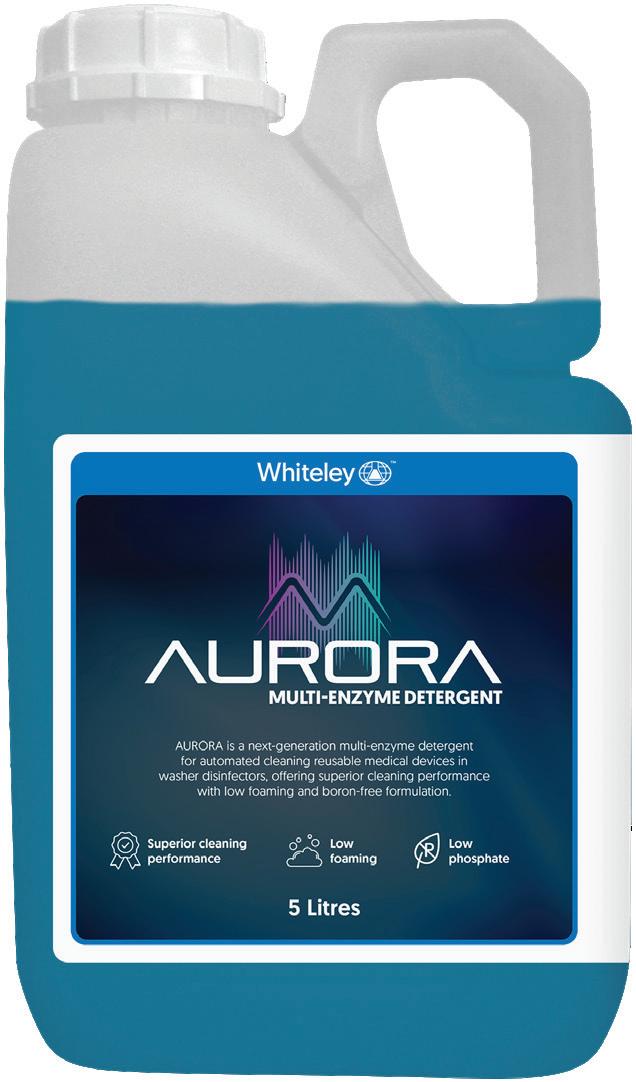
AURORA is a next-generation multi-enzyme detergent for automated cleaning of reusable medical devices in washer disinfectors, offering superior cleaning performance with low foaming and boron-free formulation. Its low phosphate, cost-effective composition makes it a more sustainable option. Formulated with protease and amylase enzymes designed specifically for medical cleaning applications.
GUIDELINES OPEN ACCESS
Nathan Peters1,2 | Marija Juraja3,4 | Nicola Isles3,5 | Ellen Woodcock6 | Elissa Kennedy-Smith7 | Cartan Costello8,9 | Jo McCann10 | Karen Mizia11,12
1Department of Anaesthesia and Perioperative Medicine, Royal Brisbane and Women's Hospital, Brisbane, Queensland, Australia | 2School of Medicine, University of Queensland, Brisbane, Queensland, Australia | 3Australasian College for Infection Prevention and Control, Hobart, Tasmania, Australia | 4Central Adelaide Local Health Network, Adelaide, South Australia, Australia | 5Healthscope Hospitals, Melbourne, Victoria, Australia | 6Department of Māori and Indigenous Health Innovation, Otago University, Christchurch, New Zealand | 7Sandringham Emergency Department, The Women's Hospital, Alfred Health, Melbourne, Victoria, Australia | 8Department of Intensive Care Medicine, Wollongong Hospital, Wollongong, New South Wales, Australia | 9University of New South Wales, Sydney, New South Wales, Australia | 10Dunedin School of Medicine, University of Otago, Dunedin, New Zealand | 11Department of Maternal Foetal Medicine, Royal North Shore Hospital, Sydney, New South Wales, Australia | 12 Australian National University, Canberra, Australian Captial Territory, Australia
Correspondence: Nathan Peters (n.peters@uq.edu.au)
Received: 13 October 2025 | Revised: 19 January 2026 | Accepted: 10 February 2026
Keywords: disinfection | equipment contamination | infection control | transducers | ultrasonography
ABSTRACT
Introduction: Ultrasound equipment is recognised as a potential source of harmful microorganisms which could lead to clinical infection. This highlights the importance of standardised infection prevention and control strategies for ultrasound practice across Australia and New Zealand. This revised guideline supersedes the similarly named 2017 guideline and was collaboratively developed by the Australasian Society for Ultrasound in Medicine (ASUM) and the Australasian College for Infection Prevention and Control (ACIPC).
Methods: A multidisciplinary working group was assembled consisting of ultrasound practitioners from varying specialty areas and infection control specialists. The guideline recommendations were informed by a review of relevant literature, national and international standards, and current Australian and New Zealand regulatory requirements. A catalyst for the revision of these guidelines was the release of the Australian Standard AS5369:2023—Reprocessing of reusable medical devices and other devices in health and non-health related facilities. Consensus was achieved through structured discussions and iterative drafting including wider stakeholder feedback.
Results: Recommendations for transducer reprocessing were based on the invasiveness of the ultrasound transducer as well as the type of human tissue it contacted during use. Particular attention is also given to the safe use of ultrasound gel, transducer covers, as well as broader considerations of standard and transmission-based precautions, including the use of aseptic technique.
Conclusion: Implementation of this guideline by ultrasound clinicians and their facilities will reduce the risk of transmission of potentially harmful microorganisms and ensure infection prevention and control practices across Australia and New Zealand are aligned with accepted national standards.
Impact Statement: This guideline outlines the Australian and New Zealand standard for infection prevention and control as it relates to ultrasound practice. This 2026 revision supersedes the similarly named 2017 guideline and was collaboratively
Abbreviations: ACIPC, Australasian College for Infection Prevention and Control; ARTG, Australian Register of Therapeutic Goods; AS/NZS4187:2014, Australian/ New Zealand Standard Reprocessing of reusable medical devices in health service organisations; AS/NZS4815:2006, Australian/New Zealand Standard Office-based health care facilities—reprocessing of reusable medical and surgical instruments and equipment, and maintenance of the associated environment; AS5369:2023, Australian Standard Reprocessing of reusable medical devices and other devices in health and non-health related facilities; ASUM, Australasian Society for Ultrasound in Medicine; AT, Aseptic technique; HLD, High level disinfection; ILD, Intermediate level disinfection; LLD, Low level disinfection; PPE, Personal protective equipment; RMD, Reusable medical device; TGA, Therapeutic Goods Administration.
This is an open access article under the terms of the Creative Commons Attribution License, which permits use, distribution and reproduction in any medium, provided the original work is properly cited. © 2026 The Author(s). Australasian Journal of Ultrasound in Medicine published by John Wiley & Sons Australia, Ltd on behalf of Australasian Society for Ultrasound in Medicine.
Australasian Journal of Ultrasound in Medicine, 2026; 29:e70038 https://doi.org/10.1002/ajum.70038
developed by the Australasian Society for Ultrasound in Medicine (ASUM) and the Australasian College for Infection Prevention and Control (ACIPC). The multidisciplinary working group used published literature, national and international standards, and relevant regulatory requirements in formulating the recommendations. This guideline should be used by clinicians and facilities using ultrasound to reduce the transmission of potentially harmful microorganisms and improve patient care.
The Australasian Society for Ultrasound in Medicine (ASUM) is the leading multidisciplinary medical ultrasound society creating healthier lives for all by driving ultrasound excellence. The Australasian College for Infection Prevention and Control (ACIPC) is the peak body for Infection Prevention and Control Professionals in the Australian and New Zealand region focused on leadership, promoting education and evidence-based practice outcomes for consumers in all disciplines of healthcare. This revised guideline has been based on a review of recently published evidence including the Australian Standard, AS5369:2023 [1]. The Ministry of Health in New Zealand has also recognised the AS5369:2023 as the most up-to- date standard and has recommended its adoption in their country. This guideline has been developed collaboratively by ASUM and ACIPC to establish accepted guidelines for infection prevention and control when ultrasound equipment is used in clinical practice across Australia and New Zealand and supersedes the similarly named 2017 guideline.
This guideline represents the best practice standards and applies to all staff who use or reprocess ultrasound equipment, as well as the facilities that they work within. The focus of this guideline is on reusable ultrasound equipment that does not require, or is not suitable for, sterilisation. Reprocessing of endoscopic ultrasound devices with working gas/water channels is addressed by other published guidelines [2]. General infection prevention and control guidance can be found in the Australian Guidelines for the Prevention and Control of Infection in Healthcare [3]. Craft groups and healthcare facilities should consider local infection prevention and control policies and undertake site specific risk assessments when applying these guidelines.
The utilisation of ultrasound in medical imaging continues to grow and evolve owing to its portability, safety, and relatively inexpensive nature [4]. The equipment used during ultrasound examinations has been recognised as a potential source of harmful microorganisms which could lead to health- care associated infections [5–8]. Therefore, it is important for operators and facilities using ultrasound equipment to ensure they adhere to the required standards of infection prevention and control.
Ultrasound examinations require direct contact between the patient and the practitioner via an ultrasound transducer. Reusable ultrasound transducers require reprocessing to prevent the
2 of 11
transference of potentially harmful microorganisms between patients. The 2017 ASUM/ACIPC ultrasound transducer reprocessing guidelines were based on the Australian and New Zealand Standards of 4187:2014 and 4185:2006 which have been revised to an Australian Standard in 2023; AS5369:2023 [9–11]. In addition to incorporating amendments consistent with the updated Australian Standard this guideline revision incorporates recently published evidence and supersedes the original 2017 ASUM/ ACIPC guideline [12]. In reviewing published evidence relating to infection prevention and control in ultrasound practice, it was recognised that there is a paucity of high-quality evidence and that recommendations made in this guideline are mostly based on lower levels of evidence (e.g., comparative studies, case series and reports, expert opinion, and consensus statements). This should be considered when applying this guideline into clinical practice as should any new evidence released following publication.
This guideline was developed using a review of published literature, existing standards, and expert consensus. It did not involve the collection of new data from human participants or animals. As such, formal ethics committee approval and informed consent were not required.
Reprocessing is a multistep process that includes cleaning, device inspection/examination, disinfection or sterilisation, and if applicable packaging and labelling [1]. Adherence to all these steps is important in ensuring ultrasound equipment is safe for ongoing use [1]. Where omissions or errors are made in the steps involved in reprocessing this invalidates the entire process and places the ultrasound equipment at higher risk of transferring potentially harmful microorganisms to the next patient [1].
Consideration should be given to the workflow and environment within which reprocessing occurs. Environmental requirements are dependent upon the type of reprocessing being undertaken and should follow the recommendations in AS5369:2023. When designing or renovating facilities, the environment must be designed to support the effective processing and reprocessing of reusable materials over single use items.
Devices must be reprocessed in a dedicated reprocessing area and/or facility, or if safe and effective to do so they may be reprocessed at the point of use. When reprocessing of ultrasound
Journal of Ultrasound in Medicine, 2026
• What is already known about this topic?
○ Equipment used during ultrasound examinations has been recognised as a source of pathogenic microorganisms which could lead to health- care associated infections
○ Reusable ultrasound transducers require appropriate reprocessing to prevent the transfer of potentially harmful microorganisms between patients
○ Infection prevention and control principles must be applied to all ultrasound equipment and to the procedural technique used to perform an ultrasound examination.
• What this paper adds?
○ The recommendations in this guideline are aligned with the requirements of Australian Standard 5369:2023 and supersede the previous version published in 2017.
○ This guideline provides clear and contemporary guidance on infection prevention and control principles as they relate to ultrasound practice.
○ It provides updated recommendations on the level of disinfection required for reprocessing different types of ultrasound transducers.
• The implications of this paper?
○ Standardisation of ultrasound transducer reprocessing practices across clinical settings, reducing variability and uncertainty in infection prevention and control.
○ Improved patient safety through clearer guidance on appropriate disinfection levels, based on transducer type, ultrasound use, and assessed infection risk.
○ Support for organisational policy development, staff training, and auditing by providing evidencebased recommendations aligned across Australia and New Zealand for ultrasound transducer reprocessing.
equipment is to occur at the point of use, this ideally should be a separate area or room to the patient treatment area. If this is not possible, reprocessing and delivering patient care should not take place simultaneously, and an adequate distance is required between the patient, the patient surroundings and the reprocessing activities to minimise possible contamination of the patient or the device [1].
Principles which apply to all ultrasound reprocessing scenarios include:
• The sufficiency of the physical environment (e.g., lighting, ventilation, sinks, bench space, workstations) and other equipment necessary for reprocessing (e.g., personal protective equipment (PPE), hand hygiene stations, waste disposal).
Australasian Journal of Ultrasound in Medicine, 2026
• Ensuring the segregation of clean and dirty areas within the reprocessing environment to minimise the risk of cross contamination between devices.
• A documentation process, relevant to the area of practice.
All ultrasound equipment must be cleaned after each use. Cleaning aims to remove contaminants to the extent necessary for further processing or for intended use [1]. For the purposes of reprocessing, ultrasound equipment can be broadly classified into components that have:
• No direct patient contact—ultrasound machine (e.g., display, interface, and cart).
• Direct patient contact—ultrasound transducer and attached cable (if present).
Cleaning ultrasound equipment should be conducted with solutions and/or wipes that are compatible with the ultrasound device in accordance with the manufacturer's instructions. Where drying is required following cleaning, this should be with lowlinting cloths or towels.
After cleaning is complete, ultrasound equipment must be visually inspected for damage or defects and, if identified, the equipment must not be used until it has been repaired or replaced. Damage or defects can prevent the effective reprocessing of ultrasound equipment leading to infection [8, 13, 14].
Ultrasound machines are transported between clinical areas and are frequently touched by clinicians, potentially facilitating transmission between patients. This includes the components of the ultrasound machine that do not have direct patient contact (e.g., display, user interface, and workstation). Resultingly, all components of the ultrasound machine are required to undergo a minimum of cleaning between patient uses. Particular attention should be given to frequently touched areas such as keyboards, touch screens, and trackballs, which should undergo thorough cleaning and disinfection between patients, in accordance with the manufacturer's recommendations.
Cleaning an ultrasound transducer and cable is always required so that subsequent processing steps can occur effectively [1, 15, 16]. For ultrasound transducers and cables cleaning should remove all visible organic (e.g., blood and other body fluids) or non- organic soiling (e.g., gel) [15, 17–19]. This should occur as soon as possible following use to minimise the risk of contaminants drying onto the device. All cleaning products used on ultrasound transducers must be included on the Australian Register of Therapeutic Goods (ARTG) as a Class I or IIb agent suitable for use on medical devices that are compatible
3 of 11
with the medical device to be cleaned. Cleaning wipes and materials should be discarded after use and must not be used for disinfection.
Once clean, ultrasound equipment which has direct patient contact (e.g., transducers) must undergo disinfection or sterilisation dependent upon the infectious risk associated with their next intended use. Disinfection is a process that inactivates viable microorganisms to a level previously specified as being appropriate for a defined purpose [1]. Sterilisation is a validated process to render a product free from viable microorganisms [1]. Ultrasound transducers are heat sensitive devices and heat or steam sterilisation techniques can cause device damage. Therefore, these sterilisation methodsare generally not recommended by equipment manufacturers. Low temperature sterilisation processes should only be utilised where this is approved by the manufacturer.
Systems suitable for the disinfection of ultrasound transducers fall into two broad categories.
• Manual systems—immersion in chemical solutions and/ or use of chemical impregnated manual disposable wipes/ sponges
• Automated disinfection systems—require the transducer to be inserted into a device or machine which then applies the disinfectant, such as chemical or Ultraviolet- C light.
The Therapeutic Goods Administration (TGA) regulates disinfectants based upon their intended use as well as their claims of effectiveness against specific microorganisms based on in vitro testing [20]. Disinfectants used on ultrasound transducers must be approved for use on medical devices and are required to be listed on the ARTG as a class IIb medical device [20]. Details about specific products listed licence class can be found by searching the ARTG website.
The level of disinfection achieved by a disinfectant system depends on the types of microorganisms it has been proven to be effective against. Some microorganisms, such as the spores arising from spore forming microorganisms, are highly resistant to some disinfectants. This is in comparison to other microorganisms which are very sensitive to disinfectants such as, larger sized lipid enveloped viruses (e.g., SARS Co-V-2, influenza, hepatitis viruses, human immunodeficiency virus) [21, 22]. Disinfectants are categorised into low-, intermediate-, and highlevels accordingly (Table 1).
Rinsing the transducer allows for the removal of any residue disinfectant agent and the requirement for this is dependent upon the disinfection system used [1]. If rinsing is required then drying must be with a single-use lint-free cloth.
4 of 11
TABLE 1 | Definition of different levels of disinfection in accordance with AS5369:2023 [1].
Level of disinfection Definition
Low level disinfection
Destroys all vegetative bacteria (except tubercle bacilli), lipid viruses, some nonlipid viruses, and some fungi, but not bacterial spores
Intermediate level disinfection
High level disinfection
Kills all microbial pathogens except bacterial endospores which is bactericidal, tuberculocidal, fungicidal (against asexual spores but not necessarily dried chlamydospores or sexual spores), and virucidal
Kills all microbial pathogens, except large numbers of bacterial endospores
Following reprocessing, ultrasound transducers should be stored to minimise contamination between uses. For ultrasound transducers that have undergone high-level disinfection, this may include storage in a dedicated sealed cabinet or using the application of a protective sterile single use storage cover. Transducer covers used for storage act as a barrier to prevent environmental contamination and must not be used on patients at the time of ultrasound examinations. Sterile item storage, handling, and release are required to meet the requirements outlined in AS5369:2023 [1].
Prior to use, the user should be satisfied that the ultrasound equipment has been adequately reprocessed for the purposes of the ultrasound examination to be undertaken. If there is uncertainty about the adequacy of the level of reprocessing prior to use, then the ultrasound transducer must not be used until the appropriate reprocessing requirements have been satisfied and can be assured.
Reprocessing requires ongoing review to ensure that the effectiveness of processes is maintained. All facilities that use ultrasound equipment are responsible for developing and monitoring reprocessing processes, policies, and procedures that are specific to their work environment and workplace. This should include periodic reassessment of reprocessing facilities and staff training, regular maintenance of reprocessing equipment, and ongoing validation of processes in accordance with the requirements of AS5369:2023.
Regular periodic audits of reprocessing should be performed (e.g., annually). Ultrasound transducers must always satisfy the reprocessing requirements before clinical use and if this cannot be assured the transducer should undergo repeat reprocessing or, if required, quarantined from clinical use until an
investigation and action plan can be undertaken to ensure reprocessing standards are being achieved [1].
All users of ultrasound equipment must be appropriately trained in all aspects of reprocessing relevant to their practice. On commencement, all new staff should undergo a formal induction and/or orientation training program which should include:
• Modes of transmission of infection; hand hygiene; workplace health and safety issues including personal protective equipment (PPE).
• Infection prevention and control principles including standard and transmission-based precautions.
• Reprocessing of ultrasound equipment, including how to:
○ Effectively clean the ultrasound machine and transducer.
○ Determine the appropriate level of disinfection.
○ Undertake the appropriate level of disinfection.
○ Store ultrasound equipment to prevent contamination between uses.
○ Complete the required documentation and record keeping.
• Correct procedure for waste management, including its safe handling and segregation into the appropriate stream.
On completion of training, staff competency in performing reprocessing activities should be objectively assessed. Staff should not use ultrasound equipment independently until this assessment has been completed and recorded. Facilities should keep records of those trained and periodic reassessment undertaken, the timing of which should be based on local policy or guidelines. Additional staff training is required with the introduction of new ultrasound equipment, reprocessing equipment, or a change in reprocessing methods.
4.5.2
A system of traceability should be used for ultrasound equipment, including loan devices, which undergo HLD or sterilisation [1]. Traceability records are intended to associate a single patient care episode with a individual reprocessing cycle. Traceability labels are commonly used for this purpose and, where used, are to be included in the patient's notes or medical record.
For ultrasound transducers undergoing sterilisation or HLD, the traceability system should be sufficient to enable identification of the individual cycle and at a minimum include:
• Transducer type and unique ID number.
• Date of cleaning and the name of competent person responsible for reprocessing.
• Identification of the disinfection process details as well as the cycle number and date of sterilisation or disinfection.
Australasian Journal of Ultrasound in Medicine, 2026
• Individual patient/client identifiers according to local facility requirements.
Multiple outbreaks have been linked to bacterial contamination of ultrasound transmission gel [5]. Common infectious microorganisms have included Burkholderia spp. [23–26], Acinetobacter spp. [27], and Pseudomonas spp. [28]. Ultrasound gel has the potential for microbial contamination and therefore, appropriate steps must be taken to minimise the risk of potential infection.
Ultrasound gel is available in single and multi-use containers and must be registered with the ARTG. Single-use products should be discarded following each patient use and have the advantage of avoiding the potential transfer of microorganisms to subsequent patients. If multi-use gel containers are to be used, they must be:
• Cleaned between patient uses.
• Discarded if there are any visible signs of gel contamination.
• Dated at the time it is opened and discarded within the timeframe specified from the manufacturer.
• Stored appropriately and protected from contamination between uses.
Heating of reusable gel bottles is not recommended as this may support microbial growth [6]. If heating is required the risk of microbial growth should be minimised by only warming single use containers or sachets immediately prior to use. Refilling of multiuse containers should not occur due to the higher risk of microbial contamination of the container and gel [6, 27]. Sterile ultrasound gel must be used during ultrasound examinations or procedures where gel could contact mucous membranes or otherwise normally sterile tissue (e.g., wounds, skin puncture sites) [29].
Ultrasound transducers have direct patient contact, and reprocessing is aimed at mitigating the risk of transmitting pathogenic microorganisms and causing infection at the site of intended use. Risk-based classification systems aim to assess and stratify this risk and then make recommendations for different reprocessing requirements.
AS5369:2023 recognises that the infectious risk is primarily determined by how invasive the device is when being used on a patient [1]. Invasive ultrasound transducers are those that penetrate inside the body, either through a body orifice or through the surface of the body [1, 30]. For the purposes of this guideline an ultrasound transducer is considered to penetrate through the surface of the body only when it is intended to be applied directly into an open wound (non-intact skin) with visible and exposed subcutaneous tissue. Open wounds
include, for example, deep dermal burns and skin ulcers. These invasive ultrasound devices carry a higher risk of causing infection than non-invasive transducers as they bypass the natural barrier and defences presented by the skin and other components of the integumentary system [16]. Non-invasive ultrasound transducers are associated with a lower risk of causing infection as they are applied onto skin thereby benefiting from its intrinsic infection protective properties. Based on this risk assessment invasive ultrasound transducers require reprocessing with high level disinfection or sterilisation and non-invasive transducers are reprocessed using low- or intermediate-level disinfection.
Spaulding's classification further delineates invasive devices into either semi- critical or critical risk classifications based upon the sterility of the human tissue encountered during use [31]. Invasive transducers inserted into areas containing sterile tissue are classified as critical whereas those that contact non- sterile tissues such as mucous membranes (e.g., rectum, vagina) or open wounds (e.g., burns, skin ulcers) are classified as semi- critical. Non-invasive ultrasound transducers are classified into the non- critical level of risk according to Spaulding's system [1] (See Table 2).
In ultrasound practice, where uncertainty regarding the reprocessing requirements exists, undertaking a local risk assessment incorporating other risk assessment methods is recommended and supported by the AS5369:2023 [1]. These scenarios may arise in different clinical settings, patient populations, or with certain procedures involving ultrasound transducers. Examples of clinical settings where a risk assessment might be required could include resource limited areas or time critical environments. To ensure patient safety in low resource settings, the recommendations set out in this guideline should be applied within the context of the available resources, including the available disinfection options. Specific patient populations, like the immunocompromised or critically ill, may also necessitate local risk assessment and
TABLE
subsequently additional risk-mitigating strategies. In some areas of ultrasound practice, such as ultrasound guided percutaneous procedures, incorporating other additional risk assessment methods may allow for a more contextualised assessment of hazards and their subsequent prioritisation [32]. Where systems, processes, and practices are demonstrably compliant with relevant standards and governance requirements, this evidence may be taken into consideration as part of the overall risk evaluation. A qualified infection prevention and control practitioner should always be engaged when undertaking local risk assessments.
Ultrasound transducer covers are manufactured single-use disposable plastic covers designed to create a physical barrier between the patient and the ultrasound transducer and cable. Transducer covers are used to:
• Prevent or minimise the contamination and soiling of an ultrasound transducer or cable during use so that subsequent reprocessing may be easier to perform; and/or
• Provide an additional protective barrier between the transducer and cable and the patient, preventing or reducing the exchange of microorganisms between them.
The incidence of transducer cover integrity loss from published data is generally reported as low [33]. The available data would suggest that commercially manufactured covers have a lower failure rate when compared to other non-purpose specific manufactured covers [33]. As such, only commercially manufactured transducer covers listed on the ARTG should be used to cover a transducer. The use of other items such as dressings, condoms, plastic bags, and gloves is not recommended [34–36]. Ultrasound transducer covers should not be used as a substitute for cleaning, disinfection or sterilisation [1].
Invasiveness of the transducer Level of risk according to Spaulding Spaulding's definition Reprocessing requirements
Invasive Critical Ultrasound transducer intended to be introduced into or have contact with the vascular system or normally sterile areas of the body
Invasive Semi- critical An ultrasound transducer that comes into contact with mucous membranes or non-intact skin
Non-invasive Non- critical An ultrasound transducer that comes into contact with intact skin but not mucous membranes
Clean as soon as possible after use. For heat or moisture sensitive devices, sterilise using low-temperature sterilisation process. Sterility to be maintained between uses. Packaged and stored to prevent environmental contamination in a designated storage area in accordance with AS5369:2023
Clean as soon as possible after use followed by low-temperature sterilisation process or high-level disinfection process according to manufacturer's instructions for use. Store to prevent environmental contamination between uses
Clean as soon as possible after use then disinfect with compatible low-level or intermediate level Class IIb disinfectant. Store to minimise environmental contamination between uses
When reusable ultrasound equipment is being used in clinical ultrasound practice, standard and transmission-based precautions should be adhered to as part of a more broadly encompassing infection prevention and control risk mitigation strategy.
As a minimum, standard precautions must be adhered to in clinical ultrasound practice. Standard precautions include hand hygiene; the use of PPE; respiratory hygiene and cough etiquette; safe use and disposal of sharps; and appropriate environmental cleaning, linen, and waste management [3].
Aseptic technique, as an element of standard precautions, aims to prevent pathogenic microorganisms, in sufficient quantity to cause infection, from being introduced into susceptible body sites by the hands of staff and from surfaces of equipment (e.g., transducers) during a procedure [3, 37]. Incorporating ultrasound transducers into a procedural aseptic technique will demand additional aseptic precautions be taken when compared to similar procedures without ultrasound guidance. These additional requirements can include the use of a sterile transducer cover, sterile gel and sterile gloves. Aseptic technique is often divided into ‘Standard’ or ‘Surgical’ where the latter is used for more technically complex procedures, of longer duration, and with large or numerous sterile areas or components [3, 37]. Correspondingly surgical aseptic technique requires more aseptic precautions such as larger critical aseptic fields, surgical hand scrub and full barrier precautions (e.g., hat, mask, eye protection, and sterile gown and gloves) [3]. More detail on aseptic technique, including procedure specific guidance, is available in the Australian Guidelines for the Prevention and Control of Infection in Healthcare [3].
Transmission-based precautions (TBP) are used in situations where standard precautions alone can be insufficient to prevent infection [3]. These precautions are in addition to the requirements of standard precautions and are applied when patients are known or suspected to be infected or colonised with certain infectious agents. Examples of TBP include contact, droplet, airborne or a combination of these. Where ultrasound equipment is required in patient care areas with TBP it would be appropriate to seek further infection prevention and control advice on how ultrasound equipment used in these scenarios should be appropriately cleaned and disinfected with products that are effective against the known or suspected pathogen.
Outlined below are the infection prevention and control recommendations for ultrasound transducers used in a selection of
Australasian Journal of Ultrasound in Medicine, 2026
common scenarios and have been based on the principles described. Given the breadth of ultrasound use in healthcare, if an area of ultrasound equipment use is not specifically addressed below, the ultrasound practitioner and facility should interpret and apply these principles accordingly using the examples below as a guide.
Non-invasive ultrasound transducers do not penetrate inside the body and are commonly used in diagnostic examinations and to aid in the performance of needle-based procedures through the surrounding skin (percutaneous procedures) [30]. Non-invasive transducers are classified as non- critical medical devices according to Spaulding's classification.
These are common ultrasound examinations that rely on the cutaneous application of an ultrasound transducer to obtain diagnostic information. Examples of these include transthoracic echocardiography, vascular, musculoskeletal, soft tissue and abdominal sonography (Figure 1).
Ultrasound-guided percutaneous procedures involve applying an ultrasound transducer onto a patient's skin and then introducing a needle through the surrounding skin towards a target region contained within the field of view of an ultrasound transducer [38]. These transducers are considered non-invasive and examples include ultrasound guided central and peripheral vascular access techniques as well as therapeutic and diagnostic injections, aspirations, and biopsies.
There is variation in international societal guidelines on the level of disinfection required of ultrasound transducers to be used in percutaneous procedures [32]. Earlier guidelines, including ASUM/ACIPIC 2017, tended to favour recommending HLD [12, 39]. More recently, LLD has been considered appropriate by some societies [40, 41]. Since the release of earlier published guidelines, there have been improvements in the variety and availability of disinfection systems for ultrasound transducers across Australia and New Zealand. Additionally, the incorporation of ARTG listing requirements for disinfectants in the AS5369:2023 supports the better differentiation and recognition of the various levels of disinfectants suitable for use on ultrasound transducers.
Current evidence suggests that the risk of infection arising from an ultrasound guided percutaneous procedure is likely to be very low, estimated to be 0.1%–0.5% [32]. When used in accordance with the manufacturer's instructions, LLD of ultrasound transducers involved in percutaneous procedures appears to present no higher infection risk compared with HLD [17, 42]. Additionally, the use of LLD for ultrasound transducers used in percutaneous procedures has also been
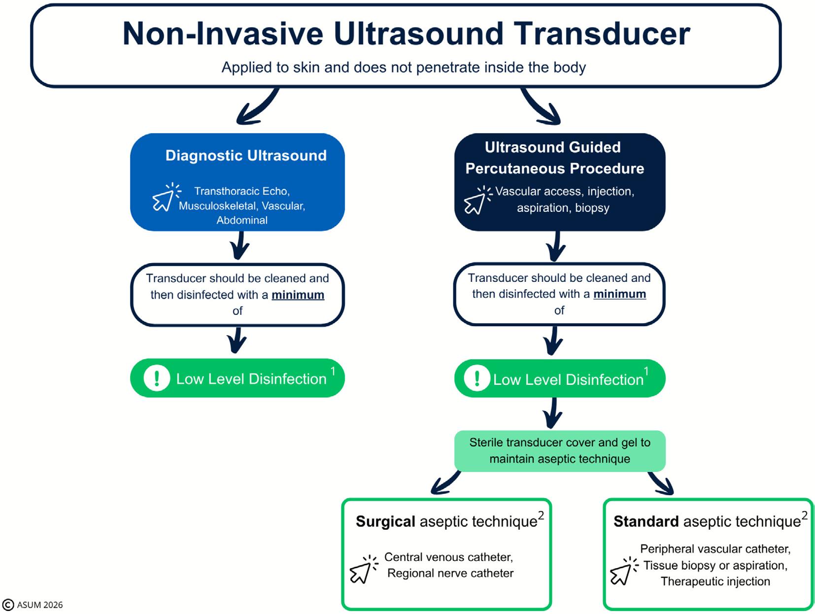
FIGURE 1 | Infection prevention and control practices required for non-invasive ultrasound transducers. 1A higher level of disinfection may be required where a documented risk assessment identifies an increased risk of pathogen transmission or infection that is not adequately mitigated by low-level disinfection. 2For advice on infection prevention measures for different procedures refer to the Australian Guidelines for the Prevention and Control of Infection in Healthcare [3].
supported by national and international societies [30, 38, 43]. As such, ultrasound transducers used in percutaneous procedures should undergo a minimum of LLD and must be incorporated into the appropriate aseptic technique required of the procedure.
To maintain asepsis during an ultrasound guided percutaneous procedure contact between the ultrasound transducer and cable with the skin puncture site, aseptic field, and sterile device being inserted must be avoided. The use of a sterile transducer cover during the performance of an ultrasound guided percutaneous procedure should always be used where contact between these components or sites could occur. In these scenarios sterile ultrasound gel as well as transducer covers of adequate length should be used to maintain procedural aseptic technique (Figure 1).
An invasive transducer is one that penetrates inside the body, either through a body orifice or through the surface of the body [1]. According to Spaulding's classification, these ultrasound transducers would be classified as critical or semi- critical based on the sterility of the tissue the ultrasound is in contact with.
Critical ultrasound transducers are required to be sterile before use as they are intended to have contact with the vascular system or normally sterile areas of the body. The use of these transducers usually mandates a surgical aseptic technique and, as
sterility of these devices is maintained during these procedures, sterile ultrasound transducer covers are not routinely required. If ultrasound gel is required, it must be single use and sterile.
Semi- critical transducers are inserted into non-sterile areas of the body lined bymucous membranes (vagina, rectum and oesophagus) and into open wounds (e.g., skin ulcers, deep dermal burns) [30, 44]. These transducers require a minimum of HLD between uses. Sterile ultrasound transducer covers are recommended when contact with mucous membranes or open wounds is anticipated. In these scenarios covers provide an additional barrier between the patient and the transducer as well as helping to minimise transducer contamination. Single use sterile ultrasound gel should also be used when transducers are classified as semi- critical (Figure 2).
The transmission of human papilloma virus (HPV) has been identified as a concern in ultrasound examinations which involve contact with mucous membranes. HPV is highly prevalent and could be transferred between patients through contact with contaminated ultrasound equipment [45, 46]. Appropriate risk mitigating steps should be taken which include, standard precautions, transducer cover use, single use ultrasound gel, and disinfection of ultrasound transducers including the handle using a minimum of high-level disinfection [46, 47].
Invasive ultrasound transducers can also be used as a guide for needle-based procedures (e.g., transrectal ultrasound guided
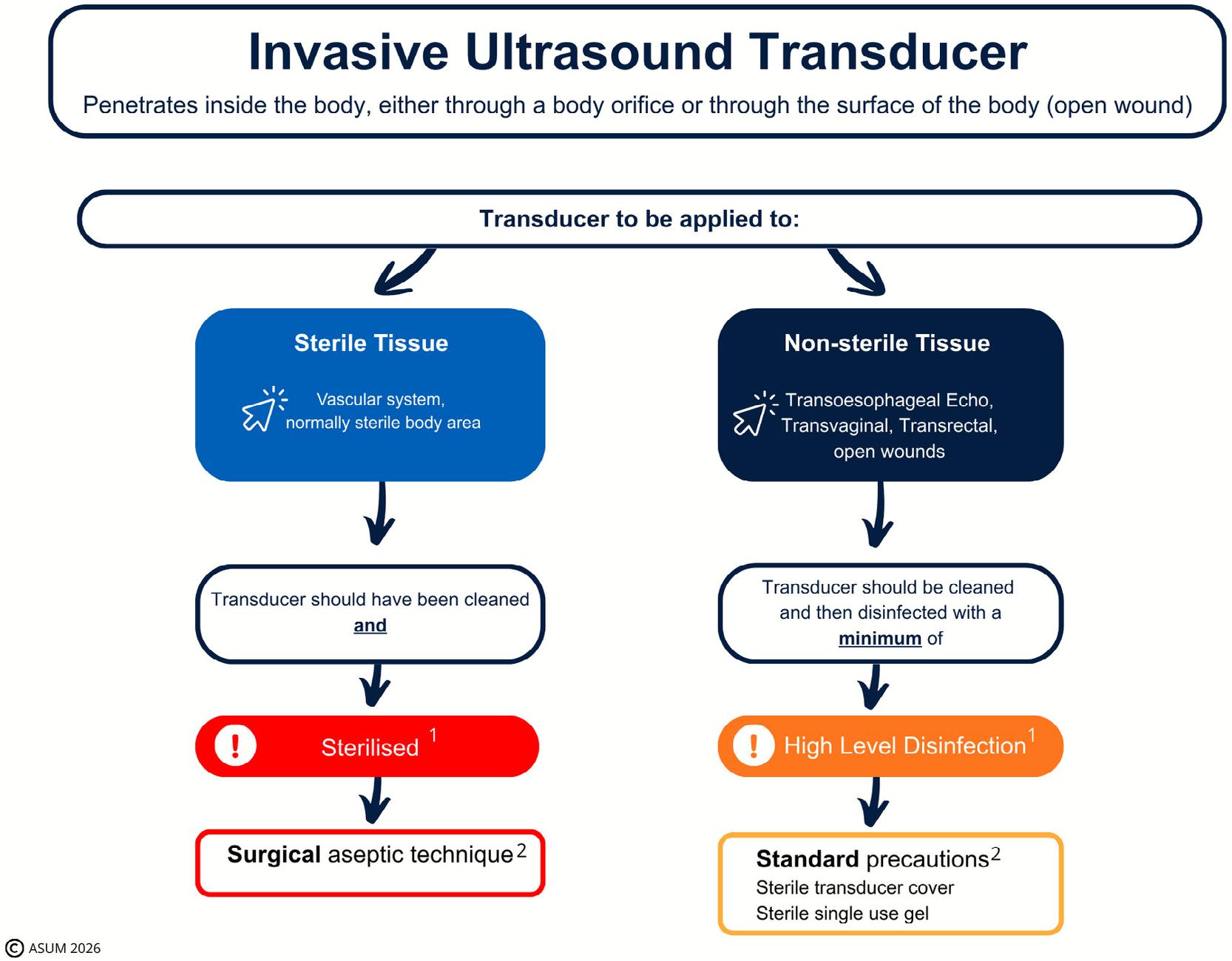
FIGURE 2 | Infection prevention and control practices required for examinations involving invasive ultrasound transducers. 1In accordance with requirements outlined in AS5369:2023. 2Refer to the Australian Guidelines for the Prevention and Control of Infection in Healthcare [3].
biopsy). In this scenario the ultrasound transducer should be reprocessed in keeping with its site of intended use and then incorporated into the aseptic technique requirements of the procedure. If a clinical scenario was to arise where a usually non-invasive ultrasound transducer is to be used in a way that meets the definition of an invasive ultrasound transducer it should be reprocessed as an invasive transducer before it is used [30].
Consideration should be given to the reduction of our carbon footprint, and where possible the aim should be to support future sustainability and preserve ecosystems. End users are encouraged to select appropriate products which also help support sustainable practices where possible [16, 48].
At the time of ultrasound equipment procurement, consideration should be given to the implications on end users and ensuring that equipment design works within infection prevention and control guidelines. This should include consideration on workflow and factors such as ease of cleaning, storage and suitability of equipment. Involvement of ultrasound practitioners is strongly encouraged in the procurement process.
Australasian Journal of Ultrasound in Medicine, 2026
Infection prevention and control practitioners and other practitioners who regularly use and reprocess ultrasound equipment should be consulted and/or involved in the development of local facility policies and procedures relating to ultrasound equipment [49]. Not only does this encourage ultrasound practitioner engagement but also allows for consideration of practice specific factors ensuring the more successful implementation of recommendations into clinical practice. Facilities must recognise and provide the additional resources required to implement and perform safe and effective ultrasound equipment reprocessing (e.g., staff time, equipment and funding).
Nathan Peters: conceptualization, methodology, writing – review and editing, writing – original draft, project administration, investigation. Marija Juraja: conceptualization, writing – review and editing, investigation, methodology. Nicola Isles: conceptualization, writing – review and editing, investigation, methodology. Ellen Woodcock: conceptualization, writing – review and editing, investigation, methodology. Elissa Kennedy- Smith: conceptualization, writing – review and editing, investigation, methodology. Cartan Costello: conceptualization, writing – review and editing, investigation, methodology. Jo McCann: conceptualization, writing – review and editing, investigation, methodology. Karen Mizia: conceptualization, writing – review and editing, investigation, methodology.
Acknowledgements
Open access publishing facilitated by The University of Queensland, as part of the Wiley - The University of Queensland agreement via the Council of Australasian University Librarians.
Disclosure
These joint guidelines were developed by a working committee representing the Australasian Society for Ultrasound in Medicine (ASUM) and the Australasian College for Infection Prevention and Control (ACIPC).
Conflicts of Interest
C.C.—received payment for 2019 presentation sponsored by Germitec. M.J.—infection control consultancy to EGO Pharmaceuticals; Haines Medical; Clean Life Medical. N.P.; M.J., N.I., E.W., E.K.-S., J.M., K.M.— no conflicts of interest.
Data Availability Statement
The data that support the findings of this study are available from the corresponding author upon reasonable request.
References
1. Standards Australia, AS5369: Reprocessing of Reusable Medical Devices and Other Devices in Health and Non-Healthcare Related Facilities (Standards Australia, 2023).
2. B. Devereaux, D. Jones, and E. Wardle, “Infection Prevention and Control in Endoscopy 2021 Melbourne 2021,” https://www.gesa.org.au/ public/13/files/ Education%20%26%20Resources/Clinical%20Practice% 20Res ources/ Endos copy% 20Inf ection% 20Con trol/ IPCE% 202021_ Feb2022update.pdf.
3. National Health and Medical Research Council, Australian Guidelines for the Prevention and Control of Infection in Healthcare (Commonwealth of Australia, 2019), https://www.safet yandquality.gov.au/sites/ defau lt/ files/ 2024- 08/austr alian - guide lines - for- the- preve ntion - andcontrol- of-infection-in-healthcare.pdf
4. S. Wang, J. A. Hossack, and A. L. Klibanov, “From Anatomy to Functional and Molecular Biomarker Imaging and Therapy: Ultrasound Is Safe, Ultrafast, Portable, and Inexpensive,” Investigative Radiology 55, no. 9 (2020): 559–572.
5. A. Angrup, R. Kanaujia, M. Biswal, and P. Ray, “Systematic Review of Ultrasound Gel Associated Burkholderia cepacia Complex Outbreaks: Clinical Presentation, Sources and Control of Outbreak,” American Journal of Infection Control 50, no. 11 (2022): 1253–1257.
6. S. C. Westerway, J. M. Basseal, A. Brockway, J. A. Hyett, and D. A. Carter, “Potential Infection Control Risks Associated With Ultrasound Equipment - A Bacterial Perspective,” Ultrasound in Medicine & Biology 43, no. 2 (2017): 421–426.
7. T. Sartoretti, E. Sartoretti, C. Bucher, et al., “Bacterial Contamination of Ultrasound Probes in Different Radiological Institutions Before and After Specific Hygiene Training: Do We Have a General Hygienical Problem?,” European Radiology 27, no. 10 (2017): 4181–4187.
8. K. de Souza Hajar, C. de Quartim Moraes Bruna, and K. Uchikawa Graziano, “Infection Transmission Associated With Contaminated Ultrasound Probes: A Systematic Review,” AORN Journal 115, no. 1 (2022): 42–51.
9. Australian/New Zealand Standard, AS/NZS4187:2014 Reprocessing of Reusable Medical Devices in Health Service Organisation (Standards Australia and Standards New Zealand, 2014).
10. Australian Commission on Safety and Quality in Health Care, “Transitioning From AS/NZS 4187:2014 to AS 5369:2023. Identifying
changes and Implementation Strategies for Health Service Organisations,” (2024), https://www.safet yandquality.gov.au/sites/default/ files/ 2024- 08/trans ition ing_ from_ as_ nzs_ 4187_ 2014_ to_ as_ 5369_ 2023.pdf.
11. Australian Commission on Safety and Quality in Health Care, “AS24/01: National Safety and Quality Health Service Standards Requirements for Reprocessing of Reusable Medical Devices in Health Service Organisations,” (2024), https://www.safet yandquality.gov.au/ newsr oom/natio nal- stand ards- updat es/as240 1- natio nal- safet y- andquality-healt h- service- standards-requi rements-reprocessing-reusablemedical- devices-health-service- organisations.
12. J. Basseal, S. C. Westerway, M. Juraja, T. van de Mortel, T. McAuley, and J. Rippey, “Guidelines for Reprocessing Ultrasound Transducers,” Australasian Journal of Ultrasound in Medicine 20, no. 1 (2017): 30–40.
13. E. Vesteinsdottir, K. O. Helgason, K. O. Sverrisson, O. Gudlaugsson, and S. Karason, “Infections and Outcomes After Cardiac Surgery—The Impact of Outbreaks Traced to Transesophageal Echocardiography Probes,” Acta Anaesthesiologica Scandinavica 63, no. 7 (2019): 871–878.
14. M. Seki, H. Machida, Y. Yamagishi, H. Yoshida, and K. Tomono, “Nosocomial Outbreak of Multidrug-Resistant Pseudomonas aeruginosa Caused by Damaged Transesophageal Echocardiogram Probe Used in Cardiovascular Surgical Operations,” Journal of Infection and Chemotherapy 19, no. 4 (2013): 677–681.
15. T. Kremer, N. J. Rowan, and G. McDonnell, “A Proposed Cleaning Classification System for Reusable Medical Devices to Complement the Spaulding Classification,” Journal of Hospital Infection 145 (2024): 88–98.
16. N. J. Rowan, T. Kremer, and G. McDonnell, “A Review of Spaulding's Classification System for Effective Cleaning, Disinfection and Sterilization of Reusable Medical Devices: Viewed Through a Modern-Day Lens That Will Inform and Enable Future Sustainability,” Science of the Total Environment 878 (2023): 162976.
17. N. Peters, F. Williamson, M. J. Bauer, et al., “Comparison of LowLevel to High-Level Disinfection in Eliminating Microorganisms From Ultrasound Transducers Used on Skin: A Noninferiority Randomized Controlled Trial,” Journal of Ultrasound in Medicine 42, no. 11 (2023): 2525–2534.
18. AIUM Official Statement, “Guidelines for Cleaning and Preparing External- and Internal-Use Ultrasound Transducers and Equipment Between Patients as Well as Safe Handling and Use of Ultrasound Coupling Gel, 2025 Revision,” Journal of Ultrasound in Medicine 44, no. 12 (2025): E1–E9.
19. S. R. Desai, L. Tang, and N. C. Hwang, “Infection Control for Perioperative Ultrasonography and Echocardiography,” Journal of Cardiothoracic and Vascular Anesthesia 36, no. 12 (2022): 4440–4448.
20. Australian Government Department of Health and Aged Care. Therapeutic Goods Administration, “Understanding Regulation of Disinfectants, Sterilants and Sanitary Products. Guidance on How Sterilants and Disinfectants Are Regulated in Australia,” (2024), https:// www.tga. gov. au/resou rces/guida nce/under stand ing-regul ation- disin fectants-sterilants-and-sanitary-products
21. G. McDonnell, Antisepsis, Disinfection, and Sterilization, 2nd ed. (ASM Press, 2017).
22. G. McDonnell and P. Burke, “Disinfection: Is It Time to Reconsider Spaulding?,” Journal of Hospital Infection 78, no. 3 (2011): 163–170.
23. E. C. Nannini, A. Ponessa, R. Muratori, et al., “Polyclonal Outbreak of Bacteremia Caused by Burkholderia cepacia Complex and the Presumptive Role of Ultrasound Gel,” Brazilian Journal of Infectious Diseases 19, no. 5 (2015): 543–545.
24. D. Solaimalai, N. K. Devanga Ragupathi, K. Ranjini, et al., “Ultrasound Gel as a Source of Hospital Outbreaks: Indian Experience and Literature Review,” Indian Journal of Medical Microbiology 37, no. 2 (2019): 263–267.
Journal of Ultrasound in Medicine, 2026
25. M. Jacobson, R. Wray, D. Kovach, D. Henry, D. Speert, and A. Matlow, “Sustained Endemicity of Burkholderia cepacia Complex in a Pediatric Institution, Associated With Contaminated Ultrasound Gel,” Infection Control and Hospital Epidemiology 27, no. 4 (2006): 362–366.
26. K. Olshtain-Pops, C. Block, V. Temper, et al., “An Outbreak of achromobacter xylosoxidans Associated With Ultrasound Gel Used During Transrectal Ultrasound Guided Prostate Biopsy,” Journal of Urology 185, no. 1 (2011): 144–147.
27. K. J. Yagnik, G. Kalyatanda, A. P. Cannella, and L. K. Archibald, “Outbreak of Acinetobacter baumannii Associated With Extrinsic Contamination of Ultrasound Gel in a Tertiary Centre Burn Unit,” Infection Prevention in Practice 1, no. 2 (2019): 100009.
28. P. Chittick, V. Russo, M. Sims, et al., “An Outbreak of Pseudomonas aeruginosa Respiratory Tract Infections Associated With Intrinsically Contaminated Ultrasound Transmission Gel,” Infection Control and Hospital Epidemiology 34, no. 8 (2013): 850–853.
29. S. C. Oleszkowicz, P. Chittick, V. Russo, P. Keller, M. Sims, and J. Band, “Infections Associated With Use of Ultrasound Transmission Gel: Proposed Guidelines to Minimize Risk,” Infection Control and Hospital Epidemiology 33, no. 12 (2012): 1235–1237.
30. Australian Commission on Safety and Quality in Health Care, “Cleaning and Disinfection of Ultrasound Transducers,” (2025), https:// www. safet yandq uality. gov. au/publi catio ns- and- resou rces/resou rcelibrary/cleaning-and- disin fection-ultrasound-transducers
31. E. Spaulding, “Chemical Disinfection and Antisepsis in the Hospital,” Journal of Hospital Research 9 (1957): 5–31.
32. N. Peters, F. Williamson, and V. Eley, “Ultrasound Transducer Disinfection for Percutaneous Procedures: A Review of the Evidence Supporting Guideline Recommendations,” Australasian Journal of Ultrasound in Medicine 27, no. 4 (2024): 242–250.
33. J. M. Basseal, S. C. Westerway, and J. A. Hyett, “Analysis of the Integrity of Ultrasound Probe Covers Used for Transvaginal Examinations,” Infectious Diseases and Health 25, no. 2 (2020): 77–81.
34. A. Prinzbach and J. Gadsden, “Practice ‘Safe Scans’: Why Ultrasound Transducer Covers Should Be Considered Best Practice,” Regional Anesthesia and Pain Medicine 50 (2024): 523–525.
35. J. Masood, S. Voulgaris, O. Awogu, C. Younis, A. J. Ball, and T. W. Carr, “Condom Perforation During Transrectal Ultrasound Guided (TRUS) Prostate Biopsies: A Potential Infection Risk,” International Urology and Nephrology 39, no. 4 (2007): 1121–1124.
36. A. Chuan, C. Tiong, M. Maley, J. Descallar, and H. Ziochos, “Decontamination of Ultrasound Equipment Used for Peripheral UltrasoundGuided Regional Anaesthesia,” Anaesthesia and Intensive Care 41, no. 4 (2013): 529–534.
37. Australian Commission on Safety and Quality in Health Care, “Principles for Aseptic Technique: Information for Healthcare Workers,” (2021), https://www.safet yandquality.gov.au/sites/default/files/ 2022- 01/ princ iples_ for_ asept ic_ techn ique- infor mation_ for_ healt hcare_workers_- december_ 2021.pdf
38. “Disinfection of Ultrasound Transducers Used for Percutaneous Procedures: Intersocietal Position Statement,” Journal of Ultrasound in Medicine 40, no. 5 (2021): 895–897.
39. C. M. Nyhsen, H. Humphreys, R. J. Koerner, et al., “Infection Prevention and Control in Ultrasound—Best Practice Recommendations From the European Society of Radiology Ultrasound Working Group,” Insights Into Imaging 8, no. 6 (2017): 523–535.
40. C. Bell, G. Sheppard, R. Andani, N. Katz, and D. J. Kim, “Canadian Association of Emergency Physicians Emergency Ultrasound Committee Best Practice Recommendations on Point- Of- Care Ultrasound Disinfection,” CJEM 26, no. 3 (2024): 145–147.
41. AIUM Official Statement, “Guidelines for Cleaning and Preparing External- and Internal-Use Ultrasound Transducers and Equipment
Australasian Journal of Ultrasound in Medicine, 2026
Between Patients as Well as Safe Handling and Use of Ultrasound Coupling Gel,” Journal of Ultrasound in Medicine 42, no. 7 (2023): E13–E22.
42. S. Adhikari, M. Blaivas, D. Morrison, and L. Lander, “Comparison of Infection Rates Among Ultrasound- Guided Versus Traditionally Placed Peripheral Intravenous Lines,” Journal of Ultrasound in Medicine 29, no. 5 (2010): 741–747.
43. Australasian College of Emergency Medicine, “Statement on Cleaning and Disinfection of Ultrasound Transducers That Are Used for Needle Based Procedures,” (2018), https://acem.org.au/getmedia/85016 5eb- 0b9b- 4aab- 82f6- da91b737e4 06/ S686_v1_ State ment_ Clean ing_ Ultrasound_Transducers.
44. B. L. Phillips and E. L. Casanova, “Using the Manufacturer's Instructions for Use and Spaulding Classification System to Assess Disinfection Practices for Ultrasound Transducers in the Perioperative Setting,” AORN Journal 118, no. 4 (2023): 232–239.
45. J. C. Lucet, I. Heard, A. Roueli, et al., “Transvaginal Ultrasound Probes Are Human Papillomavirus-Free Following Low-Level Disinfection: Cross- Sectional Multicenter Survey,” Ultrasound in Obstetrics & Gynecology 54, no. 5 (2019): 688–695.
46. S. C. Westerway, J. M. Basseal, J. Abramowicz, and C. Moran, “Recommendations for the Cleaning of Endocavity Ultrasound Transducers Between Patients,” Ultrasound in Medicine & Biology 50, no. 6 (2024): 775–778.
47. A. Ngu, G. McNally, D. Patel, V. Gorgis, S. Leroy, and J. Burdach, “Reducing Transmission Risk Through High-Level Disinfection of Transvaginal Ultrasound Transducer Handles,” Infection Control and Hospital Epidemiology 36, no. 5 (2015): 581–584.
48. Australian Commission on Safety and Quality in Health Care, “Environmental Sustainability and Climate Resilience Healthcare Module,” (2024), https://www.safet yandquality.gov.au/standards/envir onmental- susta inabi lity- and- clima te- resil ience - healt hcare - module# status- of-the-module
49. S. F. Daugherty and J. Blebea, “The Need for Participation in the Development and Critical Review of Recommendations for Infection Control Policies Involving Vascular Ultrasound,” Journal of Vascular Surgery. Venous and Lymphatic Disorders 9, no. 4 (2021): 1025–1030.
23-25 SEPTEMBER 2026 | TE PAE CHRISTCHURCH
Scan the QR Code to view the website and to register: Thank you to our Principal Sponsor:



This segment is to clarify some frequently asked questions about recognised qualifications in New Zealand:
Q1 I hold the OP3171 Level 3, do I have to complete the NZ3219 Level 4 to be a qualified technician? No you do not need to complete the new qualification if you hold the OP3171 Level 3 or NZSSA Certificate. As a graduate of these qualifications you are a qualified technician
Q2 What is meant by level 3 or level 4? These are the academic levels within the New Zealand Qualifications and Credentials Framework (NZQCF). The academic levels are not associated with remuneration steps in employment agreements
Q3. If I live in NZ and I have an overseas qualification, will it be recognised? The NZSSA Inc. has process for review of overseas qualifications. This is done on an individual basis and the person needs to follow the process There is no automatic recognition of overseas qualifications
Q4 I am recognised as a qualified technician in NZ and I am thinking of moving overseas, will my qualification be recognised? The NZSSA Inc cannot confirm your qualification will be recognised overseas If you are going overseas make sure you check with your prospective employer whether they will recognise your qualification and experience.
Q5 I am a qualified technician and now the NZSSA Inc are saying I need to be a registered technician, why do I need to do this? As a professional technicians need to be registered with their industry body. Registration is recognition of a technician as a qualified, competent professional
Q6. I have just finished my NZ3219 Level 4 Certificate, when can I apply for registration? Graduates of NZ3219 Level 4 Certificate can apply as soon as they have their Certificate Start preparing your portfolio as soon as you finish study
Q7. I have the NZ3218 Level 3 Certificate, am I a qualified technician? This certificate is for community based personnel needing some knowledge of reprocessing This certificate does not make you a qualified technician to work in a hospital reprocessing service
Ecolab Healthcare’s suite of digital plaforms are designed to bring clarity, control and efficiency to complex reprocessing environments.
Gain full visibility of surgical instruments across the entire reprocessing cycle. Improve workflow transparency, support compliance and help ensure the right instruments are available - when and where they are needed.
With a direct connection to Soluscope equipment, EP-Digital acheives full traceability of the endoscope from patient bed to the next procedure. Gain real-time insights to improve efficiency and support informed decision making.

Track every endoscope through its full lifecycle, from procedure to storage. Enhance traceability, automate documentation and strengthen patient safety with real-time data and alerts.
ECOLAB HEALTHCARE NZ
4B Pukekiwiriki Place, East Tamaki, Auckland 2013 New Zealand
NZ: 0800 425 529 www.healthcare-nz.ecolab.com Global: www.ecolab.com
Manual brushing in endoscope reprocessing: how brush design impacts endoscope cleaning efficacy
Inès Beaugelin, Ph.D., Lionel Pineau, Ph.D.
PII: S0195-6701(26)00176-3
DOI: https://doi.org/10.1016/j.jhin.2026.05.004
Reference: YJHIN 7849
To appear in: Journal of Hospital Infection
Received Date: 26 February 2026
Revised Date: 28 April 2026
Accepted Date: 6 May 2026

Please cite this article as: Beaugelin I, Pineau L, Manual brushing in endoscope reprocessing: how brush design impacts endoscope cleaning efficacy, Journal of Hospital Infection, https://doi.org/10.1016/ j.jhin.2026.05.004
This is a PDF of an article that has undergone enhancements after acceptance, such as the addition of a cover page and metadata, and formatting for readability. This version will undergo additional copyediting, typesetting and review before it is published in its final form. As such, this version is no longer the Accepted Manuscript, but it is not yet the definitive Version of Record; we are providing this early version to give early visibility of the article. Please note that Elsevier’s sharing policy for the Published Journal Article applies to this version, see: https://www.elsevier.com/about/policies-andstandards/sharing#4-published-journal-article. Please also note that, during the production process, errors may be discovered which could affect the content, and all legal disclaimers that apply to the journal pertain.
© 2026 The Healthcare Infection Society. Published by Elsevier Ltd. All rights are reserved, including those for text and data mining, AI training, and similar technologies.
Title: Manual brushing in endoscope reprocessing: how brush design impacts endoscope cleaning efficacy
Inès Beaugelin, Ph.D., Lionel Pineau Ph.D. 3
Eurofins Biotech Germande, 505 rue Louis Berton, 13290 Aix en Provence, France 4
Contact : ines.beaugelin@etfr.eurofins.com
Background: Adequate removal of organic residues during manual pre -cleaning is essential to support subsequent endoscope
disinfection, yet the effectiveness achievable with routine brushing is closely dependent on proper technique and adherence to
instructions for use.
Aim: To compare the cleaning efficacy of four commercially available endoscope brushes under recommended -use and simulated
degraded-use conditions using a standardized surrogate model.
Methods: PTFE tubes (4.2 mm inner diameter) were soiled with coagulated sheep blood according to ISO 15883 -5:2021 Annex A
and incorporated into an endoscope surrogate device. Four brushes (OLYMPUS, Boston Scientific, PENTAX, and STERIS DuoSwift®) 15 were evaluated under manufacturers’ instructions for use (IFU; three passes) and under one -pass simulated worst-case 16 conditions. Residual protein and total organic carbon (TOC) were quantified.
17
Findings: Under IFU conditions, all brushes reduced residual protein to approximately 1 µg/cm² and residual TOC to 2–4 µg/cm², 18 below ISO 15883-5:2021 alert levels (<3 µg/cm² for protein; <6 µg/cm² for TOC). Protein differences were not significant (ANOVA 19 F = 0.3844; P = 0.7647), whereas TOC differed significantly between brushes (Kruskal–Wallis H = 36.31; P < 0.000001), although 20 all values remained below the alert level. Under worst -case conditions, between-brush differences increased for protein 21 (H = 19.42; P = 0.0002) and TOC (H = 15.73; P = 0.0013). The STERIS DuoSwift® brush remained below alert levels for both protein
24
22 (mean 1.20 µg/cm²) and TOC (mean 1.99 µg/cm²), whereas the Boston Scientific brush exceeded the protein action level (mean 23 7.11 µg/cm²).
Conclusion: Under recommended-use conditions, all tested brushes achieved residual protein and TOC levels below predefined 25 alert levels. Under degraded-use conditions, performance differences became more pronounced. These findings support the
importance of adherence to IFUs.
Keywords: endoscope reprocessing; manual cleaning; brushing; total organic carbon; protein residuals; ISO 15883 -5
Introduction
Endoscopes are used in a wide range of medical procedures including exploration, tissue biopsy, or even removal of gallstones
during Endoscopic Retrograde Cholangiopancreatography (ERCP). However, endoscopy is not without risks. These temperature -
sensitive medical devices undergo reprocessing through cleaning and disinfection procedures. These procedures cannot
guarantee the complete absence of microorganisms, but aim to achieve a defined level of microbiological safety. Endoscopy -
associated infections are generally infrequent overall, but transmission events remain clinically important, particularly for
complex devices and higher-risk procedures. Endoscopes have repeatedly been linked to reprocessing deficiencies and device -
38 of contamination and biofilm formation is increased [5]
39
40
related factors[1–3]. Additionally, endoscopes are complex medical devices with intricate internal structures [4] where the risk
Polymicrobial biofilms show increased resistance to the mechanical and chemical actions of cleaning procedures used across 41 various fields, making them a challenge for cleaning and disinfection procedures, especially in the m edical field [6]. Persistence
42 of biofilms within endoscopes despite cleaning protocols have been previously described in the literature several times [7,8]. In
43 particular, dried biological residues such as blood have been shown to promote biofilm forma tion within endoscope channels,
44 within endoscope channels by providing a nutrient-rich substrate on which microbes can form biofilms which is protective from
45 disinfection processes [9]. There is broad consensus within the medical and scientific communities on the critical importance of
46 mechanically removing organic matter from endoscope channels prior to reprocessing in an AER. This mechanical action
47 typically achieved through manual brushing is widely recognized as essential for ensuring the effectivenes s of subsequent
48 disinfection steps [10,13]. Although some manufacturers claim that certain technologies can achieve effective cleaning without
49 brushing, most guidelines continue to emphasize the importance of this step, particularly for suction and biops y channels.
51
50 Moreover, Ishino et al. 2001 [14] recommends extending brushing to air and water channels to ensure a high level of disinfection.
52 In response to these needs, manufacturers have developed a variety of automatic and semi -automatic devices intended to
53 support or replace manual brushing. These include pre -treatment systems[15], automated brushing tools [16], and ball-brush
54 [17] mechanisms designed to generate mechanical action within the channels during processing cycles [16]. However, despite
56
55 these innovations, standard brushes remain the reference method for manual cleaning [17]
57
While international standards like ISO 15883-5:2021 [18] developed by the International Organization for Standardization, which 58 provides standardized performance criteria and test methods for evaluating cleaning efficacy in washer -disinfectors, provide
robust methodologies for evaluating the overall cleaning performance of endoscope reprocessing procedures, there are currentl y
no specific performance criteria or standardized methods dedicated assessing the effectiveness of the manual cleaning stage
taking place before an AER. This lack is even more concerning given that manual brushing is often questioned due to time
constraints, lack of training, or variability in technique [19]. Thus, there is a clear need for evaluation tools and protocols to ensure 63 that this critical step is both effective and consistently applied. In this study, we aimed to test the effectiveness of seve ral
endoscope channel brushes available on the market, considering the performance requirements and standards currently in place 65 (ISO 15883-5:2021[18] and AAMI ST98:2022[20] published by the Association for the Advancement of Medical Instrumentation 66 (USA), defines validation requirements for cleaning of healthcare devices). 67 68
Material and methods: 69
i. Test tubes soiling and incorporation in surrogates:
3 ml of heparinized sheep blood (ACILA, 2132007) mixed with 45 µL of protamine sulfate (ACILA, 2132007) were injected into
15cm PTFE test pieces (APPROFLON inner diameter 4.2 mm) according to ISO 15883 -5:2021 (Annex A). Test pieces were then
purged with air to avoid clot formation. Tubes were rolled for 5 minutes to spread blood evenly on the surface. After 30 minutes
of drying soiled test pieces were incorporated in the surrogate device and subjected to the tested cleaning procedure (see Figure
1). 75 76
ii. Determination of cleaning efficacy according to ”recommended” conditions: 77 The surrogate device including the contaminated test pieces was submitted to the cleaning process according to the 78 manufacturer’s instructions for use. The surrogate device was immersed in a bath containing 0.2 µm filtered water, and each 79 channel was irrigated with 50 ml of filtered water using a syringe before being brushed. Brushing of the suction channel was
performed from the control handle to the proximal end, with three passes. Brush ing of the operator channel was then carried 81 out from the control handle to the distal end, also with three passes, followed by brushing from the biopsy port to the distal end, 82 again with three passes. After each brushing step, every channel was irrigated with an additional 50 ml of filtered water using a 83 syringe. 84
iii. Determination of the cleaning efficacy in “worst-case” (“real-case”) conditions: 86
The same procedure described in ii. was applied to the surrogate device with only one passage of the brush for eac h brushing
88 pull-through extraction. For the OLYMPUS BW-412T and Boston Hedgehog™ brushes, a back-and-forth motion was used.
step. For the STERIS™ DuoSwift® and PENTAX CS6021T tri-bristled brushes, insertion was performed in one direction followed by
iv. Sampling method:
After treatment test pieces were removed from the surrogate device to be sampled. test pieces were sampled by injection of 1 0
mL water for injection (WFI) followed by 3 passages of a sampling brush (ENR05-230, Tec-Care, France) and another injection 10
93 mL irrigation of WFI. The residual amount of proteins ([Prot. assay],) and Total organic carbon ([TOC. assay]) in the 20 ml o f 94 solution collected were determined and results expressed per cm² of test piece.
95 Proteins were quantified using microBCA technique (INTERCHIM UPTIMA UP40840A with a limit of quantification of 0.5 µg/mL).
The total organic carbon (TOC) content was measured using the TOC -L analyzer (Shimadzu, Long Beach, CA, USA, limit of
quantification of 0.3 mg/L). Blank was performed usin g water for injection. Measurements were performed according to NF EN
1484 standard [21]
v. Statistical analysis:
Various statistical analysis of the different data set was used in this study:
1. Shapiro-Wilk test – To assess the normality of data distributions.
2. Levene’s test – To evaluate the homogeneity of variances across groups.
3. One-way ANOVA – To compare the means of multiple groups when the assumptions of normality and homogeneity of
variances are met.
4. Kruskal-Wallis test – To compare the medians of multiple groups when the assumptions of normality or homogeneity
of variances are not met.
5. Two-sided Mann-Whitney U test – To compare residual TOC values between IFU and simulated worst-case conditions
for each brush.
No a priori power calculation was performed because this study was designed as an exploratory comparative bench study. Final
evaluable sample sizes were n = 12 for OLYMPUS, Boston Scientific and STERIS, and n = 8 for PENTAX.
Results:
Results presented in Figure 2 show that after manual cleaning in recommended use conditions, proteins concentrations decrease
from 318 µg/cm² (mean protein concentration in positive control) to mean proteins values around 1 µg/cm² for all tested brush es.
A one-way Analysis of Variance (ANOVA) was conducted to compare the mean values obtained for the four brush types. The
ANOVA yielded an F-statistic of 0.3844 with a p-value of 0.7647, indicating no statistically significant difference in the measured
residual proteins concentration across the different brush groups.
TOC concentrations at the surface of the test pieces decrease from 30 µg/cm² (mean TOC concentration in positive control) to
mean TOC concentrations ranging between 2 to 4 µg/cm² for all tested brushes. A Kruskal -Wallis test was applied (considering
non-normal distribution of these data) and revealed a highly significant difference in performance among the brushes (H = 36.31,
p < 0.000001). These results indicate that residual TOC differed significantly between brush types under IFU conditions, wi th the
STERIS DuoSwift® brush showing the lowest TOC values in this dataset.
For the two analytes (proteins and TOC), residual bioburden after manual cleaning are lower than the alert level (< 3 µg/cm² for
proteins and < 6 µg/cm² for TOC) as defined in ISO 15883-5:2021. 125
Results presented in Figure 3 show after manual cleaning when using “worst-case” conditions (i.e. mimicking simulated degraded
condition) protein concentrations decrease from 318 µg/cm² (mean protein concentration in positive control Figure 2) to mean
residual protein values below the ISO 15883-5:2021 action level (6.4 µg/cm²) but above the alert level (3 µg/cm²) for the
OLYMPUS brush (5 µg/cm²) and the PENTAX brush (4 µg/cm²). On the opposite, for the Boston Scientific brushes, when used in
“worst case” conditions, the amount of proteins remaining on the surface after brushing remains higher than the action value
defined in ISO 15883-5:2021. Descriptive statistics revealed that the Boston Scientific brush exhibited the lowest mean cleaning
performance (mean residual proteins 7.11 µg/cm²), while the STERIS brush showed the highest (mean residual proteins 1.20
µg/cm²). A non-parametric Kruskal-Wallis test was employed to compare group medians, yielding a statistically significant
difference (H = 19.42, p = 0.0002). These results suggest that at least one brush differs significantly in performance from the
others. Among the tested brushes, the STERIS DuoSwift® brush showed the lowest residual protein values and the narrowest
dispersion under the simulated worst-case conditions.
A comparable trend was observed for TOC values, with three brushes showing residual values around 3 –4 µg/cm², whereas the
STERIS DuoSwift® brush yielded lower values. Descriptive statistics indicated that the STERIS DuoSwift ® brush had the lowest
mean residual TOC value (1.99 µg/cm²) and the smallest variability (standard deviation = 0.22) among the tested brushes under
simulated worst-case conditions. A Kruskal-Wallis test confirmed a statistically significant difference between brushes (H = 15.73,
p = 0.0013).
A statistical comparison was conducted to evaluate the cleaning performance of four endoscopic brushes OLYMPUS, Boston
Scientific, Pentax, and STERIS brushes under standard (IFU) and worst-case conditions (Figure 4). Descriptive statistics revealed
145 Scientific showing the most pronounced differences. Kruskal-Wallis tests revealed statistically significant differences in cleaning
149
that all brushes exhibited higher mean residual proteins concentration under worst -case conditions, with OLYMPUS and Boston
effectiveness between IFU and worst-case conditions for OLYMPUS (p = 3.2×10 ⁵), Boston (p = 3.2×10 ⁵), and Pentax (p = 0.0032), 147 while STERIS showed no significant difference (p = 0.453). These findings indicate that the S TERIS brush showed the smallest 148 difference between IFU and worst-case conditions among the tested brushes.
A cross-condition comparison was also conducted to evaluate the cleaning performance of the four endoscopic brushes
150 OLYMPUS, Boston Scientific, Pentax, and STERIS under standard (IFU) and worst-case conditions for residual TOC (Figure 5).
151 Descriptive statistics showed greater dispersion under worst -case conditions for the OLYMPUS and Boston Scientific brushes,
whereas differences between conditions were more limited for the Pentax and STERIS brushes. The STERIS brush showed the
lowest mean residual TOC values under both IFU and worst -case conditions (mean 2.10 µg/cm² and 1.99 µg/cm², respectively).
Two-sided Maan-Whitney U test comparisons did not reveal a statistically significant difference in residual TOC between IFU and
worst-case conditions for OLYMPUS (p = 0.073), Boston Scientific (p = 0.794), Pentax (p = 0.086), or STERIS (p = 0.349). These
findings indicate that, in this dataset, reducing brush ing from three passes to one pass had no statistically significant effect on
residual TOC within any of the tested brush groups.
In this study, we compared the cleaning performance of four commercially available brushes for manual cleaning of
thermosensitive flexible endoscope channels under standardized bench conditions. When brushes were used in accordance with
manufacturers’ instructions for use (IFUs), all tested devices achieved residual protein and TOC values below the predefined ISO
alert thresholds. Under these recommended-use conditions, protein removal did not differ significantly between brushes,
whereas TOC values differed statistically, with the STERIS DuoSwift® brush yielding the lowest residual TOC values in this dataset.
Importantly, however, the absolute TOC values for all brushes remained within the same acceptable range (approximately 2 –4
µg/cm²), well below the ISO 15883-5:2021 alert threshold of 6 µg/cm². These findings therefore indicate that, although the TOC
difference was statistically significant, its practical significance under recommended-use conditions was limited.
However, when the same brushes were tested under simulated “real -case” or degraded conditions specifically, using only one
brushing pass instead of the recommended three differences in performance became more pronounced. This scenario reflects
common deviations observed in clinical practice, often due to time constraints, lack of training, or inconsistent adherence t o
protocols. Ofstead et al.[19] reported that brushing was the least consistently performed step in the endoscope reprocessing
workflow, with compliance observed in only 43% of cases. The time spent on brushing varied significantly across sites, rangin g
from 45 seconds to 3 minutes, which could correspond to either a single pass or the recommended three passes. These
observations are concerning when one considers that this stage is so critical [13]
Under the suboptimal conditions, only the STERIS DuoSwift® brush consistently reduced both protein and TOC levels below the
alert levels. When residual TOC was compared directly between IFU and simulated worst -case conditions, no statistically
significant within-brush difference was observed for any of the tested brushes. Compared with residual protein, TOC therefore
appeared less sensitive to the effect of reducing brushing from three passes to one pass in the present bench model. The Pentax
Tri-bristled brush also performed adequately for TOC but failed to meet the protein level. In contrast, the Olympus and Boston
brushes showed residual protein concentrations on the surface of the lumen tubes exceeding the action level, indicating a marked
reduction in cleaning efficacy when brushing was not performed according to IFUs. The present data therefore show a
3739.STE.23.P1-V3
reproducible advantage for the STERIS DuoSwift® brush across the tested conditions, particularly under simulated degraded use
186 However, this difference cannot be attributed to a single factor on the basis of the present study design. A plausible explanation
187 is that the observed performance reflects a combination of brush -related and procedural factors. Brush architecture may
influence lumen-wall contact and soil removal, while the mode of use may also contribute, since the pull-through configuration
used for the STERIS DuoSwift® differs from the “way-and-back” use of some of the other tested devices. In addition, operator –
device interaction cannot be excluded, as some brush designs may be inherently less sensitive to variations in handling under
degraded-use conditions[22,23]. These considerations should nevertheless remain interpretative, because the study was not
specifically designed to disentangle the respective contributions of brush design, brushing modality, and user-dependent factors.
The possible interaction between brush design and brushing technique therefore warrants confirmation in future studies
conducted independently of brush-manufacturer sponsorship.
These findings are consistent with recent research emphasizing the importance of brush design in manual endoscope cleaning. A
2025 study published in BMC Gastroenterology demonstrated that a disposable double-headed nylon brush significantly reduced
bacterial load measured by ATP compared to conventional brushes, especially in colonoscopes with 3.8 mm channels [24]
More recently, a simulated contamination study published in the Journal of Hospital Infection (2025) showed that shaft
206
composition, bristle material, and the number of passes can all significantly influence brushing efficacy, further supporting the
view that brush-related and procedural variables jointly affect endoscope channel cleaning performance[25]
Similarly, an in vitro study in Infection Control & Hospital Epidemiology (2022)[26] found notable differences in cleaning efficacy
among three brush types, highlighting the role of mechanical action and material composition in achieving optimal results. These
studies reinforce the notion that brush design can substantially influence cleaning outcomes, particularly under non -ideal
conditions.
Our results further corroborate this perspective, showing that all tested brushes when used correctly contributed to a
measurable reduction in residual protein and total organic carbon (TOC) levels. This confirms that brush design not only impacts
microbial removal but also plays a key role in eliminating organic residues, which are critical indicators of cleaning performance.
A limitation is that the present study focused on blood-soiled PTFE surrogate channels representative of the tested pathways and
did not assess biofilm formation or cleaning performance in other endoscope channels, such as air/water or channel junctions.
Previous original studies have shown that biofilm may develop in multiple endoscope channel types during clinical use and that
this may be influenced by reprocessing practices, including manual cleaning and drying conditions [27,28]. This untested factor
could affect the ecological validity of the present model and may influence the generalizability of the findings to routine clinical
reprocessing. In addition, the study was exploratory and based on unequal group sizes, with a smaller number of observations
for the PENTAX brush, which limits the precision of between-brush comparisons and should be taken into account when
interpreting differences in performance.
The generalizability of these findings should also be considered in light of the surrogate model used. Although the PTFE test pieces
with an internal diameter of 4.2 mm are aligned with ISO 15883-5:2021 Annex A and provide a standardized basis for comparative
testing, they do not reproduce the full diversity of endoscope channel configurations encountered in clinica l practice. In
particular, narrower instrument channels, such as those of bronchoscopes, may generate different brushing contact conditions,
while ultrasound gastroscopes present more complex lumen architecture and channel transitions than those represente d by a
straight PTFE surrogate. The present results should therefore be interpreted primarily as a controlled comparative assessment
under standardized bench conditions, rather than as a direct prediction of performance across all flexible endoscope types.
Nevertheless, the ecological value of the model lies in its ability to reduce device -related variability and allow differences in brush
performance to be examined under reproducible conditions.
Further research is needed to confirm these exploratory bench findings under conditions that more closely reflect clinical use. In
particular, independent studies using clinically relevant endoscope geometries, including narrower and more complex channel
configurations, would help determine the extent to which the present comparative results apply across device types. Additional
work using mature biofilm or cyclic build-up models, as well as studies designed to quantify operator-related variability, would
also be valuable to clarify the respective contributions of device design, brushing technique, and user-dependent factors to
cleaning performance.
All four evaluated brushes achieved acceptable cleaning performance under recommended -use conditions, with residual protein
and TOC values remaining below predefined alert thresholds. Under simulated degraded-use conditions, performance differences
became more apparent, with the STERIS DuoSwift® brush showing the most consistent results across the tested conditions.
These findings provide comparative evidence that, within a standardized bench model, manual brushing can effectively reduce
organic residuals when performed in accordance with manufacturers’ instructions for use, while also showing that performance
may be more sensitive to brush-related and procedural factors under degraded-use conditions.
The results should nevertheless be interpreted in light of the study’s limitations, including the use of PTFE surrogate chann els,
unequal group sizes, and the absence of assessment of mature biofilm remova l or more complex endoscope channel
configurations
Ethics statement: This study did not involve human participants, patient material or animals and therefore did not require ethics
committee approval.
Conflict of interest statement: This study received financial support from STERIS, the manufacturer of one of the evaluated
brushes. In addition, the article processing charges were funded by STERIS. This financial relationship is declared as a pote ntial
competing interest. Role of the funder and safeguards for analytical independence: The sponsor had no role in the execution of
the experiments, raw-data acquisition, statistical analysis, interpretation of the results, or drafting of the manuscript. All testing
was performed independently by Eurofins Biotech Germande using predefined methods and recognized standards. Analytical
endpoints and acceptance criteria were defined before testing.
Funding: Study costs and article processing charges were funded by STERIS.
Declaration of generative AI and AI-assisted technologies in the manuscript preparation process
During the preparation of this work the author used COPILOT to identify repetitions and to locate relevant sections where
reviewer-requested information could be incorporated especially in the discussion section. After using this tool/service, the
author reviewed and edited the content as needed and take full responsibility for the content of the published article.
[1] van der Ploeg K, Klaassen CHW, Vos MC, Severin JA, Mason-Slingerland BCGC, Bruno MJ. A search strategy for detecting
duodenoscope-associated infections: a retrospective observational study. Journal of Hospital Infection 2024;147:56–62.
https://doi.org/10.1016/j.jhin.2024.02.015.
[2] Wang P, Xu T, Ngamruengphong S, Makary MA, Kalloo A, Hutfless S. Rates of infection after colonoscopy and
osophagogastroduodenoscopy in ambulatory surgery centres in the USA. Gut 2018;67:1626–36.
https://doi.org/10.1136/gutjnl-2017-315308. 273
276
278
[3] van der Ploeg K, Haanappel CP, Voor in ’t holt AF, de Groot W, Bulkmans AJC, Erler NS, et al. Unveiling 8 years of
duodenoscope contamination: insights from a retrospective analysis in a large tertiary care hospital. Gut 2024;73:613 –
21. https://doi.org/10.1136/gutjnl-2023-330355.
277 61. doi: 10.1016/S1096-2867(03)00073-2.
[4] Shumway R, Broussard JD. Maintenance of gastrointestinal endoscopes. Clin Tech Small Anim Pract. 2003 Nov;18(4):254-
[5] Alfa MJ. Biofilms on instruments and environmental surfaces: Do they interfere with instrument reprocessing and
https://doi.org/10.1016/j.ajic.2019.02.027.
285
surface disinfection? Review of the literature. Am J Infect Control 2019;47:A39 –45.
[6] Peterson BW, He Y, Ren Y, Zerdoum A, Libera MR, Sharma PK, et al. Viscoelasticity of biofil ms and their recalcitrance to
mechanical and chemical challenges. FEMS Microbiol Rev 2015;39:234–45. https://doi.org/10.1093/femsre/fuu008.
[7] Pajkos A, Vickery K, Cossart Y. Is biofilm accumulation on endoscope tubing a contributor to the failure of clea ning and 284 decontamination? Journal of Hospital Infection 2004;58:224–9. https://doi.org/10.1016/j.jhin.2004.06.023.
287
286 1999;284:1318–22. https://doi.org/10.1126/science.284.5418.1318.
[8] Costerton JW, Stewart PS, Greenberg EP. Bacterial Biofilms: A Common Cause of Persistent Infections. Science (1979)
[9] Ofstead CL, Smart AG, Holdsworth JE, Gantt BM, Lamb LA, Bush KM. Unseen threats: Lumens 2.0 study reveals the hidden 288 challenges of cleaning lumened surgical instruments. Am J Infect Control 2025;53:537 –47.
290
289 https://doi.org/10.1016/j.ajic.2025.02.003.
291 2019;47:A10–6. https://doi.org/10.1016/j.ajic.2019.02.029.
[10] Alfa MJ. Medical instrument reprocessing: current issues with cleaning and cleaning monitoring. Am J Infect Control
292
293 Control 2023;12:95. https://doi.org/10.1186/s13756-023-01290-4.
[11] Maillard J-Y, Centeleghe I. How biofilm changes our understanding of cleaning and disinfection. Antimicrob Resist Infect
294
295 2013;21:15–27.
[12] Pineau L., De Philippe E. Evaluation of endoscope cleanliness after reprocessing: A clinical-use study. Zentralsterilisation
296
[13] Ribeiro MM, Graziano KU, Olson N, França R, Alfa MJ. The polytetrafluoroethylene (PTFE) channel model of cyclic-buildup 297 biofilm and traditional biofilm: The impact of friction, and detergent on cleaning and subsequent high -level disinfection.
299
298 Infect Control Hosp Epidemiol 2019:1–9. https://doi.org/10.1017/ice.2019.306.
301
[14] Ishino Y, Ido K, Koiwai H, Sugano K. Pitfalls in endoscope reprocessing: brushing of air and water channels is mandatory
for high-level disinfection. Gastrointest Endosc 2001;53:165–8. https://doi.org/10.1067/mge.2001.112195.
[15] Mevel F, Rahmi G, Sornet A, Hillion A-M, Ronceray C, Cellier C, et al. Evaluation of a novel automated pre-cleaning
method for endoscopy. Journal of Hospital Infection 2025;163:82–6. https://doi.org/10.1016/j.jhin.2025.06.011.
[16] van der Ploeg K, Bruno MJ. The potential benefits of an automated flexible endoscope chann el brushing system for
manual cleaning of endoscopes. Endoscopy 2023;55:643–4. https://doi.org/10.1055/a-2065-0196.
[17] Chung KH, Chae JD, Choe W, Lee HY, Oh IH, Son BK. Efficacy of a novel channel -cleaning ball brush for endoscope 306 reprocessing: a randomized controlled trial. Clin Endosc 2022;55:674–82. https://doi.org/10.5946/ce.2021.210. 307
308 cleaning efficacy. 2021.
[18] ISO 15883-5: 2021 Washer-disinfectors - Part 5: Performance requirements and test method criteria for demonstrating
309
310 2010;33:304–11. https://doi.org/10.1097/SGA.0b013e3181e9431a.
[19] Ofstead CL, Wetzler HP, Snyder AK, Horton RA. Endoscope Reprocessing Methods. Gastroenterology Nursing
311
312
[20] AAMI ST98 Cleaning validation of health care products. ANSI/AAMI 2022.
313 organique dissous (COD). European Standard 1997.
[21] NF EN 1484: 1997 - Analyse de l’eau - Lignes directrices pour le dosage du carbone organique total (TOC) et carbone
314
[22] Okamoto N, Sczaniecka A, Hirano M, Benedict M, Baba S, Horino Y, et al. A prospective, multicenter, clinical study of 315 duodenoscope contamination after reprocessing. Infect Control Hosp Epidemiol 2022;43:1901 –9. 316 https://doi.org/10.1017/ice.2021.525.
317
318 Instrum Technol 2023;57:1–7. https://doi.org/10.2345/0899-8205-57.1.1.
[23] Garcia NB, de Oliveira AC. Criteria for the Use, Processing, and Disposal of Flexible Endoscope Cleaning Brushes. Bi omed
319
322
[24] Shen R, Wu Y, Lv J, Liu Q, Yao W, Chen S, et al. A prospective, quasi-experimental study on the efficacy of a novel double-
headed endoscope cleaning brush for cleaning flexible endoscopes. BMC Gastroenterol 2025;25:90.
https://doi.org/10.1186/s12876-025-03680-x.
[25] Li JY, Hu WQ, Zhou CY, Ge YX, Gu Q. Comparison of brushing efficacy for gastroscopes using different brush types under 323 simulated contamination conditions. Journal of Hospital Infection 2025;162:160–6. 324 https://doi.org/10.1016/j.jhin.2025.04.029.
325
327
[26] Colman S, Vanzieleghem T, Leroux-Roels I. In vitro comparison of 3 different brushes for manual cleaning of endoscopes.
Infect Control Hosp Epidemiol 2022;43:1076–8. https://doi.org/10.1017/ice.2021.170.
[27] Wu R, Xi H, Qi K, Dong W, Nie X, Li Z. Correlation between the growth of bacterial biofilm in flexible endoscopes and 328 endoscope reprocessing methods. Am J Infect Control 2014;42:1203–6. https://doi.org/10.1016/j.ajic.2014.07.029.
[28] Primo MGB, Tipple AFV, Costa D de M, Guadagnin SVT, Azevedo AS, Leão -Vasconcelos LSN de O, et al. Biofilm
accumulation in new flexible gastroscope channels in clinical use. Infect Control Hosp Epidemiol 2022;43:174 –80.
https://doi.org/10.1017/ice.2021.99.
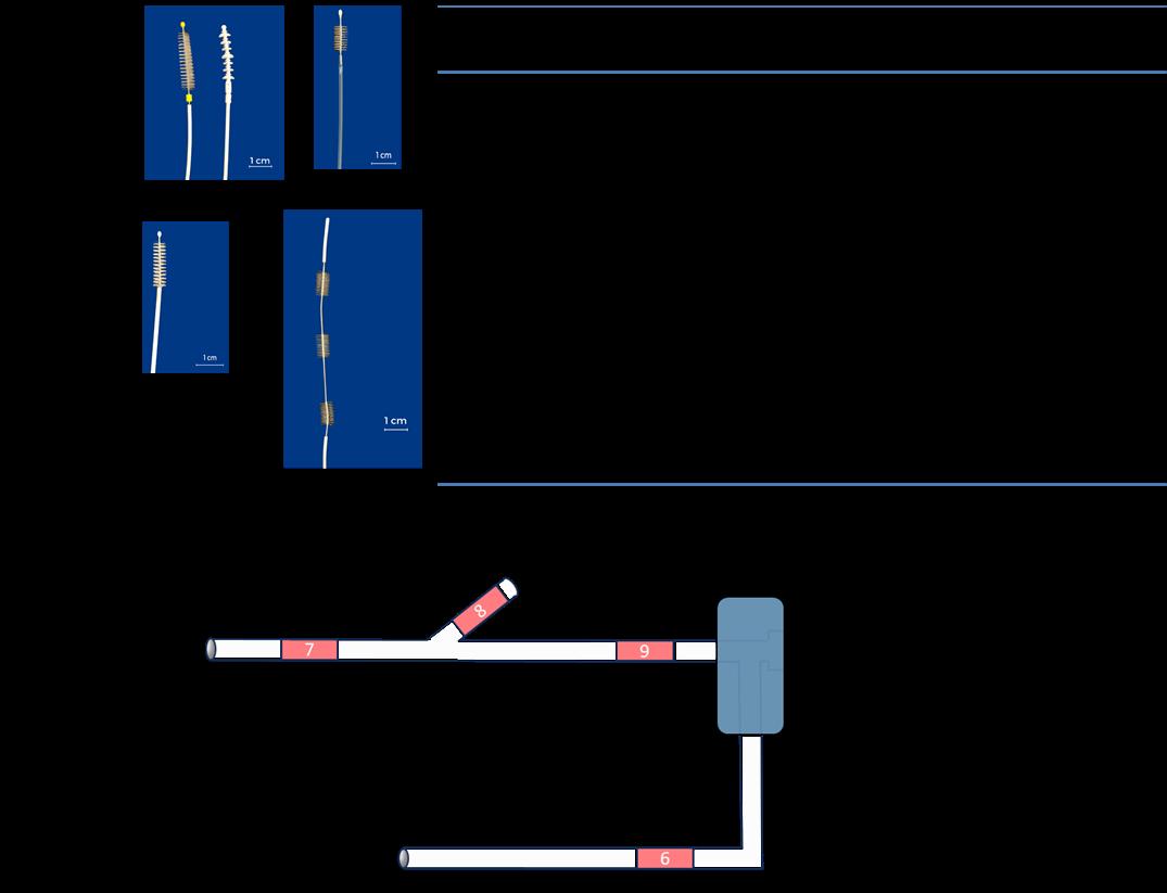
Figure 1: 1A. Tested brushes descriptions and characteristics and B. surrogate B2 schematic representation 2 1B. Surrogate device according to ISO 15883-4:2018 (Block B2): A: suction valve cylinder. B: suction port. C: distal end. D: biopsy
port. 6, 7, 8, and 9: positions of test pieces used for cleaning testing. 4
Results: 5
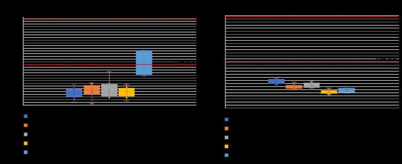
Figure 2 : Proteins and TOC concentration after brushing in recommended use conditions (three-pass).
Alert and action levels are those defined in ISO 15883-5:2021
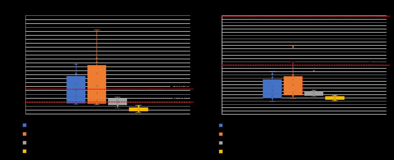
Figure 3: Proteins and TOC concentration after brushing in “worst-case” (“real-case”) conditions (one-pass).
Alert and action levels are those defined in ISO 15883-5:2021
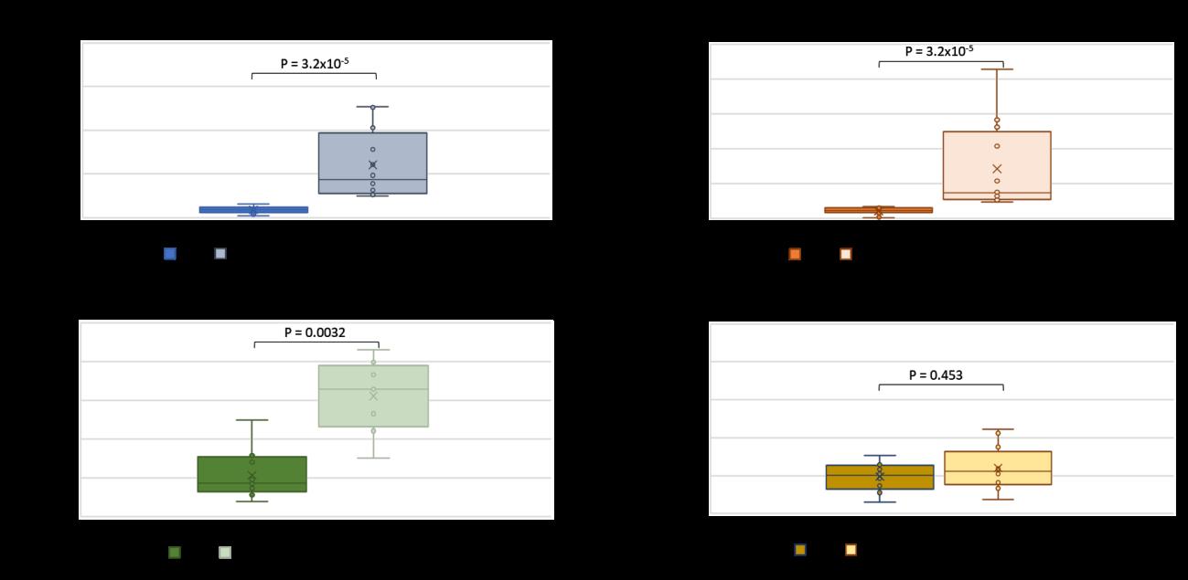
Figure 4: Comparison of the residual amount of proteins when brushes are used either following IFUs (three-pass) or in simulated
“real-case” conditions (“worst-case”: one-pass)). Proteins concentrations after manual brushing in µg of proteins per cm². n = 12
for OLYMPUS, Boston Scientific and Steris brushes, n = 8 for Pentax brush.
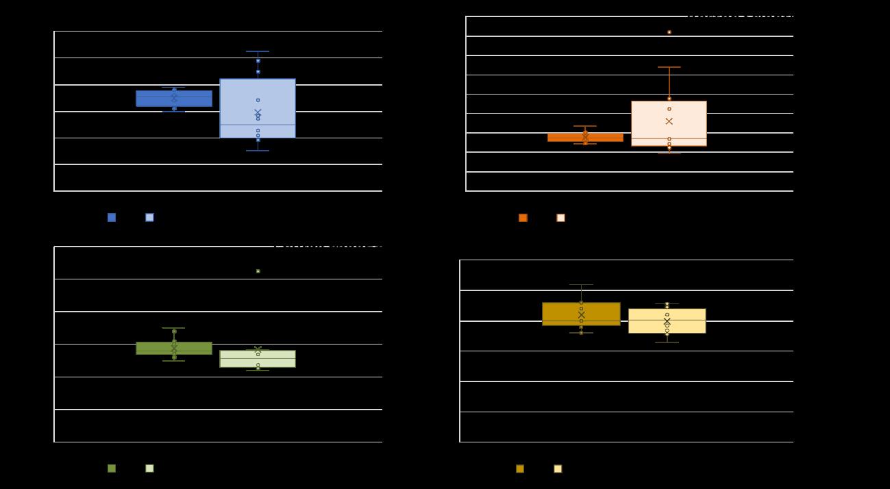
Figure 5: Comparison of the residual amount of TOC when brushes are used either following IFUs (three -pass) or in simulated 17 “real-case” conditions (“worst-case”: one-pass)). Proteins concentrations after manual brushing in µg of proteins per cm². n = 12 18 for OLYMPUS, Boston Scientific and Steris brushes, n = 8 for Pentax brush.
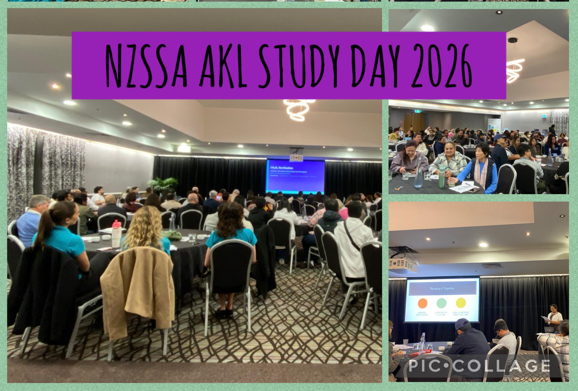
On Saturday, 6 June 2026, the NZ Sterile Sciences Association (Inc.) Auckland Study Day was held at the Waipuna Hotel & Conference Centre, bringing together nearly 100 keen and motivated Sterilisation Technicians from a wide range of healthcare facilities.
Attendees enjoyed a full day of professional development, shared learning, and connection with colleagues across the sterile sciences community.
The programme opened with a presentation from Mel Slattery of Multigate, who delivered an insightful overview of Ethylene Oxide (ETO) sterilisation. Mel explained how the process functions, its applications, and the specific considerations required when using ETO within hospital environments. As ETO is not commonly
used in most CSSDs, the session offered valuable context on its benefits, risks, and the role it plays in specialised reprocessing.
Suzanne Barnes from ASP followed with a focused exploration of the validation process for lowtemperature sterilisers in alignment with AS/NZS 5369:2023 and ISO 22441. She outlined the essential steps involved in conducting a compliant validation, the specialised equipment required, and the critical performance parameters that must be met. Her session provided clarity on how validation is structured, what evidence is necessary, and how teams can ensure ongoing compliance in clinical reprocessing environments.
Surface cleaning within Sterile Sciences Departments was the next topic, presented by Janee Botma from Whiteley. Janee highlighted the importance of maintaining clean,
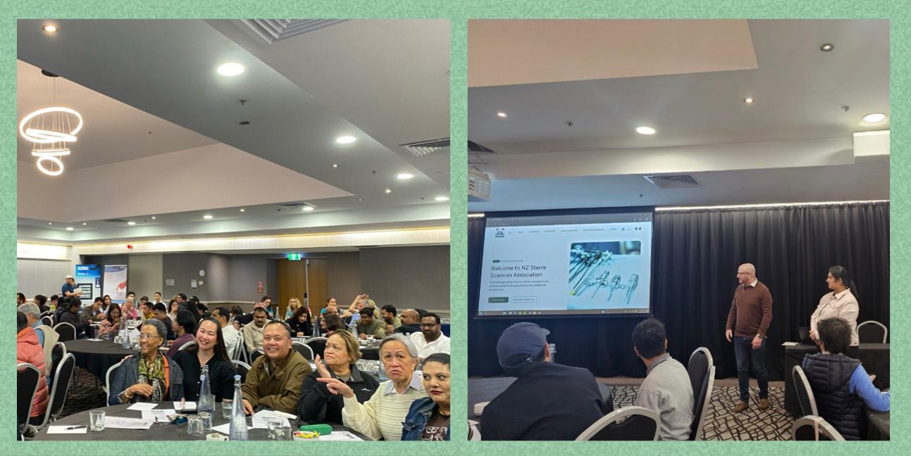
contaminationfree workspaces and explained the cleaning requirements essential for eliminating harmful microorganisms. She also discussed how to select the most appropriate cleaning tools and products to support safe and compliant reprocessing practices.
Providing experience from within the profession, Ashika Devi from Allevia then shared an engaging session on the value of targeted education in strengthening both compliance and capability within sterilisation departments. She demonstrated how structured learning supports the successful implementation of AS/NZS 5369:2023, helping teams build confidence, consistency, and competence in meeting the standard’s requirements. The final learning session of the day was delivered by Murray Hey from Orthomedics, who provided a comprehensive overview of validation and routine checks for rigid containers. Murray outlined the essential inspection points, common faults that compromise container integrity, and the steps required to confirm that rigid containers remain fit for ongoing use in sterile processing environments.
The Study Day also offered an opportunity for the NZSSA Executive to update members on several exciting developments, including the launch of the new NZSSA website, the refreshed membership application process, updated educational requirements for NZSSA registration, and the outstanding quality and number of applicants for this year’s Scholarship Awards.
A warm thankyou goes to all who attended, presented, and contributed to the success of this event. Providing meaningful educational opportunities is a core purpose of the NZSSA, and Study Days like this one continue to be

held around the country for the benefit of all members. The Association looks forward to welcoming both new and familiar faces at the 50th NZSSA Conference in Christchurch this September.
Ngã mihi,
Aileen Derby
NZSSA Executive Member
Charanjeet Sidhu
NZSSA Executive Member
Antony Owens
NZSSA Executive Member
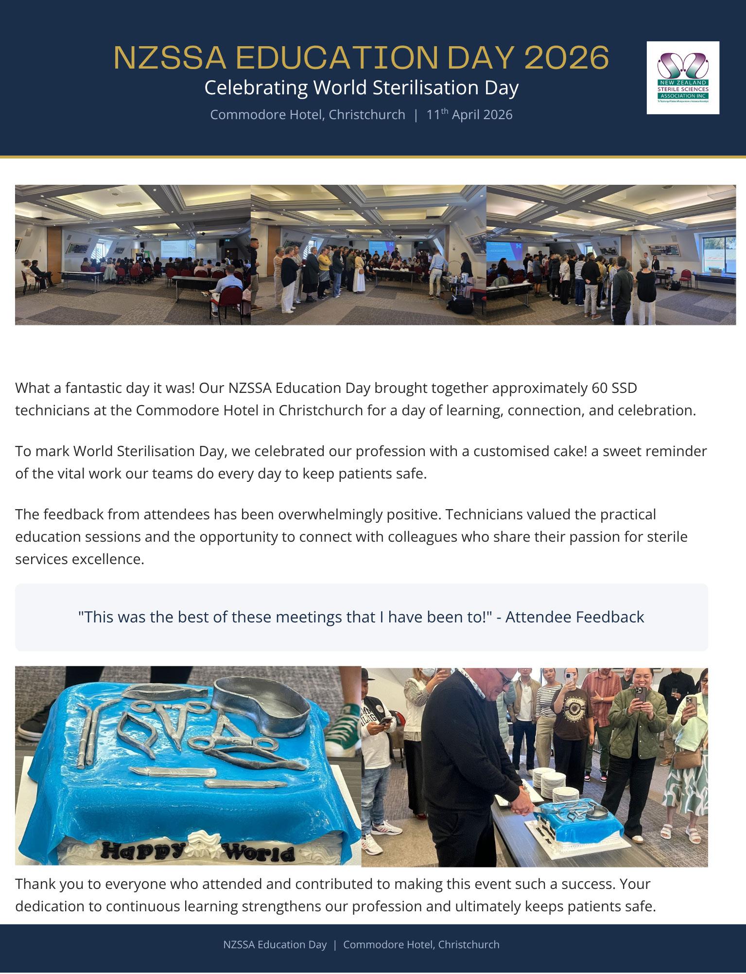
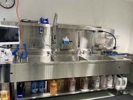
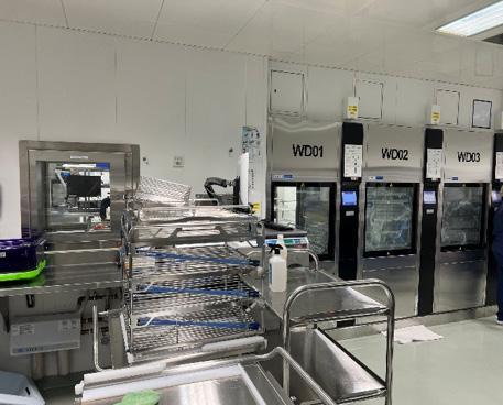
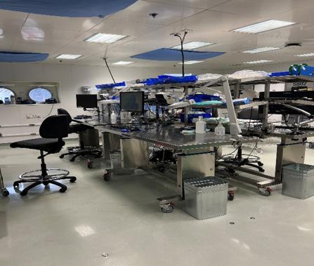
Justine Wheatley, IPC Clinical Nurse Specialist
Some of my favourite moments volunteering as an Infection Prevention and Control nurse aboard a Mercy Ship in Freetown, Sierra Leone, happened in the Sterile Services Department. Every audit, the team lit up and walked me through everything, proud of a set-up that was genuinely state of the art. What amazed me most was the water: the hospital ship negotiates with the government for a raw untreated supply, filters every drop taking at least three days to get the quality needed so it is clean enough to drink, let alone reprocess a surgical instrument. Turning on a tap will never feel ordinary to me again.
Water quality was tested regularly, wash cycles were validated, and every washer-disinfector and steriliser was tracked and logged with the same discipline I would expect in any world-class hospital in a high resource
setting. The packing and assembly area was just as impressive: an orderly, gleaming space where trays were checked, wrapped and prepared with real care, and where the team happily talked me through their process step by step whenever I had a question during an audit. That openness, that willingness to be seen and to keep improving, is exactly what good IPC practice looks like in action.
If you are a sterile services technician wondering whether there is a place for you in humanitarian work, the answer is a very happy yes. This crew would welcome you with open arms and be proud to show you how they make SSD the heart of safe care for all their patients who have nowhere else to turn. It was one of the highlights of my time on board, and I would recommend it to anyone in our profession without hesitation.
The NZSSA Inc. Executive was very impressed with quantity and quality of applications for the 2026 scholarships. It is very pleased to announce the following registered technicians as this years recipients.
Natalie Manley
Jaison Jacob
NZSSA Conference, Christchurch
Anita Castelino
Crystal Cruz
Denise Wylie
Jessica Chadfield
Josini Jose
Sophia Samuel
Sue Granthan
FSRACA Congress, Gold Coast Australia
Ula Rademeyer
Kimberley Nitschke
WFHSS Congress
Annamarie Tee
By Kylie Joy
Possibly the most annoying rhetorical question that a student can ask their teacher or tutor is the one that starts with the word: “Why?” It usually makes any educator wonder about the reasoning behind the subject being taught. So, the question asked as a technician in Sterile Sciences is “Why do we have standards anyway?”
To provide context for reasoning, the World Bank (2025) emphasises that international standardisation is a crucial element for the functioning and growth of the global economy. Its absence would significantly disrupt various industries and economic activities. So, without standardisation of processes and equipment, the global economy would cease to exist and thus, all industries would grind to a halt.
A delve into recent history provides a brief overview of how ISO standards emerged:
The year is 1946, World War 2 had just finished, and nations set to work to rebuild shattered infrastructure. Twenty-six countries, including the United Kingdom, United States, Soviet Union and China (who were allies in World War 2), recognised the need for consistency in the quality of building products. A charter was signed by these 26 nations, agreeing to join forces to produce quality goods with minimal waste of resources and
reduced costs associated with defective products. Thus, the International Organisation for Standardisation commenced operations on 23 February 1947 with the adoption of ISO Recommendations (ISO/R) in manufacturing. Standardisation meant that fair trade agreements could be signed with the assurance that goods and services met set acceptance criteria (British Assessment Bureau, 2025).
In the 1990s, Sterile Services recognised that they also needed their own standards. These standards revolutionised how reprocessing of medical devices would occur. Standards allowed reassurance for the customer, e.g. patients, that a set sterility assurance level (SAL) was being met. AS/NZS 4187:1994 became the foundation for what is now accepted as the “norm” in the New Zealand Sterile Sciences industry (Whiteley, 2026).
This year marks the 50th annual conference since the inception of the New Zealand Sterile Sciences Association in 1974. In recognition of this milestone, the 50th annual conference theme is: “Aim High, Fly High”.
By striving for the highest levels in process standardisation, our industry positions itself to achieve new heights, surpassing what appears as the limit on the horizon. Through continued commitment to excellence, we ensure ongoing progress and innovation in sterile sciences.
NZSSA Inc. registration requires 20 hours education over 3 years. This is not difficult if you plan ahead and make the most of opportunities.
Ongoing education is not the learning you do as part of your orientation or developing your skills as a technician That is a natural part of becoming a sterile sciences technician.
Ongoing eduction is:
Ongoing education is about maintaining current knowledge and skills and expanding so you continuously grow as a professional.
Start preparing your portfolio of education from the time you start working When you have qualified you will then have the content ready.
Sources of ongoing education
Department in-services
Webinars
Projects
Regional meetings
Conference
Online learning modules
Qualifications
Journal article
Research articles
Presentation
Create a member login so you have full access to member only pages. These pages have links to online learning opportunities
NZSSA Inc Professional Development

Semester 2 starts on Monday 20 July and the team at Toi Ohomai Sport and Health team are looking forward to welcoming you. Each course has a kaiako (teachers) to support ākonga (students) throughout their journey along with the wider Toi Ohomai services. Remember to reach out, Rachel, Emma, Lizzy and Alison are there for you .

Need to get ready for your sterilising technology course?
Here is the answer for you! Toi Ohomai has a FEES FREE computer certificate designed to build your skills and prepare you for today’s world and study.
Click on this link: NZ Certificate in Computing (Foundation User) level 2
Key contacts:
Alison Stewart, Programme Coordinator; alison.stewart@toiohomai.ac.nz
Priscilla Hamilton, Administrator : priscilla.Hamilton@toiohomai.ac.nz

Available online at www.sciencedirect.com
journal homep age: www.el sevier.com/l ocate/jhin

S. Zeng *
Department of Disinfection Supply Center, West China School of Medicine, Sichuan University, Sichuan University affiliated Chengdu Second People’s Hospital, Chengdu Second People’s Hospital, Chengdu, Sichuan Province, 610021, China
A R T I C L E I N F O
Article history:
Received 12 January 2026
Accepted 28 March 2026
Available online 9 April 2026
Keywords:
Medical device cleaning
Water hardness
Cleaning agent ratio
Interaction
Response surface optimization

S U M M A R Y
Objective: The aim of the study was to investigate the effects of water hardness and cleaning agent ratio on the cleaning efficiency of 304 stainless steel medical devices and their interaction mechanisms, providing a theoretical basis for optimizing medical device cleaning processes.
Methods: Single-factor experiments and an L25(52) orthogonal design were employed to study the impact of five levels of water hardness (50 250 mg/L) and cleaning agent ratios (1:50 1:250) on cleaning efficiency Residual soil was quantified using ultraviolet-visible (UV-Vis) spectrophotometry, surface morphology was observed via scanning electron microscopy, and cleaning efficiency was evaluated through surface tension measurements and dynamic contact angle analysis. Response surface methodology was utilized to optimize the cleaning process parameters.
Results: As water hardness increased, cleaning efficiency significantly decreased (P < 0.05). At a water hardness of 50 mg/L, the cleaning efficiency was highest (97.5%); it dropped to 65.0% at 250 mg/L. The cleaning agent ratio exhibited an optimal value, with a 1:100 ratio achieving a cleaning efficiency of 95.0%. Analysis of variance (ANOVA) revealed that both water hardness (F = 50.23) and cleaning agent ratio (F = 30.45) significantly influenced cleaning efficiency (P < 0.001), and there was a significant interaction effect (F = 3.67, P = 0.002). Under low hardness water conditions (50 100 mg/L), the effect of cleaning agent ratio on cleaning efficiency was minimal; however, under high hardness conditions (200 250 mg/L), a higher concentration of cleaning agent was necessary to maintain cleaning efficiency. Response surface optimization indicated that a water hardness of 80 mg/L and a cleaning agent ratio of 1:120 yielded the best cleaning performance.
Conclusion: Water hardness significantly affects cleaning efficiency by influencing the effectiveness of surfactants, and there is a notable interaction between water hardness and cleaning agent ratio. Cleaning agent ratios should be appropriately adjusted based on water hardness conditions to achieve optimal cleaning efficiency Specifically, under low hardness conditions (50 100 mg/L), cleaning agent ratios between 1:50 and 1:150 can achieve cleaning efficiency above 95%, while under high hardness conditions (200 250 mg/L), more concentrated ratios (1:50 1:100) are required to maintain acceptable
* Corresponding author. Address: No. 10, Qingyun Street, Jinjiang District, Chengdu City, Sichuan Province, 610017, China. E-mail address: zsy67838134@126.com
https://doi.org/10.1016/j.jhin.2026.03.034 0195-6701/ª 2026 Published by Elsevier Ltd on behalf of The Healthcare Infection Society
cleaning efficiency. The findings of this study provide an important practical guidance for enhancing the quality of medical device cleaning and reducing operational costs.
ª 2026 Published by Elsevier Ltd on behalf of The Healthcare Infection Society.
With the rapid advancement of medical technology and the increasing variety of medical devices, the application of medical devices in clinical diagnosis, surgical operations, and daily care has become more widespread. Cleaning, disinfection, and sterilization of medical devices, as a crucial aspect of their lifecycle management, not only affect the efficiency of device reuse but also directly impact patient safety and treatment outcomes [1]. In medical practice, inadequate cleaning of devices may lead to the residue of pathogenic micro-organisms, causing cross-infection, increasing the risk of patient infections, and even resulting in increased patient morbidity and mortality [2]. It is well established that ineffective cleaning can shield micro-organisms from subsequent disinfection or sterilization processes, rendering them ineffective and posing a significant risk to patient safety.
The complexity of cleaning medical devices lies in the multiple types of soil involved, including blood, body fluids, organic substances, protein deposits, and micro-organisms. These types of soil are not only diverse but also have varying physical and chemical properties, posing significant challenges to the cleaning process [3]. In the cleaning process, water quality, as the basic medium, significantly affects cleaning efficiency Water hardness is primarily determined by the concentration of calcium (Ca2+) and magnesium (Mg2+) ions. These alkaline metal ions interact with the active components in cleaning agents during the cleaning process, affecting the detergent’s ability to remove soil and its stability [4]. High hardness water not only reduces the cleaning efficiency of surfactants but may also lead to scale deposition, increasing equipment maintenance costs and shortening the lifespan of devices [5].
The impact of water hardness on the cleaning process has been widely studied across various research fields. Studies have shown that the presence of calcium and magnesium ions in hard water can form complexes with non-ionic surfactants, reducing the effective concentration of surfactants in water and thereby weakening their soil-removing ability [6]. Additionally, hardness ions can react with organic and inorganic substances in the soil to form insoluble precipitates, further increasing cleaning difficulty [7]. In the cleaning of medical devices, this phenomenon is particularly significant because device surfaces often harbour complex biological soil. Cleaning agents need to possess strong soil-removing capabilities and broad applicability to handle different types of soil [8].
The selection and ratio of cleaning agents are another key factor affecting cleaning efficiency. Commercially available general-purpose medical device cleaning agents typically contain multiple components, such as non-ionic surfactants (1 5%), alkaline components (sodium hydroxide 0.1 5%, carbonates 1 10%, phosphates 1 5%), acidic components (phosphoric acid 0.1 5%, citric acid 0.1 5%), chelating agents (ethylenediaminetetraacetic acid [EDTA] 0.1 2%, sodium citrate 0.1 5%), enzymes (proteases, lipases 0.01 1%), and other
additives (defoamers 0.01 0.5%, preservatives 0.01 0.5%), which work synergistically to achieve efficient removal of soil [9]. Non-ionic surfactants are widely used in the cleaning of medical devices due to their good soil-removing ability and low foaming characteristics. They can effectively disperse and dissolve soil, reducing the adhesion of soil on device surfaces [10]. Chelating agents form stable complexes with metal ions in water, reducing the inhibitory effect of hardness ions on surfactant performance, thereby enhancing the overall soilremoving efficiency of cleaning agents [11]. The addition of auxiliary detergents further enhances the stability and soil-removing effect of the cleaning solution, helping to maintain the physical and chemical balance during the cleaning process [12].
However, the cleaning performance of a detergent can vary considerably depending on its dilution ratio in practical applications. Determining the optimal ratio to adapt to different water quality conditions still requires in-depth research. Excessively high cleaning agent concentrations may lead to excessive foam, affecting cleaning efficiency, while too low cleaning agent concentrations may not sufficiently remove soil, reducing cleaning efficiency Additionally, process parameters such as cleaning temperature and time also significantly affect cleaning efficiency. Under high-temperature conditions, the activity of many chemical cleaning agents (e.g. alkaline or enzymatic components) is enhanced, typically leading to stronger soil-removing abilities. However, excessively high temperatures may denature enzymatic components or damage heat-sensitive device materials, affecting device lifespan [13]. Conversely, enzymatic cleaners have an optimal temperature range, and temperatures outside this range can significantly reduce their efficacy.
In practical applications, medical device cleaning often faces the influence of various complex environmental factors, such as diverse types of soil, making the stability and controllability of cleaning efficiency urgent issues to be addressed. To tackle this challenge, many studies have attempted to systematically investigate the effects of various factors on cleaning efficiency through experimental design and statistical analysis methods and to establish corresponding mathematical models to guide practical operations. For example, orthogonal experimental design, as an efficient experimental method, can comprehensively evaluate the effects of multiple factors and levels on cleaning efficiency with fewer experiments, demonstrating significant advantages [14]. Through reasonable experimental design, not only can experimental resources be conserved but the accuracy and reliability of data analysis can also be improved, providing a scientific basis for optimizing cleaning processes [15].
This study aims to use systematic experimental design to systematically investigate the effects of water hardness and cleaning agent ratio on the cleaning efficiency of 304 stainless steel medical devices and the interaction between them, providing a scientific basis for optimizing medical device cleaning processes.
Experimen tal materials and instruments
Water sample preparation
High-purity deionized water (conductivity ≤0.1 μS/cm, 25 ◦ C) was used as the base water sample in this experiment. Analytical-grade calcium chloride (CaCl2 2H2O, purity ≥99.0%, China National Pharmaceutical Group Chemical Reagent Co., Ltd., Shanghai, China) and magnesium chloride (MgCl2 6H2O, purity ≥98.0%, China National Pharmaceutical Group Chemical Reagent Co., Ltd., Shanghai, China) were used to adjust water hardness. While other divalent cations such as manganese, iron, and zinc can contribute to overall water hardness, calcium and magnesium are the predominant ions and are primarily responsible for the negative effects on surfactant performance in cleaning processes. Therefore, this study focused on these two ions to establish a foundational understanding of the interaction. By precisely preparing mixed salt solutions of different concentrations, the water sample hardness was adjusted to five gradients: 50, 100, 150, 200, and 250 mg/L (as CaCO3). In central sterile supply departments, soft water and purified water are typically used, with soft water hardness standards ≤150 mg/L (as CaCO3), according to GB 5749-2006 (Standards for Drinking Water Quality), and purified water hardness ≤10 mg/L (as CaCO3), according to WS507-2016 (Regulation for cleaning and disinfection technique of flexible endoscope). The hardness adjustment process strictly followed the relevant provisions in GB/T 5750.4-2006 ‘Standard Methods for Examination of Drinking Water - Part 4: Determination of Hardness.’ A HACH DR3900 water quality analyser (Hach Company, Colorado, USA, Performance: measurement accuracy ±0.5 mg/L) was used to verify the hardness of the prepared water samples. Each level was measured three times, and the average value was taken as the final hardness data. To ensure the stability of the water quality used in experiments, preparation of all water samples was completed within 24 h before use and stored in polyethylene containers at 4 ◦ C away from light.
Selection and preparation of cleaning agents
This study used a single, commercially available, neutral pH, general-purpose medical device cleaning agent as the experimental cleaning agent. The key components include non-ionic surfactants (e.g. alcohol ethoxylates, 1 5%), a chelating agent (tetrasodium EDTA, <1%), and proteolytic enzymes (<0.1%). The cleaning agent-to-water volume ratios were set at five gradients: 1:50, 1:100, 1:150, 1:200, and 1:250. The cleaning solution was prepared by volumetric method using an IKA RCT Basic magnetic stirrer (IKA Werke, Staufen, Germany, Performance: speed accuracy ±2 revolutions per minute [rpm]) at room temperature (25±0.5 ◦ C). Specifically, the required volume of the cleaning agent was accurately measured, and the corresponding hardness water was slowly added to reach the set volume. The mixture was stirred at 300 rpm for 30 min to ensure complete dissolution of the cleaning agent. Each ratio solution was prepared at a volume of 500 mL, allowed to stand for 10 min before use to observe the uniformity and stability of the solution. All prepared cleaning solutions were used within 24 h to prevent possible component changes during storage.
Three hundred and four stainless steel medical device samples were selected as the research objects, with sample specifications of 25 mm × 20 mm × 1 mm, surface area of 5 cm2, and surface roughness Ra (average roughness) value of 0.8 μm (measured using a TR200 handheld roughness meter). To ensure the reliability of the experimental results, all samples underwent strict pretreatment. The pretreatment process included the following steps: first, cleaning with anhydrous ethanol (purity ≥99.9%) in an ultrasonic cleaner for 10 min to remove surface oil and impurities; then rinsing repeatedly with deionized water three times, each for 2 min, to ensure the cleanliness of the sample surface; placing the cleaned samples in a class 100 laminar flow clean bench to air-dry naturally for 24 h; and finally weighing using a precision electronic balance (accuracy 0.1 mg) and recording the initial weight. All pretreated samples were numbered and individually sealed in selfsealing bags to prevent contamination and scratches.
Following pretreatment, an artificial test soil was applied to the samples to simulate clinically used medical devices. The artificial soil was formulated based on a modified version of a standard recommended by EN ISO 15883-1 (washer disinfector performance requirements and their accessories) [7], comprising defibrinated sheep blood (80% v/v), egg yolk (10% v/v), and a mixture of amino acids (5% w/v each of glycine, L-cysteine, and L-glutamic acid) in a phosphate buffer. The soil was prepared fresh daily and used within 2 h. A volume of 10 μL of the artificial soil was evenly spread over the entire surface of each precleaned sample using a micropipette. The soiled samples were then dried in a laminar flow hood at 35±2 ◦ C for 1 h to ensure adhesion. After drying, the samples were weighed again (W2) to determine the initial soil load (W1) (W2 W1). This standardized soiling procedure was strictly followed for all samples prior to the ultrasonic cleaning experiments.
The main instruments and equipment used in this study and their technical parameters are as follows:
Ultraviolet-visible (UV-Vis) Spectrophotometer (Agilent 8453, Agilent Technologies, Santa Clara, California, USA): This instrument was the core device used for quantitatively measuring residual soil in the experiment. It has a wavelength range of 190 1100 nm, wavelength accuracy of ±0.5 nm, a measurement range of 0 3.0 Abs and is equipped with 1-cm quartz cuvettes. Prior to the experiment, the instrument was calibrated using a standard potassium dichromate (K2Cr2O7) solution to ensure the accuracy of the measurement results.
Precision Constant Temperature Water Bath (Thermo Scientific Precision 6000, Thermo Fisher Scientific, Waltham, Massachusetts, USA): This device was used to control the temperature environment during the cleaning process. It has a temperature control range from ambient temperature +5 ◦ C to 100 ◦ C, temperature stability of ±0.1 ◦ C, and temperature control accuracy of 0.1 ◦ C. It is equipped with a digital display and a microcomputer temperature control system, allowing precise temperature adjustment and constant temperature control. A calibrated standard thermometer was used for temperature verification during the experiment.
Ultrasonic Cleaner (Branson 5800, Branson Ultrasonics, Brookfield, Connecticut, USA): This device was the key equipment for achieving efficient cleaning. It operates at a frequency
of 40 kHz, with a maximum power of 550 W and a temperature control range from room temperature to 69 ◦ C. The cleaning tank features a specially designed oscillation system that generates a uniform ultrasonic field, ensuring consistency of the cleaning effect. The device is equipped with a digital timer and power adjustment device, allowing a precise adjustment of cleaning parameters according to experimental needs.
Single-factor experiments
This study first conducted single-factor experiments to determine the independent effects of water hardness and cleaning agent ratio on cleaning efficiency. When studying the effect of water hardness, the cleaning agent ratio was fixed at 1:100, with five hardness gradients set at 50, 100, 150, 200, and 250 mg/L, each level repeated three times. During the experiments, other parameters were strictly controlled: temperature maintained at 60±0.5 ◦ C, ultrasonic cleaning time at 10±0.5 min (in accordance with standard requirements that ultrasonic cleaning should not exceed 10 min), and solution pH controlled at 7.0±0.2. In the study of the effect of cleaning agent ratio, the water hardness was fixed at 100 mg/L, and five ratio gradients were set at 1:50, 1:100, 1:150, 1:200, and 1:250, with experimental conditions kept consistent with the water hardness study. This systematic single-factor experimental design clearly reveals the influence patterns of each factor on cleaning efficiency
Orthogonal experiments
Based on the results of the single-factor experiments, an L25(52) orthogonal table was used to design multi-factor experiments, systematically studying the effects of two factors: water hardness (A) and cleaning agent ratio (B) on cleaning efficiency and their interaction. The experiment set five levels: water hardness at 50, 100, 150, 200, and 250 mg/L; cleaning agent ratios at 1:50, 1:100, 1:150, 1:200, and 1:250. Each set of experimental conditions was repeated three times, and analysis of variance (ANOVA) was used to determine the significance of each factor and their interaction. Other parameters were strictly controlled to remain constant to ensure the reliability of the experimental results.
The process of preparing cleaning solutions was divided into two steps: adjusting water hardness and preparing cleaning solution. First, a mixed stock solution of 1000 mg/L CaCl2 and MgCl2 was prepared by accurately weighing according to a molar ratio of 3:1 and dissolving in high-purity deionized water Using grade A volumetric flasks, the solution was diluted stepwise to the required hardness. After each dilution, the water hardness was verified using a water hardness meter, with an allowable error controlled within ±2%. For the preparation of cleaning solutions, the cleaning agent was accurately measured using a pipette, and the prepared hardness water was slowly added to the designated volume. A magnetic stirrer was used to stir at 300 rpm for 30 min until the solution became clear and transparent. The prepared cleaning solutions were to be used immediately. If short-term storage was necessary, they should be kept in a dark environment at 4 ◦ C and used within 24 h.
Cleaning process procedure
The execution of the cleaning process strictly followed standard operating procedures. First, the initial weight of the sample was accurately measured using a precision balance (accuracy 0.1 mg), and the surface state of the sample before cleaning was recorded using a digital microscope (Dino-Lite AM4113T). The constant temperature water bath was preheated to 60±0.5 ◦ C for no less than 30 min, during which a standard thermometer was used for regular calibration. The prepared cleaning solution was poured into the ultrasonic cleaner, set to a power of 400 W, frequency of 40 kHz, and cleaning time controlled at 10±0.5 min. During cleaning, the cleaning solution volume was maintained at 500 mL, and samples were completely immersed. After cleaning, the samples were immediately rinsed three times with deionized water at 25±2 ◦ C, each rinse lasting 5 min. Finally, the samples were gently wiped with 100-grade dust-free cloth and placed on a clean workbench to air-dry naturally. The drying environment temperature was controlled at 25±2 ◦ C and relative humidity maintained at 45±5%.
Cleaning efficiency was evaluated using a combination of residual soil measurement and surface morphology analysis. Following the standardized soiling procedure described in section 1.1.3, the cleaning efficiency was evaluated. To quantify the residual soil on the devices after ultrasonic cleaning, a surrogate measure was used. The soiled samples were subjected to the cleaning process. After cleaning, the remaining soil on each device was thoroughly re-suspended by immersing the device in 50 mL of deionized water and subjecting it to mild ultrasonication for 2 min. This rinsate, containing the residual soil, was collected, filtered using a 0.45-μm filter membrane to remove particulates, and stored at 4 ◦ C for testing. Ten millilitres of this soil-containing filtrate was placed in a colorimeter tube, 1.0 mL of 1.0 mol/L nitric acid was added, and the solution was allowed to react at room temperature (25 ◦ C) for 30 min. This step ensures that any proteinaceous material in the soil is sufficiently protonated for detection. The absorbance of the solution was measured at 600 nm using a UV-Vis spectrophotometer with a 1-cm quartz cuvette, using deionized water as a blank control. At 600 nm, the turbidity caused by the suspended soil particles, primarily coagulated proteins and lipids, is measured. This method provides a relative quantification of residual organic soil. The residual soil amount was calculated based on a pre-established standard curve (0 50 mg/L) prepared using the same artificial test soil. Cleaning efficiency (%) was calculated using the following formula: Cleaning Efficiency (%) = ([Initial Soil Load Residual Soil Load / Initial Soil Load) × 100 where the Initial Soil Load (W2 W1) was determined gravimetrically and the Residual Soil Load was quantified via UV-Vis spectrophotometry against a standard curve.
To further characterize the cleaning solutions under different ratios, surface tension and foam stability were measured. Surface tension of the prepared cleaning solutions (at 25±0.5 ◦ C) was determined using the Wilhelmy plate method with a calibrated tensiometer (DCAT 11, DataPhysics Instruments, Germany). A platinum iridium plate was used, and the instrument was calibrated against ultrapure water (72.8 mN/m
at 20 ◦ C) before each measurement session. Each sample was measured five times, and the average value was recorded. Foam stability was assessed using a simple shake test. Fifty millilitres of the cleaning solution was placed in a 100-mL graduated cylinder and vigorously shaken manually for 30 s. The initial foam volume was recorded immediately after shaking, and the foam decay, i.e. the time for the foam volume to reduce by half, was measured and reported as foam duration.
Statistical analysis of experimental data was performed using SPSS 25.0 software. ANOVA was conducted on the singlefactor experimental results, with a significance level set at α = 0.05, and multiple comparisons were performed using the least square difference (LSD) method. Orthogonal experiment data were processed using range analysis, and ANOVA was used to determine the significance of each factor and their interaction effects. All experimental data were expressed as mean ± standard deviation (mean ± SD, n=3).
Effect of water hardness on cleaning efficiency
Residual soil amount analysis
Under a fixed cleaning agent ratio (1:100), the effect of different water hardness conditions on the cleaning efficiency of medical devices is shown in Table I. As water hardness increased, the residual soil amount showed a significant
Table I
Evaluation indicators of cleaning efficiency under different water hardness conditions*
Water hardness (mg/L)
Residual soil amount (mg/L) Cleaning efficiency (%) Surface stain coverage (%) Solution turbidity (NTU)
50 2.5±0.2a 97.5±0.3a 3.2±0.3a 1.2±
Data are expressed as mean ± standard deviation (N = 3). Different superscript letters in the same column indicate significant differences (P < 0.05).
NTU, nephelometric turbidity unit.
Table II
upward trend (P < 0.05), and cleaning efficiency correspondingly decreased. At a water hardness of 50 mg/L, the residual soil amount was the lowest (2.5±0.2 mg/L), with a cleaning efficiency of 97.5%. When water hardness increased to 250 mg/ L, the residual soil amount significantly increased to 35.0±1.5 mg/L, and cleaning efficiency dropped to 65.0%. ANOVA results indicated that water hardness had a significant impact on cleaning efficiency (F(4, 25) = 45.67, P < 0.001).
Cleaning efficiency analysis
Under a fixed water hardness of 100 mg/L, the effect of cleaning agent ratio on the cleaning efficiency of medical devices is shown in Table II The results indicate that when the cleaning agent ratio varied from 1:50 to 1:250, the cleaning efficiency first increased and then decreased. At a cleaning agent ratio of 1:100, the best cleaning efficiency was achieved, with the lowest residual soil amount (5.0±0.3 mg/L) and a cleaning efficiency of 95.0%. Notably, at a cleaning agent ratio of 1:50, although the cleaning agent concentration was highest, the cleaning efficiency was not significantly better than that of the 1:100 ratio (P > 0.05). This may be due to excessive foam caused by too high a cleaning agent concentration, which adversely affects cleaning efficiency.
Surface activity characteristic analysis
The surface activity characteristics under different cleaning agent ratios are shown in Figure 1 As the cleaning agent concentration decreased (ratio increased from 1:50 to 1:250), the surface tension of the solution gradually increased and foam stability decreased. Surface tension measurements showed that at a 1:100 ratio, the solution exhibited ideal surface activity (35.8±0.6 mN/m), ensuring sufficient soil-removing capability without excessive foam production. The critical micelle concentration (CMC) determination at this ratio indicated that cleaning agent molecules could form a stable monolayer on the device surface, facilitating soil removal.
Interaction between water hardness and cleaning agent ratio
Analysis of orthogonal experiment results
Using the L25(52) orthogonal design to examine the interaction between water hardness and cleaning agent ratio, the results are shown in Table III ANOVA indicated that both factors significantly influenced cleaning efficiency (P < 0.001), and there was a significant interaction effect (F(16, 80) = 3.67, P = 0.002). Among them, the influence of water hardness (F =
Evaluation indicators of cleaning efficiency under different cleaning agent ratios*
1:200
Data are expressed as mean ± standard deviation (N = 3). Different superscript letters in the same column indicate significant differences (P < 0.05).
1. Effects of cleaning agent ratios on solution properties. A: Surface tension variation curves under different ratios. B: Foam stability decay curves over time. C: Relationship between cleaning efficiency and surface tension. D: Comparison of critical micelle concentration (CMC) values under different ratios.
50.23) was greater than that of cleaning agent ratio (F = 30.45).
By establishing the response surface for the interaction between water hardness and cleaning agent ratio (Figure 2), the synergistic effects between the two factors can be visually observed. Under low hardness water conditions (50 100 mg/ L), the influence of cleaning agent ratio on cleaning efficiency was relatively small, and good cleaning efficiency (cleaning efficiency >95%) was achieved within the ratio range of 1:50 1:150. However, under high hardness water conditions (200 250 mg/L), the influence of cleaning agent ratio significantly increased, requiring higher concentrations of cleaning agents (1:50 or 1:100) to maintain good cleaning efficiency. This interaction characteristic indicates that, in practical applications, the amount of cleaning agent should be appropriately adjusted based on water hardness conditions.
Discussion
This study systematically explored the effects of water hardness and cleaning agent ratio on the cleaning efficiency of medical devices and revealed the interaction mechanisms between them through single-factor experiments and orthogonal experimental design.
The experimental results indicated that water hardness significantly affected the cleaning efficiency, with residual soil amount increasing and cleaning efficiency decreasing as water hardness increased. This phenomenon is consistent with previous research findings. In high hardness water, the presence of calcium and magnesium ions forms complexes with surfactants, reducing the effective concentration of surfactants and thereby weakening their soil-removing ability [16]. Additionally, high hardness water leads to scale deposition, increasing equipment maintenance costs and shortening device lifespan [17]. In this study, as water hardness increased from 50 mg/L to 250 mg/L, cleaning efficiency decreased from 97.5% to 65.0%, indicating a significant negative impact of water hardness on cleaning efficiency The impact of high hardness water on the cleaning process is not only reflected in the reduced soilremoving ability of surfactants but also may cause the formation of insoluble precipitates with organic and inorganic substances in the soil, increasing cleaning difficulty [18]. In the cleaning of medical devices, the device surfaces often harbour complex biological soil, including blood, body fluids, proteins, etc., requiring strong soil-removing ability and broad applicability Notably, the impact of water hardness on cleaning efficiency showed different trends within different hardness ranges. Under low hardness water conditions (50 100 mg/L), cleaning efficiency was high, and residual soil amount was low, indicating that within this range, the negative impact of water
Table III
Data are expressed as mean ± standard deviation (N = 3). Different superscript letters in the same column indicate significant differences (P < 0.05).
hardness on cleaning efficiency was small. However, as hardness further increased, cleaning efficiency significantly decreased, indicating that additional measures are needed under high hardness water conditions to maintain cleaning efficiency. This is consistent with related research results that appropriately adjusting the cleaning agent ratio can effectively improve cleaning efficiency [19]. The cleaning agent ratio is another key factor affecting cleaning efficiency. The experimental results showed that, under a fixed water hardness of 100 mg/L, the cleaning agent ratio significantly affected cleaning efficiency When the cleaning agent ratio was 1:10, the cleaning efficiency was highest and the residual soil amount was lowest. As the cleaning agent ratio increased, cleaning efficiency first increased and then decreased, indicating an optimal ratio range.
This result is consistent with those of related studies, indicating that nonionic surfactants have optimal soil-removing capabilities and low foaming characteristics within an appropriate ratio, suitable for cleaning the surfaces of medical devices [20]. Additionally, the presence of chelating agents can form stable complexes with metal ions in water, reducing the inhibitory effect of ions on surfactant performance, thereby enhancing the overall soil-removing efficiency of cleaning agents [21]. In this study, at a cleaning agent ratio of 1:10, the synergistic effect of nonionic surfactants and chelating agents reached the optimal state, ensuring sufficient soil-removing capability without excessive foam production, maintaining the stability of the cleaning solution. Although high cleaning agent concentrations theoretically should have stronger soilremoving abilities, the experimental results showed that their
cleaning efficiency was not significantly better than that of the 1:10 ratio. This may be due to excessive foam caused by too high a cleaning agent concentration, which affects the permeability and contact efficiency of the cleaning solution, thereby reducing cleaning efficiency. This is consistent with studies indicating that excessively high cleaning agent concentrations may lead to excessive foam, affecting the stability of cleaning efficiency [22].
Orthogonal experiment results showed that there is a significant interaction between water hardness and cleaning agent ratio, and the influence of water hardness on cleaning efficiency was greater than that of cleaning agent ratio. This means that under different water hardness conditions, the influence of cleaning agent ratio on cleaning efficiency varies, necessitating adjustments in cleaning agent amounts based on specific water quality conditions. Under low hardness water conditions (50 100 mg/L), the influence of cleaning agent ratio on cleaning efficiency was small, and good cleaning efficiency (>95%) could be achieved within the ratio range of 1:5 1:15. This is because in low hardness water, the concentration of calcium and magnesium ions is low and does not significantly affect the soil-removing ability of surfactants, so even with changes in the cleaning agent ratio, cleaning efficiency remains high. This is consistent with studies finding that under low hardness water conditions, the influence of cleaning agent ratio on cleaning efficiency is small and cleaning efficiency remains relatively stable [23].
However, under high hardness water conditions (200 250 mg/L), the influence of cleaning agent ratio significantly increased, requiring higher concentrations of



(90, 95) (85, 90) (80, 85) (75, 80) (70, 75) (65, 70) (60, 65) (55, 60) Efficiency (%)
cleaning agents (1:5 or 1:10) to maintain good cleaning efficiency. This is because in high hardness water, the concentration of calcium and magnesium ions is high, forming complexes with surfactants and reducing their effective concentration, thereby weakening their soil-removing ability Therefore, increasing the cleaning agent ratio can compensate for the decrease in effective surfactant concentration, enhancing soil-removing capability and maintaining cleaning efficiency This is consistent with related studies indicating that increasing the cleaning agent ratio in high hardness water conditions can effectively improve cleaning efficiency [24].
The presence of interaction indicates that water hardness and cleaning agent ratio do not act independently during the cleaning process but rather interact with each other, jointly determining the final cleaning efficiency. This interaction characteristic is of important practical significance, suggesting that cleaning agent amounts need to be adjusted under different water quality conditions to achieve optimal cleaning efficiency. For example, in hospitals or medical institutions, water quality conditions may vary due to factors such as region and season, making it particularly important to develop flexible cleaning agent usage schemes. This study further revealed the mechanism by which cleaning agent ratio affects cleaning efficiency through the analysis of surface activity characteristics. The experimental results showed that as the cleaning agent ratio increased (from 1:50 to 1:250), the surface tension of the solution gradually increased and foam stability decreased. This indicates that the concentration of the
cleaning agent significantly affects the surface activity characteristics of the solution.
At a 1:10 ratio, the solution exhibited ideal surface activity, with a surface tension of 35.8±0.6 mN/m and moderate foam stability, ensuring sufficient soil-removing capability without excessive foam production, which affects cleaning efficiency. This is consistent with studies indicating that non-ionic surfactants can form stable monolayers at appropriate ratios, aiding in soil removal [25].
Foam stability has an important impact on the cleaning process. Moderate foam can enhance the permeability and contact efficiency of the cleaning solution, promoting the dispersion and dissolution of soil. However, excessive foam can reduce the fluidity of the cleaning solution, affecting its full contact with soil and thus decreasing cleaning efficiency. This phenomenon was verified in this study; at a cleaning agent ratio of 1:5, although the cleaning agent concentration was high, excessive foam may have adversely affected the cleaning efficiency, resulting in no significant improvement over the 1:10 ratio [26]. Furthermore, the determination results of CMC showed that at a cleaning agent ratio of 1:10, the cleaning agent molecules could form a stable monolayer on the device surface, facilitating soil removal. This indicates that at this ratio, the soil-removing ability and stability of surfactants reached an optimal balance, further enhancing cleaning efficiency This is consistent with studies indicating that the presence of chelating agents can enhance the surfactant’s surface activity and improve soil-removing efficiency [27].
In addition to water hardness and cleaning agent ratio, process parameters such as cleaning temperature and time also significantly affect cleaning efficiency Under hightemperature conditions, cleaning agents typically have stronger soil-removing abilities, effectively dissolving and dispersing soil. However, excessively high temperatures may damage device materials, shortening device lifespan. Therefore, optimizing cleaning process parameters, especially selecting appropriate cleaning agent ratios and controlling temperature under different water hardness conditions, is key to achieving efficient cleaning.
Studies have shown that appropriately increasing cleaning temperature can significantly enhance cleaning efficiency, but it needs to be controlled within a range that does not damage device materials [28]. In this study, the cleaning temperature was strictly controlled at 60±0.5 ◦ C, a temperature that could ensure the soil-removing ability of the cleaning agent without damaging 304 stainless steel medical device samples, ensuring the reliability and reproducibility of the experimental results.
Although this study achieved important results, there are still some limitations. First, while calcium and magnesium are the principal components of water hardness, other ions like manganese, iron, and zinc were not included in the experimental design. Their potential impact on cleaning efficacy, possibly through different interaction mechanisms with detergent components (e.g. catalysing decomposition or forming different precipitates), warrants further investigation. Second, only 304 stainless steel medical device samples were used, without involving devices of other materials, limiting the generalizability of the results. Different materials may have different tolerances and reactivities to cleaning agents, so future research should expand to more types of medical devices to improve the broad applicability of the results. Third, the experimental conditions were relatively simplified; in actual medical device cleaning processes, more complex factors may exist, such as the diversity of soil types and the complexity of water quality parameters. These factors may have additional impacts on cleaning efficiency. Future research should consider more complex experimental conditions to better simulate real application scenarios. Additionally, this study mainly focused on the effects of water hardness and cleaning agent ratio on cleaning efficiency under fixed ultrasonic conditions, without delving into other factors that may influence cleaning efficiency, such as cleaning time, temperature variations, and cleaning solution flow rate, or comparing different cleaning modalities (e.g. flow/pressure-based systems). Future research can adopt multi-factor experimental designs to systematically study more factors affecting cleaning efficiency and their interactions, thereby comprehensively optimizing cleaning process parameters.
In summary, this study, through systematic experimental design and data analysis, systematically explored the independent effects and interaction of water hardness and cleaning agent ratio on the cleaning efficiency of medical devices. The results showed that both water hardness and cleaning agent ratio significantly affect cleaning efficiency, and there is a significant interaction between them. Under low hardness water conditions (50 100 mg/L), the influence of cleaning agent ratio on cleaning efficiency was small, with ratios from 1:50 to 1:150 achieving cleaning efficiency above 95%. Under high hardness water conditions (200 250 mg/L), appropriately increasing the cleaning agent concentration (using ratios of
1:50 1:100) could significantly improve cleaning efficiency and maintain it above 80%.
CRediT authorship contribution statement
S. Zeng: Writing review & editing, Writing original draft, Methodology, Formal analysis, Data curation, Conceptualization.
Ethical approval
All research is in strict accordance with and adheres to the Declaration of Helsinki. The study was approved by the Ethics Committee of Chengdu Second People’s Hospital.
The datasets used and/or analysed during the current study are available from the corresponding author on reasonable request.
Funding sources
No funding was received.
Conflict of interest statement
None declared.
[1] Josephs-Spaulding J, Singh OV. Medical device sterilization and reprocessing in the era of Multidrug-Resistant (MDR) bacteria: issues and regulatory concepts. Front Med Technol 2021;2:587352.
[2] Kremer T, Rowan NJ, McDonnell G. A proposed cleaning classification system for reusable medical devices to complement the Spaulding classification. J Hosp Infect 2024;145:88 98.
[3] McLaughlin M, Moussourakis M, Phillips J. Selection and Validation of Detergents and Surfactants for Aqueous Critical Cleaning. In: Surfactants in precision cleaning. Elsevier; 2022. p. 273 310
[4] Monteiro DR, Gorup LF, Takamiya AS, Ruvollo-Filho AC, de Camargo ER, Barbosa DB. The growing importance of materials that prevent microbial adhesion: antimicrobial effect of medical devices containing silver. Int J Antimicrob Agents 2009; 34:103 10.
[5] Sakurai K, Hiratsuka M, Nakamori H, Namiki K, Hirakuri K. Evaluation of sliding properties and durability of DLC coating for medical devices. Diam Relat Mater 2019;96:97 103
[6] Alfa MJ. Medical-device reprocessing. Infect Control Hosp Epidemiol 2000;21:496 8.
[7] European Committee for Standardization. EN ISO 158831:2009+A1:2021 Washer-disinfectors - Part 1: general requirements, terms and definitions and tests. Brussels: CEN; 2021.
[8] Panta G, Richardson AK, Shaw IC. Quality of water for reprocessing of medical devices in healthcare facilities in Nepal. J Water Health 2021;19:682 6
[9] Kremer TA, Felgar J, Rowen N, McDonnell G. Validation of the device feature approach for reusable medical device cleaning evaluations. Biomed Instrum Technol 2023;57:143 52
[10] Bahreini R, Doshmangir L, Imani A. Influential factors on medical equipment maintenance management: In search of a framework. J Qual Maint Eng 2019;25:128 43
[11] Kim JM, Lee WR, Kim JH, Seo JM, Im C. Light-induced fluorescence-based device and hybrid mobile app for oral hygiene
management at home: development and usability study. JMIR Mhealth Uhealth 2020;8:e17881
[12] Zhu Y, Xu Y, Ling Z, Zhao C, Xu A, He F. The biofilm removal effect and osteogenic potential on the titanium surface by electrolytic cleaning: An in vitro comparison of electrolytic parameters and five techniques. Clin Oral Implants Res 2024;35:454 66
[13] Danby SG, Brown K, Wigley AM, Chittock J, Pyae PK, Flohr C. The effect of water hardness on surfactant deposition after washing and subsequent skin irritation in atopic dermatitis patients and healthy control subjects. J Invest Dermatol 2018;138:68 77.
[14] Rehman MAU, Bastan FE, Haider B, Boccaccini AR. Electrophoretic deposition of PEEK/bioactive glass composite coatings for orthopedic implants: A design of experiments (DoE) study. Mater Des 2017;130:223 30
[15] Rowan NJ, Kremer T, McDonnell G. A review of Spaulding’s classification system for effective cleaning, disinfection and sterilization of reusable medical devices: Viewed through a modern-day lens that will inform and enable future sustainability. Sci Total Environ 2023;878:162976.
[16] Albert DE. Methods for verifying medical device cleanliness. In: Developments in surface contamination and cleaning. William Andrew Publishing; 2015. p. 109 28.
[17] World Health Organization. Guidelines for drinking-water quality: incorporating the first and second addenda. Geneva: WHO; 2022.
[18] Knetsch MLW, Koole LH. New strategies in the development of antimicrobial coatings: the example of increasing usage of silver and silver nanoparticles. Polymers 2011;3:340 66.
[19] MacNeill AJ, Hopf H, Khanuja A, Alizamir S, Bilec M, Eckelman MJ. Transforming the medical device industry: road map to a circular
economy: study examines a medical device industry transformation. Health Aff 2020;39:2088 97
[20] Guan A, Li Z, Phillips KS. The effects of non-ionic polymeric surfactants on the cleaning of biofouled hydrogel materials. Biofouling 2015;31:689 97.
[21] Petrova E, Serdechnova M, Shulha T, Lamaka SV, Wieland DCF, Karlova P. Use of synergistic mixture of chelating agents for in situ LDH growth on the surface of PEO-treated AZ91. Sci Rep 2020;10:8645
[22] Hoover J, Drosnock MA, Carfaro C, Kremer TA. Cleaning challenges: Can extended soil dry times be reversed? Biomed Instrum Technol 2023;57:44 51
[23] Pradhan P, Costa L, Rybski D, Lucht W, Kropp JP. A systematic study of sustainable development goal (SDG) interactions. Earth’s Future 2017;5:1169 79
[24] Muraliraja R, Selvan RAS, Selvakumar A, Franco M, Tamilarasan TR, Sanjith U, et al. A review of electroless coatings on non-metals: Bath conditions, properties and applications. J Alloys Compd 2023;960:170723
[25] Desrousseaux C, Sautou V, Descamps S, Traore ´ O. Modification of the surfaces of medical devices to prevent microbial adhesion and biofilm formation. J Hosp Infect 2013;85:87 93
[26] Bureiko A, Trybala A, Kovalchuk N, Starov V. Current applications of foams formed from mixed surfactant polymer solutions. Adv Colloid Interface Sci 2015;222:670 7
[27] Chelazzi D, Bordes R, Giorgi R, Sekine FH, Ogura T, Baglioni P. The use of surfactants in the cleaning of works of art. Curr Opin Colloid Interface Sci 2020;45:108 23
[28] Walsh LJ. Current challenges in environmental decontamination and instrument reprocessing. Int Dent J 2024;74:S455 62
Martin Bird (President)
CSSD Manager
Dunedin Hospital
Dunedin
martin.bird@southendhb.govt.nz
Carla Coetzee (Secretary)
SSD
Health NZ Hawkes Bay
Hastings
Email: secretary@nzssa.co.nz
Aileen Derby
CSSD Manager Counties Manukau District
Auckland aileen.derby@tewhatuora.govt.nz
Charanjeet Kaur Sidhu
CSSD Quality Lead Clinical Counties Manukau District
Auckland Charanjeet.Sidhu@TeWhatuOra.govt.nz
Jenny Carston
Team Leader CSU
Tauranga Hospital Tauranga Jenny.Carston@bopdhb.govt.nz
Shelley Morrison
CSSD
University of Otago Faculty of Dentistry Dunedin 9054
shelley.vivian@otago.ac.nz
General Enquiries
Donna Belle Dador (Vice President)
National Sterile Services Adviser Southern Cross Healthcare Christchurch donna.dador@southerncrosshospitals.co.nz
Shelagh Thomas CSSD
Hutt Valley Capital, Coast and Hutt Valley shelagh.thomas@huttvalleydhb.org.nz
Antony Owens
Team Leader
Southern Cross Healthcare
Southern Cross Gillies Hospital Auckland antony.owens@schl.co.nz
John Barnacott
Manager Sterile Services Unit
Mid Central Palmerston North Hospital Palmerston North John.Barnacott@midcentraldhb.govt.nz
Alison Stewart (Treasurer)
NZSSA Treasurer
28 Brighton Street, Island Bay Wellington 6023 Email: accounts@nzssa.co.nz
secretary@nzssa.co.nz
Overseas qualification and membership inquiries accounts@nzssa..co.nz
Complaints complaints@nzssa.co.nz
Registration Assessors –Charanjeet Sidhu
Maureen Scott
Donna Dador
Shelagh Thomas
Charanjeet.Sidhu@TeWhatuOra.govt.nz Maureen.Scott@waikatodhb.health.nz donna.dador@southerncrosshospitals.co.nz shelagh.thomas@huttvalleydhb.org.nz