14 minute read
INFLAMMATORY NODULES


Dr Patrick Treacy asks, what is causing dermal filler inflammat ory nodules?
Hyaluronic acid (HA)--based dermal fillers are injectable viscoelastic gels frequently used globally in aesthetic medicine to enhance youthfulness, replenish diminished volume, or improve areas of structural insufficiency.
The foremost objective of employing HA-based fillers is to secure an aesthetic improvement or rectify signs of soft tissue ageing, ensuring both the results’ longevity and the procedure’s safety.
While these fillers have been traditionally seen as immune system-neutral, the formulations of many products on the market have undergone significant alterations, especially in production.
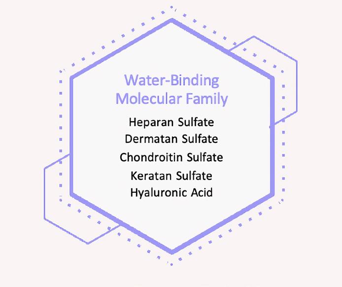
Hyaluronic acid (HA), a polysaccharide prevalent in the skin’s extracellular matrix, possesses a notable capacity to bind and retain water molecules. This property is crucial for maintaining skin hydration and imparting volume.
Structurally, HA comprises repeated disaccharide units of D-gluconic acid and D-N-acetylglucosamine, interconnected by β-1,4 glycosidic bonds. Beyond enzymatic degradation by hyaluronidase, HA is subject to breakdown by various factors, including mechanical stress, reactive oxygen species, fluctuations in temperature and pH, and ultrasonic waves. 1
Numerous hyaluronic acid (HA) fillers are available, each with unique properties like elastic modulus (G’), viscous modulus (G”), cohesivity, particle size, and HA concentration. These characteristics influence the suitability of a filler for specific procedures or skin types, its behaviour under mechanical stress once injected, and its dissolution response to hyaluronidase.
Extensive research has been conducted on the rheological properties of these fillers to guide clinicians in selecting the most appropriate filler. The elastic modulus (G’) is a crucial parameter for evaluating HA fillers because it encapsulates several critical aspects, such as HA concentration and crosslinking degree. Higher crosslinking and HA concentration generally result in a higher G’ value. Fillers with a lower G’ are typically softer, while those with a higher G’ are firmer. However, G’ is not an infallible measure; for instance, variations in G’ values for the same filler have been observed to range widely, with differences up to 7.4 times due to different methods of experimentation. 2

When injected, the immune system recognises all fillers as foreign entities. This typically triggers an inflammatory response around the implant, which is a standard reaction by the host body, leading eventually to the breakdown and absorption of biodegradable fillers. The extent of the body’s response can vary, ranging from minimal macrophage infiltration to an intense foreign-body granulomatous reaction accompanied by fibrosis. Different fillers elicit different reactions; for example, calcium hydroxylapatite tends to provoke a macrophage-dominant response, whereas hyaluronic acid (HA) is more likely to cause a lymphocytic infiltrate. The severity of this immune reaction depends on how immunetolerant the injected material is, which is influenced by various factors. These include the composition and volume of the substance injected, the shape and size of the particles, their ability to biodegrade, and, in the case of HA fillers, aspects such as HA concentration, the level of crosslinking, and the specific techniques used for HA structuring and crosslinking. 3
Modifying HA dermal fillers
Given its relatively brief half-life of less than one to four days in the skin, HA in dermal fillers is often chemically modified to enhance its stability and prolong cosmetic effects. Dermal fillers are histologically classified as either “volumisers,” which elicit a minimal cellular response, or “stimulators,” which induce a strong cellular reaction.
Hyaluronic acid (HA) fillers vary in properties such as crosslinking, gel particle size, and concentration. Extensive crosslinking is believed to enhance longevity due to increased resistance to degradation by native hyaluronidase. Similarly, larger gel particle sizes, due to their reduced total surface area, are more resistant to enzymatic degradation. However, larger particle sizes and increased concentration enhance the hydrophilic nature of the product, resulting in more tissue swelling post-procedure. Higher concentration, larger particle size, and greater crosslinking extend the product’s longevity and theoretically heighten the risk of adverse reactions.
Experience with higher molecular weight HA fillers (like SubQ, Voluma, and Macrolane) suggests they may be problematic regarding capsulation and delayed onset nodules. The most common modification technique involves crosslinking HA chains using 1,4-butanediol diglycidyl ether (BDDE), which forms ether bonds with HA’s hydroxyl groups. This crosslinking process preserves the β-1,4 glycosidic bonds’ accessibility for cleavage by hyaluronidase. These changes aim to enhance durability and tailor the fillers for particular uses.
Do these modifications cause problems?
Historically, HA fillers have been considered to be immunologically inactive due to their universal presence in living organisms and their nonspecificity to tissues. However, the crosslinking technologies employed by manufacturers to enhance the durability and adjust the characteristics of each HA filler differ significantly.
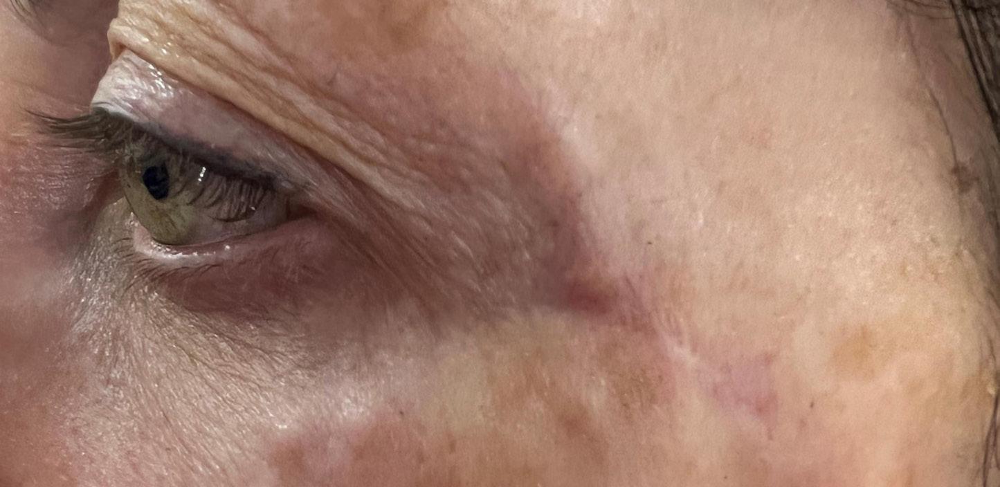
It is unclear how much the HA molecule can be altered before it becomes unrecognisable as native HA. However, it is plausible that extensive modifications might heighten the likelihood of an inflammatory response. 4 Additionally, the inherent properties of the filler or its breakdown products could potentially trigger inflammation. However, these modifications, possibly in conjunction with other variables like the patient’s immune condition, events that activate the immune system, and bacterial contamination, can sometimes result in nodules appearing late after the treatment in a subset of patients. 5 Some of these lumps that appear several months after injecting hyaluronic acid dermal fillers are typically considered to be inflammatory rather than infective. These delayed inflammatory nodules usually present as firm to hard lumps at or near the filler injection sites. Inflammatory nodules resulting from hyaluronic acid dermal fillers often present with symptoms like pain, tenderness, or redness. The timing of their appearance, often weeks to over a year post-injection, with the most common onset around four months, is a critical factor in differentiating them from infective processes, which usually manifest more acutely. They are generally named “delayed onset nodules” (DON), a term which encompasses various conditions such as lumps, masses, nodules, areas of hardening, delayed hypersensitivity reactions, biofilms, sterile abscesses, and granulomas. There is often a significant overlap among these conditions, which is mirrored in their treatment approaches. To move away from relying on histological categories that are generally not very informative for clinical practice, the UK-based Aesthetic Complications Expert Group prefers to use the term DON. It’s important to note that a granuloma is, in fact, a specific diagnosis based on tissue examination under a microscope, and therefore, only a lump or nodule should be identified as a granuloma with histological proof. 6 Personally, I feel all currently available fillers in the market are capable of inducing adverse inflammatory reactions, which can manifest both early and late after administration. This phenomenon became particularly poignant during the Covid pandemic when the vaccination program.
What is the prevalence of the problem?
The exact prevalence of these reactions remains unclear but is presumed to be considerable. Predominantly, the late-onset adverse effects are of an inflammatory and immunemediated nature. Common clinical manifestations include oedema, granulomas, sarcoid-like conditions, and panniculitis. Although less frequent, systemic granulomatous and autoimmune diseases, as well as acute hypersensitivity reactions, have been observed. Implanted, injected, and blood-contact biomaterials universally elicit a spectrum of adverse responses, emerging either early or late and varying from localised to systemic implications. The majority of fillers function primarily as adjuvants rather than direct T-cell activators within a context of genetic susceptibility. In terms of the immunological aspects of HA fillers, recent in vitro research by Hee and Messina (2022) provides insights. This study focused on the adaptive immune system, specifically investigating whether T-cell activation (a necessity for initiating an immunological reaction) was triggered by short, uncrossed HA fragments and uncrosslinked oligosaccharides (medium-size HA), as well as formulated Hylacross and VyCross HA (Juvéderm) dermal filler. The background to this study involves understanding that a local immune and inflammatory response occurs following antigen exposure, attracting leukocytes that engulf the antigen. The digested antigen is then presented to T cells, which become sensitised and activated, subsequently releasing cytokines and chemokines that can cause tissue damage.
What is causing the problem?
From current literature, it is recognised that there is a prevalent belief that small HA fragments can induce inflammation within the body’s native tissues. These small molecules, including degraded native HA, are known to be involved in activating and advancing the immune response. The enzymatic breakdown of native HA is regulated by growth factors, cytokines (such as TGF, PDGF, EGF), and other proteins like kinases, which are specific to different cells and tissues. These growth factors influence the expression of HAS enzymes at the transcriptional level. HAS enzymes play a crucial role in both physiological and pathological processes in the body. 4 Furthermore, it’s known that native HA undergoes degradation via several pathways, primarily through enzymatic or specific means - predominantly via hyaluronan acid synthetase (HAS) enzymes. In humans, three HAS enzymes are better understood as key in this process, each existing in various isoforms. 5 In a soon-to-be-published laboratorybased study, a novel protocol involving multiple doses of hyaluronidase was utilised to degrade hyaluronic acid (HA)-based fillers. 6 This method facilitated the real-time observation of the viscoelastic properties of the fillers under nearly static conditions. Each filler underwent degradation via 20 sequential applications of hyaluronidase, administered at fiveminute intervals until the gel’s rigidity (G’) was reduced to 30 Pa or below. There were notable variations in the ease of degradation among different classes of fillers, influenced by their design and manufacturing technologies. Vycross fillers exhibited the highest resistance to degradation, while the Cohesive Polydensified Matrix filler was more easily degraded. 7-9
No clear link was found between the gel degradation characteristics and individual factors like HA concentration, HA chain length, or the extent of modification in each filler when these elements were considered in isolation. However, a broader correlation was observed with specific physicochemical properties. The manufacturing technology emerged as a critical determinant of a filler’s reversibility. Understanding these differential degradation profiles of available commercial fillers enables clinicians to choose products with a higher safety margin, thanks to their enhanced reversibility.
Could Pro-inflammatory cytokines (PICs) give us the answer?
Pro-inflammatory cytokines play a pivotal role in the body’s immune response and can be deeply involved in the process of inflammation, including the formation of inflammatory nodules following hyaluronic acid (HA) filler injections. These cytokines, such as interleukins (IL-1, IL-6), tumour necrosis factor-alpha (TNF-α), and interferon-gamma (IFN-γ), are signalling proteins released by cells, particularly immune cells, in response to various stimuli, including infection, trauma, or inflammation. In the context of HA filler injections, these cytokines could potentially contribute to the development of inflammatory nodules through several mechanisms:
• Triggering inflammation: Pro-inflammatory cytokines can initiate and amplify inflammatory processes. They recruit immune cells to the site of the HA filler, which can lead to the formation of nodules.
• Collagen degradation: Collagen, a primary structural protein in the skin, can be degraded by enzymes like matrix metalloproteinases (MMPs), which are regulated by pro-inflammatory cytokines. This degradation can affect the structural integrity of the skin and contribute to nodule formation.
• Immune system activation: Pro-inflammatory cytokines can activate various components of the immune system. This activation can lead to an exaggerated immune response to the HA filler, contributing to nodule formation.
• Foreign body reaction: The body may recognise the HA filler as a foreign material, leading to an inflammatory reaction mediated by cytokines.
As for their role as biological markers, pro-inflammatory cytokines could potentially be used to understand and predict the occurrence of inflammatory nodules. 10-11 Elevated levels of these cytokines in the vicinity of HA filler injections might indicate an ongoing inflammatory process, suggesting a higher risk of nodule formation. However, the use of cytokines as reliable biomarkers for predicting HA filler-associated nodules requires further research, as many factors, including individual variability and the specific properties of the HA filler used, can influence the body’s response.
How to Treat Hyaluronic Acid Dermal Filler Inflammatory Nodules
The treatment of these complications is not yet established through rigorously designed research. The management of both acute and systemic reactions often presents challenges, necessitating the use of anti-inflammatory and, in some cases, immunosuppressive treatments. I usually advise treating these with antibiotics and steroids in combination. My protocol is as follows. I am aware that some physicians may disagree with my more liberal use of steroids, but I have the advantage of having dealt with hundreds of patients with good results.
1. Initial treatment should be with an antibiotic, either a macrolide (e.g., clarithromycin 500mg twice daily or doxycycline 100mg twice daily) as well as oral steroids (Dexamethasone 4mg daily x 5/7 considered)
2. If there has been no significant improvement after one week and the DON results from injection with hyaluronic acid dermal filler, then hyaluronidase mixed with diluted concentration of an intralesional steroid injection should be used
3. I recommend graduated injections of either DepoMedrone 0.1ml starting with 20mg/mL or Triamcinolone acetonide 0.1mL starting with a 10mg/mL concentration and then increasing concentration to 20mg/mL and 40mg/mL at four weekly intervals
4. When administering intralesional steroids, there is a small risk of post-treatment soft tissue atrophy, and the patient should be made aware of this effect
5. If there has still not been any significant improvement, consider punch biopsy, anaerobic and aerobic cultures, and the use of


1. 5FU or Methotrexate at 10mgs weekly for three months
2. Consider surgical excision as a last option
Using Methotrexate in dermal filler inflammatory nodules
During the Covid-19 pandemic, there were reports of individuals with cosmetic fillers experiencing adverse effects following their vaccination. It was believed that these side effects might be attributed to Type IV hypersensitivity, leading to the formation of foreign body granulomas. Initially, treatments such as antihistamines, 5-fluorouracil, hyaluronidase, and intralesional steroids were used. Despite these efforts, a group of patients did not respond to these standard treatments.
The author assessed the effectiveness of the immunosuppressant drug methotrexate in treating delayed onset nodules associated with the Covid vaccine. This was specifically in 23 patients who had moderate facial disfigurement and were not responding to the usual treatment methods. All these patients showed a positive response to a three-month course of methotrexate, at a dosage of 10mg, resulting in the resolution of their nodules. 13
Conclusion
The correlation between delayed-onset nodules and the use of any hyaluronic acid (HA) filler product represents a significant safety concern. However, the absence of comprehensive epidemiological data complicates the assessment of risk factors and the development of strategies for their mitigation. Addressing and effectively managing delayed-onset nodules post-HA filler injection is hampered by an incomplete understanding of their underlying causes. The production process may influence the degradation, but because the underlying cause is multifaceted, it adds complexity to identifying the specific mechanisms that lead to inflammation. The intricacy of any biological process typically makes initial investigation reliant on in vitro systems to attempt to decipher the process of an in vivo problem and its stages. I suggest we should be trying to identify these through measuring pre-inflammatory cytokines (PICs) when faced with such a patient presentation.
References
1. Plast Reconstr Surg Glob Open. (2022). Jun; Treatment of Delayed-onset Inflammatory Reactions to Hyaluronic Acid Filler: An Algorithmic Approach David K. Funt, MD, FACS
2. Rheologic and Physicochemical Properties Used to Differentiate Injectable Hyaluronic Acid Filler Products Fagien, Steven M.D.; Bertucci, Plastic and Reconstructive Surgery 143(4)
3. Alijotas-Reig J, Fernández-Figueras MT, Puig L. Late-onset inflammatory adverse reactions related to soft tissue filler injections. Clin Rev Allergy Immunol. 2013;45:97–108.
4. Lee JM, Kim YJ. Foreign body granulomas after using dermal fillers: pathophysiology, clinical appearance, histologic features, and treatment. Arch Plast Surg. 2015;42:232–239.
5. Marinho et al. (2021). Hyaluronic Acid: A Key Ingredient in the Therapy of Inflammation. Biomolecules, 11, 1518
6. J Clin Aesthet Dermatol. (2016). Nov; Management of Delayed Onset Nodules Martyn King, MD, Stephen Bassett, MD
7. Fallacara et al., 2018. Hyaluronic Acid in the Third Millennium Polymers, 10(7), 701
8. Cyphert et al. (2015). Size Matters: Molecular Weight Specificity of Hyaluronan Effects in Cell Biology International Journal of Cell Biology Volume 2015,8 pages
9. Comparison of Hyaluronidase-Mediated Degradation Kinetics of Commercially Available 4 Hyaluronic Acid Fillers In Vitro Jimmy Faivre PhD; Kevin Wu, PharmD; Mélanie Gallet, BSc Downloaded from https://academic.oup.com/asj/advance-article/ doi/10.1093/asj/sjae032/7609151 on 25 February 2024
10. Berdiaki et al. Hyaluronan and Reactive Oxygen Species Signaling—Novel Cues from the Matrix. Antioxidants 2023, 12, 824
11. Journal of Immunology and Regenerative Medicine In vitro inflammatory and immune response to uncrosslinked hyaluronic acid (HA) and HA fillers Christopher K. Hee, Darin J. Messina
12. Lemperle G, Gauthier-Hazan N, Wolters M, et al.. Foreign body granulomas after all injectable dermal fillers: part 1. Possible causes. Plast Reconstr Surg. 2009;123:1842–1863.
13. https://issuu.com/im-aesthetics/docs/prime_journal_nov_dec_2022/s/17360163