2 minute read
INNOVATIVE RESEARCH IN CARDIOVASCULAR CARE


Georgia Heart Institute is actively contributing to three National Institute of Health (NIH) studies, all of which have received funding from the NIH. This impressive list of projects is a collective effort between the doctors and clinical researchers at Georgia Heart Institute and other researchers around the country to address areas in cardiac imaging, coronary artery disease management, and heart attack prevention.
Collaborators: Univ. of Utah
Grant: NIH R01
Description: Wall Stress in CAD
BIOMECHANICAL INDICES FOR CORONARY LESION RUPTURE RISK AND LESION PROGNOSTICATION
Using advanced cardiac imaging and analysis, this study focuses on wall stress and if determined as an accurate predictor, can calculate not only if a patient will have a heart attack but also when one might occur. This study will help further advance efficient and proactive interventional treatment for coronary artery disease (CAD) and even prevent heart attacks all together. Lucas Timmins, PhD, from the University of Utah, collaborates with the Georgia Heart Institute clinical research team to delve further into better understanding two questions: why certain plaques within coronary arteries rupture and what causes these plaques to develop.
Collaborators: Cornell Univ.
Grant: NIH R01
Description: 3D Printing for FFR
PATIENT-SPECIFIC CORONARY HEMODYNAMICS BY 3D PRINTING
Simon Dunham, PhD, at Weill Medical College of Cornell University, assists Georgia Heart Institute’s research team to understand the limitations of a new coronary artery disease diagnostic test for CT Fractional Flow Reserve (FFR). This study utilizes 3D printing, creating models of patients’ arteries to corroborate CT FFR models. These 3D printed models can recreate a patient’s blood flow which offers a more in-depth and accurate observation to validate CT and identify treatment plans for patients with suspected or confirmed ischemia. This effort can enable researchers to develop innovative diagnostic imaging, advance equipment and technology and develop procedures to improve effectiveness.
Collaborators: Harvard & Cornell
Grant: NIH R01
Description: WSS for MACE
INTEGRATING CORONARY ATHEROSCLEROSIS WITH PHYSIOLOGIC FEATURES FOR OPTIMIZED RISK STRATIFICATION
This study focuses on key characteristics of CAD and the anatomy of plaque in coronary arteries. Peter Stone, MD, from Harvard Medical School joins Georgia Heart Institute’s research team for this study. Using CT imaging, researchers can further their understanding on the various predicters of heart attacks, calculate a biomarker, and investigate wall shear stress in coronary arteries of patients. This study also takes a closer look at additional characteristics of CAD, beyond wall shear stress, that should be considered when determining increased risk of heart attack. These characteristics include: FFR, plaque burden, plaque phenotype, particle resident time, axial plaque stress, and plaque structural stress. Keeping all the above listed factors in mind, more precision in a predictive structure can be used in patient care, especially for patients who have not been previously diagnosed with CAD.
Georgia Center for Cardiovascular Biomechanics and Data Modeling (GCCBM)
Biomechanics of Atherosclerosis
Collaboration on three NIH R01 Studies
Machine Based
Learning CV Imaging
Valvular Heart Disease
Heart Failure Device Biomechanics
Shear Stent Restoration
Valvular Heart Disease
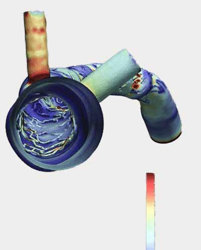
In addition to the three NIH research projects at Georgia Heart Institute, the Georgia Center for Cardiovascular Biomechanics & Modeling (GCCBM) located at the Gainesville campus, highlights three areas of study within technology-based research: machine-based learning for CV imaging, device biomechanics, and biomechanics of atherosclerosis. With the utilization of Optical Coherence Tomography (OCT), the GCCBM can investigate, measure, and determine certain predictors of major cardiovascular events. Particularly, the GCCBM has developed the DeepIVUS platform that automatically segments IVUS images with capabilities to measure lumen, plaque, and plaque burden areas within coronary arteries.
Learn more about cardiovascular clinical research initiatives at georgiaheartinstitute.org/research