COLUMBIA CROWN
A LOOK INTO THE MINDS OF OUR STUDENTS & FACULTY

A LOOK INTO THE MINDS OF OUR STUDENTS & FACULTY
ESTHER YU
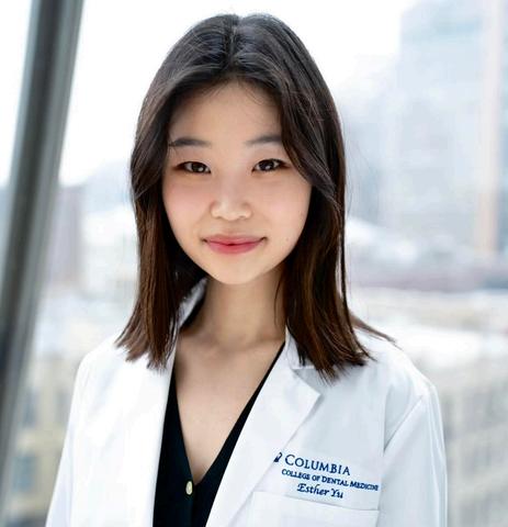
Dear Readers,
Welcome to this edition of our magazine. This issue brings together conversations and reflections on the future of dentistry from the perspective of Columbia College of Dental Medicine students Through interviews with faculty and articles exploring topics such as orthognathic surgery, AI, sleep medicine, radiology, and digital dentistry, our writers aimed to better understand where the field is headed and the people helping shape it.
Many of these pieces began simply with curiosity. As students, we are constantly surrounded by faculty whose work is influencing dentistry in meaningful ways, yet we rarely have the chance to step outside the classroom and ask broader questions about their experiences, philosophies, and hopes for the future of the profession. This publication became an opportunity to do exactly that.
I’d like to extend my sincere thanks to our incredible Editorial Committee co-chairs, Anna Jonczyk and Samuel Schwartz, for their dedication and leadership over the past several months in bringing this publication to life I’m also deeply grateful to our student authors for their thoughtful interviews and compelling writing, and to the faculty who generously shared their time and insight with us.
Thank you for reading, and I hope you enjoy these conversations as much as we enjoyed creating them
Sincerely,
Esther Yu Editor-in-Chief, Class
of ‘27
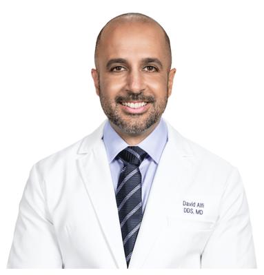
The way we talk about orthognathic surgery in dental school is often straightforward: it is “jaw surgery to fix the bite and correct imbalances between the upper and lower jaws.” Yet in my conversation with Dr. David Alfi, an oral and maxillofacial surgeon known for advancing digitally planned, custom jaw surgery, it became clear that this definition is no longer sufficient. In his view, the field is undergoing a paradigm shift that moves from occlusion-first thinking to airway-centered diagnosis, from manually bent plates to digitally engineered precision, and from symptom management to root-cause medicine.
Dr Alfi’s perspective is compelling not simply because it is technological, but because it connects surgical innovation to broader questions that students are increasingly asking: What does it mean to treat a patient rather than a symptom? How do we keep our education relevant in a profession evolving at an exponential pace?
Dr. Alfi began his academic path at the University of Southern California. While selecting a major, he “stumbled upon the study of gerontology,” describing it not as the study of the elderly, but of aging itself. In retrospect, he sees that framework as directly relevant to his current practice: the goal is not simply to correct anatomy, but to intervene early and prevent the downstream physiologic consequences of poor breathing and inadequate sleep.
That interest in long-term health carried with him to Columbia College of Dental Medicine, where he “fell in love with surgery.” Like many Columbia students, he was influenced by the integration of medical school coursework across body systems, as well as early exposure to oral and maxillofacial surgery (OMFS) in the predoctoral curriculum.
Orthognathic surgery stood out to him early He acknowledged that historically it was “a terrible procedure” for patients and trainees alike, characterized by long operating times, unpredictable movements, prolonged recovery, and the era of wiring patients shut Yet despite those limitations, the transformations were undeniable When indicated, jaw surgery produced profound functional and physiological changes Witnessing those outcomes left a lasting impression that ultimately shaped his decision to pursue the specialty
After completing dental school at Columbia, Dr Alfi chose to remain at Columbia College of Dental Medicine to pursue his OMFS residency During this time, he trained in traditional orthognathic planning using stone models, which are physical casts of a patient’s upper and lower jaws made from dental stone Surgeons would manually cut and reposition these models in the laboratory to simulate surgical movements and fabricate acrylic splints to guide jaw placement during the operation This process often required 10 to 14 hours of lab work per case and, even then, left room for variability
In the operating room, surgeons would secure the repositioned jaws with titanium plates that had to be manually bent and adapted intraoperatively. Because these plates were shaped by hand, even small differences in contouring could subtly alter the final jaw position, contributing to unpredictability.
After completing his training, Dr. Alfi moved to Houston in 2012 to immerse himself in virtual surgical planning (VSP), which was just emerging at the time. Digital planning enabled surgeons to simulate movements on a computer rather than on stone models, dramatically increasing precision and reproducibility.
In parallel with the rise of VSP, Dr. Alfi incorporated 3D-printed custom titanium guides and plates into his practice. Although these guides had been used in other surgical fields, such as oncologic reconstruction, they were not widely adopted in orthognathic surgery at the time. Instead of manually bending plates during surgery, custom plates are engineered preoperatively to match the exact planned skeletal movements, significantly reducing intraoperative variability
Dr. Alfi first applied this approach to a particularly complex case at the Texas Medical Center one that traditionally would have required a bone graft from the patient’s hip, a 10–12 hour surgery, and an external incision through the neck Instead, he performed what became the first fully custom double jaw surgery completed entirely through an intraoral, minimally invasive approach He refers to this case as “Patient Zero,” shown in Figure A, as its outcome reshaped his surgical philosophy and served as a proof of concept for what digitally driven, custom jaw surgery could achieve
These technological advances also changed the logic of treatment sequencing. Traditionally, patients underwent 6 to 12 months of orthodontics before surgery to “set up” the bite, followed by surgery, and then additional 6 to 12 months of orthodontic finishing. This orthodontics-first model was designed to reduce uncertainty by first aligning the teeth into their ideal postoperative positions, so that jaw movements during surgery could be guided by a stable and predetermined bite relationship. With digital planning and custom guides, many of those variables can now be visualized and accounted for preoperatively, reducing or eliminating the need for pre-operative orthodontics. The shift is not simply procedural; it reflects how technological precision has expanded what is safely and logically possible in orthognathic care

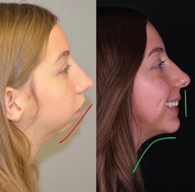
Dr Alfi returned repeatedly to a theme that resonates across modern healthcare: we are trained to manage symptoms rather than investigate root causes In dentistry, we learn to replace a missing tooth with a flipper, partial denture, bridge, or implant, yet we often do not ask why the tooth was lost in the first place or whether the same forces will compromise the replacement In medicine, he observed a similar pattern in the pharmacologic management of hypertension, where clinicians become experts in selecting medications without fully addressing upstream contributors.
He illustrated this through concrete dental examples. Gingival recession may be managed with hygiene counseling, frequent scaling, and grafting, but if the underlying drivers are mouth breathing and traumatic occlusion, the disease process persists. Wisdom teeth are removed appropriately in many patients, yet the more fundamental question of why there is insufficient space in the first place often points back to inadequate jaw development. Orthodontic relapse becomes predictable when crowding is treated purely as a tooth-position issue rather than as a limitation of skeletal space. Lifelong retainers then become a compensatory solution for anatomy that was never fully corrected.
It is within this broader critique that he situates orthognathic surgery. In his view, jaw surgery should not be limited to aligning teeth or correcting occlusion. Instead, it represents an opportunity to address structural limitations that affect airway volume, tongue position, and breathing mechanics By repositioning the jaws to create space not only for the dentition but also for the tongue and upper airway, orthognathic surgery can serve as a corrective intervention for patients with sleep-disordered breathing and related systemic consequences
To evaluate airway compromise, Dr. Alfi described two primary tools. The first is cone beam computed tomography, which allows clinicians to visualize and estimate airway volume in three dimensions. He noted that color-mapped imaging, often simplified for patient understanding as blue representing larger airway space and red representing constriction, can illustrate structural limitations in a tangible way as shown in Figure B.
The second, which Dr. Alfi considers essential, is the sleep study. Airway risk is not merely an anatomic measurement but a physiologic event that occurs during sleep, when muscle tone decreases and airway collapse may occur He emphasized the strong associations between obstructive sleep apnea and chronic conditions such as hypertension, mood disorders, and neurovascular disease, arguing that sleep evaluation should become routine rather than exceptional
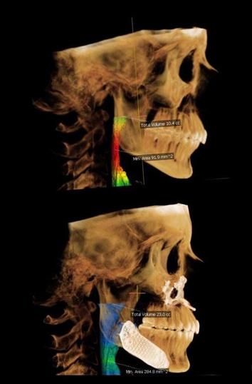
While orthognathic surgery can dramatically improve airway structure and function, Dr Alfi is careful to note that it remains a corrective measure Like many interventions in dentistry and medicine, it addresses the consequences of a developmental process that has already gone off course Surgery restores structure after misalignment of the jaws and airway has already occurred
If jaw size and airway compromise have developmental roots, the more important question becomes whether we can intervene earlier Dr Alfi’s answer is yes, and he emphasizes that timing is critical He describes reduced jaw development as multifactorial but strongly influenced by modern environmental changes, including softer diets, decreased chewing demands, bottle feeding, reduced breastfeeding, and altered early oral function Comparative evidence from anthropologic skull studies, indigenous populations, and animal models supports the idea that craniofacial growth responds to functional demands.
He references functional matrix theory, originally proposed by Columbia’s own Dr. Melvin Moss, as a conceptual foundation for this understanding. Growth is shaped by function and environmental pressure. Clinically, this translates into prioritizing nasal breathing, correcting chronic mouth breathing, and addressing tongue posture early in life He emphasizes collaboration with myofunctional therapists, noting that proper tongue posture acts as a significant driver of maxillofacial development
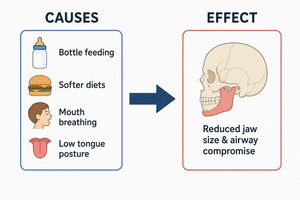
Dentistry is not merely restoring teeth. It is positioned at the front line of identifying airway-related risk long before hypertension, neurocognitive decline, and other systemic sequelae become established
Dr. Alfi observes that private practices have adopted many of these strategies more rapidly than academic centers, suggesting that educational curricula must evolve to reflect emerging evidence He contends that airway considerations remain underemphasized even within leading institutions Clinical findings that are routinely treated in isolation, including crowding, impactions, fractured teeth, gingival recession, and periodontal disease, may in fact represent structural manifestations of underlying airway dysfunction and maladaptive oral posture. If this framework holds, the implications are not subtle. Dentistry is not merely restoring teeth. It is positioned at the front line of identifying airway-related risk long before hypertension, neurocognitive decline, and other systemic sequelae become established.
Orthognathic surgery also occupies a unique intersection between function and facial aesthetics. When asked about the increasingly common claim that jaw surgery is pursued for “health” as a proxy for appearance, Dr Alfi offered a nuanced view Most patients present explicitly seeking improved breathing and sleep, while others primarily want to look better and may frame breathing concerns as part of that justification Clinically, however, he sees most of these patients as fundamentally the same The anatomic features that contribute to a weak chin or midface deficiency often correlate with functional compromise, and careful evaluation frequently uncovers symptoms consistent with obstructive sleep apnea or related dysfunction
He acknowledged that true body dysmorphia in physiologically healthy patients exists but described it as rare. More often, when the face appears structurally retrusive, it reflects a genuine anatomic limitation with physiologic consequences. In his words, “a healthy face is a cosmetic face,” meaning that esthetic improvement and functional correction often share the same structural foundation. This reframing calls for an integrated assessment of skeletal anatomy, airway function, and long-term health risk
“A HEALTHY FACE IS A COSMETIC FACE”



When asked what Columbia students should carry forward, Dr. Alfi’s message was both respectful and direct. Value the education, respect the professors, but remain intellectually independent. Zoom out. Ask difficult questions. Look beyond managing symptoms and search for underlying causes. The most important skill developed in professional school, he emphasized, is not mastery of static information but the ability to continually learn. What is taught today will evolve, particularly as technology reshapes diagnostics and treatment planning at an accelerating pace Curiosity, critical thinking, and the willingness to reassess long-held assumptions are the qualities that endure As dentistry increasingly intersects with airway health, sleep medicine, and systemic disease, the clinicians who will lead the field are those prepared to challenge convention, integrate emerging evidence, and pursue solutions that make patients not simply restored, but genuinely healthier


“Why are you studying with medical students? Aren’t you in dental school?” I frequently get this question asked by family and friends. My answer is simple: “Columbia School of Dental Medicine gets it right.”
Oral health does not exist in isolation from systemic disease. As dentistry continues to expand beyond traditional procedural care, the role of the dentist is evolving alongside it. A strong medical foundation in physiology and pathology allows for a deeper understanding of how systemic conditions manifest in the oral cavity, ultimately supporting more comprehensive and integrated patient care
One of the clearest examples of this intersection between dentistry and medicine is sleep medicine. Dr. Brittany A. Klein, an oral medicine specialist, works closely with otolaryngologists, pulmonologists, neurologists, and sleep physicians to provide oral appliance therapy for obstructive sleep apnea. Her work reflects a broader shift toward a multidisciplinary model of care, one in which dentistry plays a more active role in managing systemic conditions.
Dr Klein emphasizes the value of practicing within a unified medical system “Before that, patients were being sent out to community dentists, which made care more disjointed and harder to coordinate,” she explains “Delivering care within one system improves communication between providers and strengthens continuity of care ”
Oral appliances offer a noninvasive treatment option for obstructive sleep apnea by repositioning the lower jaw during sleep. The condition occurs when the airway collapses often due to relaxation of throat muscles, enlarged structures such as the tongue or tonsils, or obesity. As Dr. Klein notes, “When patients fall asleep, muscles relax, and gravity works against them ” By advancing the mandible forward, these appliances help maintain airway patency Typically, they consist of upper and lower trays connected by adjustable components that allow for controlled mandibular advancement
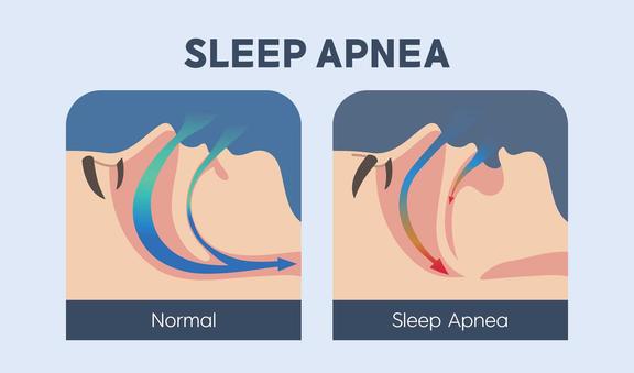
While the core principle remains unchanged, appliance design continues to evolve. Traditional devices are made from acrylic and metal, often incorporating telescoping arms for incremental adjustment. Designs such as dorsal fin and bar-style systems remain widely used for their durability and compatibility with insurance requirements.
Recent advances in materials and digital fabrication have significantly improved these devices Dr Klein highlights the growing use of nylon-based appliances: “They’re very durable, biocompatible, and rarely cause allergic reactions ” Newer 3D-printed designs are thinner and more flexible, improving comfort and potentially increasing patient compliance Because they are digitally fabricated, these appliances can also be reproduced or adjusted more efficiently
Artificial intelligence has not yet transformed appliance fabrication, but it is beginning to influence other areas of dental practice. Dr. Klein already uses AI-assisted tools for administrative tasks such as reviewing prior authorizations and documentation. Still, insurance remains a major barrier. Oral appliances are classified as durable medical equipment and billed through medical rather than dental insurance, creating logistical challenges that can limit patient access despite growing demand.
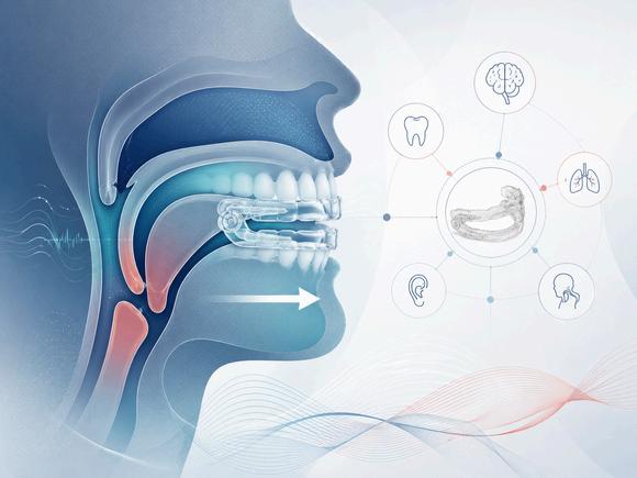
“Dental providers are in a good position to screen for obstructive sleep apnea. We routinely visualize the airway and often see patients more frequently than physicians.”

Dental sleep medicine underscores dentistry’s expanding role in systemic health. “Dental providers are in a good position to screen for obstructive sleep apnea,” Dr. Klein notes. “We routinely visualize the airway and often see patients more frequently than physicians ” This places dentists in a unique position to facilitate early detection and referral, reinforcing their role within interdisciplinary care teams
For students entering a rapidly evolving profession, Dr Klein stresses the importance of strong foundational training paired with critical engagement in emerging tren continues to integrate with disciplines, fields like dental illustrate how innovation, c technology are reshaping Increasingly, dentists are not providers, but also essential comprehensive, patient-centered

Technology has become both the driving force of innovation and a defining feature of modern education. Dental schools have made significant efforts to integrate technology into their curricula, now using tools such as 3D scanners, haptic simulators, and digital workstations This modern shift represents a fundamental change in how future dentists learn, practice, and think about their profession
Throughout history, the digitalization of dental education has progressed in waves Early innovations, such as digital radiography and electronic recordkeeping, have evolved into advanced technologies such as virtual reality (VR), computer-aided design and manufacturing (CAD/CAM), and precision data analytics These developments have not only transformed clinical practice but have also reshaped how institutions prepare their students. Dental education, traditionally a hands-on curriculum, emphasizing manual dexterity and clinical experience, has expanded beyond mannequins and instructor-guided repetition. With digital imaging, 3D printing, and laser technologies redefining treatment, schools have worked to update their curricula accordingly. The New York University College of Dentistry chronicles this evolution in the “Technology in Dentistry Through the Ages,” tracing dentistry’s journey from simple hand tools to the digital and robotic era. What began as a craft defined by manual skills has become a profession increasingly fueled by science and engineering.
Dental schools such as Columbia University, NYU, and the University of Connecticut have each developed distinct approaches to integrating technology into their teaching, reflecting a shift toward more data-driven and digitally proficient education.
At Columbia University’s Center for Precision Dental Medicine, technology is foundational to both teaching and patient care The Center promotes “precision dentistry,” combining clinical training with digital analytics Every operatory is equipped with dental chairs that collect quantitative data on student and patient interactions timing, posture, and procedural details which then feed into a real-time feedback system Faculty can monitor performance and tailor guidance accordingly The Center also incorporates intraoral scanning, CAD/CAM fabrication, and digital imaging, ensuring that students engage with digital workflows throughout their clinical training Columbia’s approach prepares graduates to enter a technologically advanced dental landscape with confidence
NYU Dentistry has also integrated technology while preserving the profession’s historical context. The NYU Virtual Museum of the History of Dentistry offers an interactive digital f l i d ti t ’ i tifi d lt l d l t hi hli hti h f th

The University of Connecticut School of Dental Medicine new tools. As reported by ADA News, the school uses VR students to practice procedures in a fully immersive simulators replicate drilling, cutting, and polishing, givi sense of working on live patients without the risk or s smooths the transition between preclinical and clinica jumping directly from mannequin-based exercises to real refine their skills in a controlled virtual setting. Early resu with students reporting higher engagement, improved s increased confidence before entering clinical rotations.

The incorporation of new technology into dental education offers numerous advantages. Interactive platforms, virtual case studies, and digital simulations make learning more engaging According to Western University of Health Sciences, 3D models and videos make complex material more vivid and accessible Digital systems also improve assessment: VR simulators and smart dental chairs can track hand movement, pressure, and efficiency, generating objective performance data Online platforms such as NYU adVANCE offer instant feedback, allowing faculty to quickly identify areas for improvement Technology supports self-paced, individualized learning while building confident clinicians ready for real-world practice
However, the drawbacks of emerging technologies must also be considered. Implementing digital systems such as VR simulators, CAD/CAM equipment, or data-enabled operatories involves substantial cost, and many institutions struggle to keep pace. There is also the risk of over-reliance on technology. Despite their sophistication, digital tools cannot fully replicate real-life patient care. Interpersonal skills communication, empathy, and emotional intelligence are essential to the patient-provider relationship, and technology cannot replace them. Finally, the increased use of digital systems raises concerns about privacy and data security. With vast amounts of student and patient information being collected, institutions must ensure responsible data management and transparency. Dental education has embraced new technologies to improve student training. Today’s challenge is balancing innovation with the human elements that technology cannot teach. By integrating digital tools alongside strong interpersonal training, dental schools can prepare a generation of practitioners who are not only technologically capable but also empathetic, ff i d d l nnected to their patients

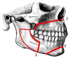
INTRODUCTION: WHAT IS ORTHOGNATHIC SURGERY?
Orthognathic surgery, or corrective jaw surgery, is used to treat discrepancies in the alignment of the upper and lower jaws. These imbalances can affect chewing, speaking, and breathing, and facial aesthetics. The procedure repositions the upper jaw, lower jaw, or both to improve function and achieve facial harmony
Candidates may present with severe overbites, underbites, crossbites, difficulty chewing, speech impairment, sleep apnea, jaw pain, or noticeable facial asymmetry While many patients seek treatment for functional concerns, others pursue surgery primarily for cosmetic reasons.
The goals of modern orthognathic surgery extend beyond occlusion to include airway optimization, improved sleep quality, and overall facial balance. Proper jaw alignment supports more efficient mastication, clearer speech, reduced snoring, and relief from temporomandibular joint discomfort. These functional gains are often accompanied by increased confidence and improved social wellbeing. Common procedures include Le Fort I osteotomy (upper jaw), bilateral sagittal split osteotomy (lower jaw), and genioplasty (chin). Recent advances in three-dimensional imaging and digital surgical planning have increased precision and opened the door for alternative approaches, such as the surgery-first method


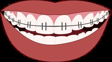
The conventional orthodontics-first approach has been the standard for decades and remains widely practiced. Treatment begins with a comprehensive diagnostic workup, including radiographs, photographs, bite analysis, and, in some cases, three-dimensional imaging. This is followed by a preoperative orthodontic phase designed to align the teeth and prepare the arches for predictable surgical repositioning.
Orthodontic treatment typically lasts between 12 to 24 months. During this period, braces correct dental compensations and reveal the underlying skeletal discrepancy. This step is critical, as attempting surgery without proper alignment could compromise surgical control and the final occlusion. Once preparation is complete, detailed preoperative planning, often using digital models, is performed to simulate jaw movements and predict outcomes.
Surgery is performed under general anesthesia and usually lasts two to five hours. Incisions are made intraorally to avoid visible scarring. Depending on the case, the maxilla, mandible, or both may be repositioned, with genioplasty added when needed to optimize facial balance.
Recovery occurs gradually. Swelling and discomfort peak during the first week, when patients typically follow a liquid or soft diet
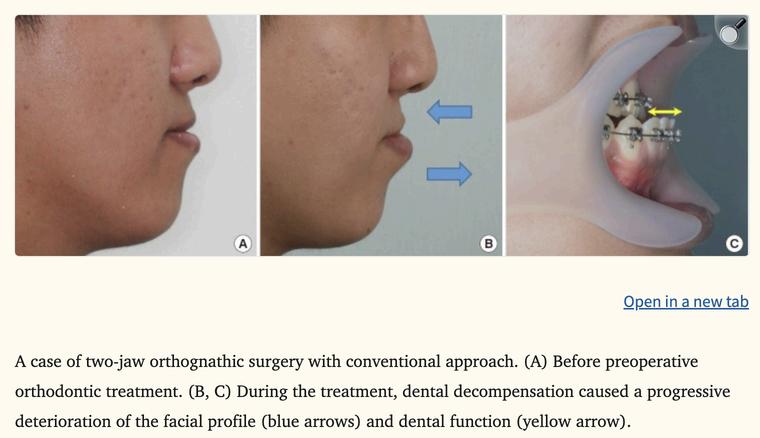
Figure:A case of two-jaw orthognatic surgery with conventional approach. (A) Before preoperative orthodontic treatment. (B,C) During the treatment, dental decompensation caused a progressive deterioration of the facial profile (blue arrows) and dental function (yellow arrow) Source: Adapted from Seo HJ and Choi YK, 2021
Over time, swelling subsides and normal function returns Complete bone
healing generally takes three to six months, during orthodontic treatment continues to refine the bite
Despite its effectiveness, this approach has limitations The presurgical orthodontic phase can temporarily worsen facial aesthetics and reduce functional occlusion as compensations are removed Because this phase may last up to two years, patients may experience difficulty chewing, frustration, and psychosocial effects such as decreased confidence. These challenges have motivated the development of alternative methods.
The surgery-first approach reverses the traditional sequence by performing jaw surgery before orthodontic treatment. In this method, skeletal discrepancies are corrected at the outset, resulting in immediate improvements in facial aesthetics and function, followed by orthodontic refinement.
Careful planning is essential in the surgery-first approach. Advanced imaging techniques–including three-dimensional imaging, computed tomography, and virtual surgical planning–are used to simulate jaw movements and predict outcomes. These tools also help create precise surgical guides and occlusal splints, ensuring accurate repositioning of the jaws.
During surgery, the jaws are repositioned to correct skeletal imbalances and facial asymmetry Patients frequently experience immediate functional benefits, including better chewing and increased airway space, which may alleviate symptoms of sleep apnea
After surgery, orthodontic treatment fine-tunes tooth positioning and stabilizes the occlusion Unlike the orthodontics-first method, where teeth are initially moved to decompensate, post-surgical tooth movement typically follows the corrected skeletal framework, often shortening overall treatment time.
This approach offers several advantages. It eliminates the lengthy presurgical orthodontic phase, reduces total treatment time, and provides immediate aesthetic improvement, which can enhance patient confidence. Functional benefits also occur earlier, improving overall quality of life. It is best suited for patients with mild to moderate skeletal discrepancies and minimal dental crowding, though careful case selection is essential.
However, limitations remain. Predicting the final occlusion can be more challenging, particularly in complex cases or when extractions are required. Surgical precision is critical, as there is no presurgical orthodontic phase to compensate for minor discrepancies. Patients with severe crowding, significant overbite, temporomandibular joint disorders, multiple missing teeth, or marked asymmetry may be less ideal candidates
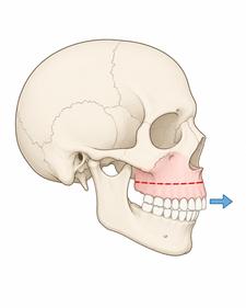
Advances in digital planning and three-dimensional modeling have significantly improved the accuracy, safety, and predictability of orthognathic surgery While the orthodonticsfirst approach remains the traditional standard, the surgery-first approach represents a more patientcentered alternative, offering earlier functional and aesthetic benefits.
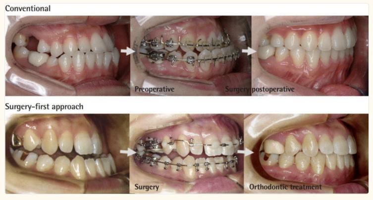
Figure: Comparison between the conventional approach (top) and surgery-first approach (bottom) In the conventional approach, braces are used before surgery to prepare the teeth, while in the surgery-first approach, surgery happens first and the bite is corrected afterward with orthodontic treatment. Source: Adapted from Choi JW and Lee JY, 2021.
Current evidence suggests that postoperative stability is comparable between the two approaches when applied to appropriately selected Class II, Class III, and asymmetrical cases The surgery-first method reduces treatment duration, avoids the drawbacks of prolonged presurgical orthodontics, and delivers immediate improvements in facial appearance.
With careful patient selection and precise digital planning, it is a safe, effective, and highly satisfying treatment modality. Future research should focus on long-term outcomes and the standardization of digitally driven protocols to further optimize results for both patients and clinicians.
SOURCES:
CHOI, JONG-WOO, AND JANG-YEOL LEE “CURRENT CONCEPT OF THE SURGERY-FIRST ORTHOGNATHIC APPROACH.” ARCHIVES OF PLASTIC SURGERY, VOL. 48, NO. 02, MAR. 2021, PP. 199–207, HTTPS://PMC.NCBI.NLM.NIH.GOV/ARTICLES/PMC8007470/, HTTPS://DOI.ORG/10.5999/APS.2020.01305. ACCESSED 5 DEC. 2025.
SEO, HYUNG JOON, AND YOUN-KYUNG CHOI. “CURRENT TRENDS IN ORTHOGNATHIC SURGERY.” ARCHIVES OF CRANIOFACIAL SURGERY, VOL. 22, NO. 6, 1 DEC. 2021, PP. 287–295, HTTPS://PMC.NCBI.NLM.NIH.GOV/ARTICLES/PMC8721433/, HTTPS://DOI.ORG/10.7181/ACFS.2021.00598.ACCESSED 5 DEC. 2025.
VIEIRA DE NAZARÉ, GABRIEL HENRIQUE , ET AL. “SURGERY-FIRST APPROACH IN ORTHOGNATHIC SURGERY: LITERATURE REVIEW.” RESEARCH, SOCIETY AND DEVELOPMENT, VOL. 11, NO. 13, 4 OCT. 2022, RSDJOURNAL.ORG/RSD/ARTICLE/VIEW/35093/29598, HTTPS://DOI.ORG/10.33448/RSDV11I13.35093.. ACCESSED 5 DEC. 2025.
When people hear “new wave of dentistry,” they often imagine dramatic technological breakthroughs or robotics replacing clinicians. But after speaking with Dr. Andrew Inagaki of Williamsburg Endodontics, a different narrative emerged: endodontics hasn’t been completely reinvented. Rather, it’s being refined in the new wave of dentistry.
This is mostly because root canal therapy has long been a highly successful procedure. For initial cases, success rates have historically hovered around 90%, and today, they remain remarkably similar despite all the advances made over the past years What has changed, however, is not necessarily the outcome, but the efficiency, precision, and philosophy behind how we get there, says Dr Inagaki
Dr Inagaki trained in California, completed an AEGD and GPR before specializing at Lutheran Medical Center (now NYU Langone), and now practices in Williamsburg while serving as a postgraduate faculty member at NYU For him, adapting to new technology has always been part of the profession Staying current requires continuous learning: attending the AAE annual conference, reading the Journal of Endodontics, teaching, and engaging with colleagues “It never ends,” he reflected

“The most important step is disinfecting the canal system”
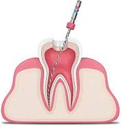
“100% irrigation mindset,”which recognizes that files alone cannot clean the complex anatomy within every root canal system.
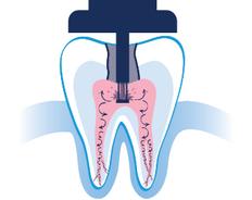
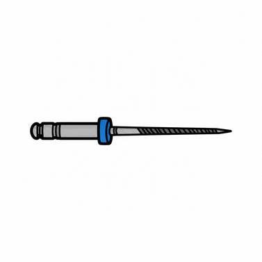
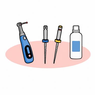
Endodontics has always evolved alongside technology. Nickel-titanium files have improved over time, becoming more flexible and resistant to fracture. Imaging has advanced. Materials have changed. But according to Dr. Inagaki, the most significant shift in recent years hasn’t been about better shaping. Rather, it’s been about better cleaning. “The most important step is disinfecting the canal system,” he emphasized. There has been a growing realization in the field that success depends less on how we shape canals and more on how effectively we disinfect them. Enhanced irrigation systems, including technologies like GentleWave and laser-assisted irrigation, along with devices such as the Odion (pressurized saline irrigation), are centered around maximizing disinfection The philosophy has shifted toward what he described as a “100% irrigation mindset,” which recognizes that files alone cannot clean the complex anatomy within every root canal system Every tooth is unique There is no universal recipe That variability is exactly why irrigation (and not just instrumentation) has become such an important shift in modern endodontics

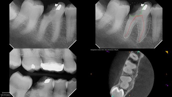
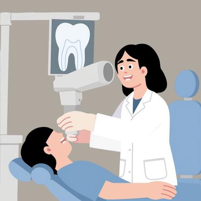
Cone beam computed tomography (CBCT) has become the standard of care for retreatments, surgical cases, and complex anatomy. While CBCT has existed for years, image clarity has significantly improved. Reduced radiation doses, decreased beam hardening, and fewer artifacts around crowns allow clinicians to diagnose with greater confidence. Endodontics was among the first specialties to adopt digital radiography because intraoperative imaging is essential Dr Inagaki noted that research increasingly supports using CBCT when there is diagnostic uncertainty At the same time, he emphasized discernment Technology is a tool, not a replacement for clinical judgment
Artificial intelligence is beginning to enter dentistry, primarily in clinical documentation and radiographic interpretation. While AI may assist in identifying pathology, Dr. Inagaki expressed caution about overreliance. In healthcare, the margin for error is small. A missed diagnosis is unacceptable. History has demonstrated that premature adoption of emerging technologies can compromise outcomes rather than improve them. For example, bonding gutta-percha to dentin once seemed promising but ultimately lowered success rates. It was a reminder that not every innovation improves outcomes. The “new wave” requires critical thinking.
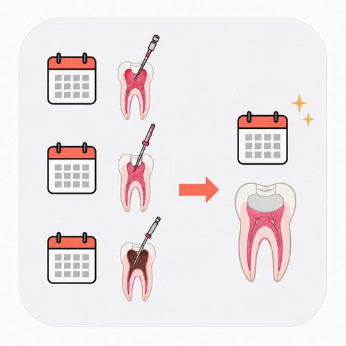
One undeniable improvement in outcome that we have seen is efficiency. Root canals that once required three or four appointments can now often be completed in a single visit Tools are easier to handle, more ergonomic, and more predictable These advances have improved the clinician’s workflow and enhanced patient comfort while maintaining success rates In that sense, endodontics demonstrates something important: progress does not always mean higher percentages. Sometimes it means delivering the same high success rate more comfortably, more precisely, and more efficiently.
So the new wave of endodontics isn’t about dramatic disruption. It’s about thoughtful refinement: recognizing that a field already built on such high success can still evolve through improved techniques, materials, and mindset.

Dentistry is a rapidly evolving field, in large p to technological advances Artificial intelligenc particular, is impacting many aspects of dental facilitating office administration, enhancing di and improving clinical decision-making W potential transformative impact on dentistry educators are evaluating and strategizing should be incorporated into dental curricula. offers clear opportunities to support learning raises important questions about how sk developed, how responsibility is taught, and w human judgment should continue to p healthcare. Dr. Dana Wolf, Associate Profes Senior Associate Dean for Predoctoral Acade Student Affairs, shared her perspective on h being incorporated into education and what remain at the core of dental training.

A central idea emphasized by Dr. Wolf is t foundational diagnostic, clinical, and critica experience remain essential, and those are s effective when it helps confirm thinking or im student When it comes to diagnostics, the key question is not whether AI should be used at all, but when it makes sense to use it. Students still need to learn how to identify caries, interpret radiographs, and work through diagnoses independently, with AI serving as a secondary tool rather than the primary driver of decisions.
AI can be especially helpful when it comes to managing large amounts of information Dental school comes with an overwhelming volume of material, and AI tools that assist with searching the literature or organizing research can support evidence-based decision-making. According to Dr. Wolf, these tools can help students process information more efficiently and stay current with evidence-based practices However, AI tools need to be used responsibly AI-generated information is not always accurate, and without strong foundational knowledge, it can be difficult to recognize errors or limitations. In this sense, AI makes a solid understanding of core concepts even more important.
Another major issue that emerged is responsibility AI use is already happening, whether or not it is formally taught, and that reality presents new challenges. Students may not always consider data security or privacy when using these tools, which is especially concerning in a healthcare setting. Entering information into platforms that are not secure can have serious ethical and professional consequences Rather than attempting to restrict AI use entirely, Dr Wolf emphasized the importance of teaching students how to use these tools responsibly by clearly defining where e curriculum and what boundaries need to be in place.


AI also has the potential to change how feedback is delivered in dental education. Presenting work to faculty can be stressful, especially early in training, and that stress can interfere with learning. AI tools could help reduce these barriers by offering preliminary feedback in a lower-pressure setting Receiving early feedback on a preparation or practicing a patient interaction before presenting to faculty may help students feel more prepared and confident, while still keeping faculty mentorship central to the learning process.
As AI becomes more integrated into dentistry, Dr. Wolf made it clear that core skills are not going away. Communication and personal interaction play a major role in patient care, from explaining treatment plans to managing expectations and building trust. While AI may assist students in practicing how to structure these conversations, it cannot replace the human side of dentistry. Patients value feeling comfortable, understood, and reassured, and those experiences depend on real human interaction
This connects to broader questions about technology in healthcare. Dentistry is not the only field facing these changes. Across healthcare, professionals are being asked to think carefully about what technology can support and what should remain human Empathy, communication, and trust are just as important as technical skill, and those are qualities AI cannot replicate.
Empathy, communication, and trust are just as important as technical skill, and those are qualities AI cannot replicate.
From a faculty perspective, reactions to AI are mixed There are concerns about academic integrity and the possibility that students could rely too heavily on AI instead of developing essential skills. At the same time, there is optimism about what AI could offer. Tools that reduce time spent on repetitive tasks may allow educators to focus more on teaching, mentorship, and discussion as they navigate how to integrate AI in ways that truly support education.
Overall, Dr. Wolf’s perspective highlights that AI is not about replacing dentists or educators, but about changing how dental education is delivered When used thoughtfully, AI can support diagnostics, improve feedback, and lower barriers to learning At the same time, it makes protecting core skills, such as judgment, communication, and human connection, more important than ever As dentistry continues to evolve, the future of dental education will depend less on how advanced technology becomes and more on how intentionally it is used to train capable, thoughtful, and compassionate clinicians.


y g g y g g Hospital, and later returned to NYU for his endodontics specialty training He was drawn to the field of endodontics because he loved being able to quickly relieve patients’ pain, and with the help of a few great mentors and some lucky timing, he found himself on the path to endodontics This also meant he entered the specialty right as major technological changes were taking off, giving him a unique and insightful view of how the field was and has evolved
One of the most transformative innovations has been the refinement of cone-beam computed tomography (CBCT) CBCT, first introduced in the U S market in 2001, is a 3D imaging technology that provides dentists with a full volumetric view of the teeth, bone, and surrounding structures It is essentially a detailed map that reveals anatomy traditional X-rays can’t capture Early CBCT units had limited field-of-view options and produced images with higher radiation and lower clarity Today’s systems, however, generate much sharper, more focused scans while delivering significantly less radiation These high-resolution images give endodontists a true three-dimensional look at root anatomy and can even help estimate canal lengths more precisely than traditional twodimensional films Dr Chubak notes that this level of detail and diagnostic accuracy has made the field considerably more precise


Another area of endodontic treatment that has seen major is crucial because it helps ensure the root canal system is and over the last decade the available technologies h vibrating or sonic activators have been around for many y option at just a few cents per use In the mid-2010s, mor began gaining popularity, using fluid dynamics and acousti that files and traditional irrigants can’t reach These system per-use cost, often over $100, which impacts how readily office incorporated an endodontic laser more recently, ado it first came to market. This laser uses water-based acti that’s deeper than standard techniques. Despite the spec these tools aim for the same outcome: a more effective d traditional syringe irrigation could reliably achieve.
Pairing the huge advancements in imaging and irrigation, D instruments themselves have undergone their own transf sterilized and reused are now single-use, pre-sterilized, a flexible, and far less prone to breaking Bioceramic sealer promote better healing, have further improved treatment o
Some emerging tools are still finding their place AI platfo s l e ag ocat ca a aly e C C scans and highlight lesions and potential issues, and while they’re still early in development, they’re improving quickly 3D-printed guides and robotic navigation systems are also being explored, mostly for difficult, highly calcified cases rather than routine treatment Microscopes remain standard, though newer stereoscopic systems where clinicians operate using a 3D display may eventually become more common once training catches up

With so much new technology emerging, endodontists stay current by attending AAE conferences, reading journals like the Journal of Endodontics, and learning from vendor fairs and sales reps. The challenge isn’t just staying up to date but deciding what’s actually useful. Dr. Chubak explained that in his practice, decisions about adopting something new are made collectively. The group considers patient benefit, cost, practicality, and safety.
Despite all these advancements, the area where Dr. Chubak wishes to see the most improvement is surprisingly simple: anesthesia. Lower molars, especially those considered “hot,” with inflamed pulps, remain notoriously difficult to numb due to multiple nerves. Even with tools like the Wand or Dentapen that deliver anesthetic at a greater pressure in a slower and more controlled manner than the human hand could, achieving full numbness isn’t always guaranteed for some patients. A more predictable anesthetic technique for these challenging cases, he says, would be a true game-changer for the specialty
As we wrapped up the conversation, Dr Chubak reflected on the broader picture: dental school offers the foundation, but lifelong learning defines the profession Technologies may to advance, but it is the clinician’s adaptability and commitment to ongoing education shape the “new wave” of endodontics

Elizabeth Nazginov
Dr. Vicky Evangelidis Sakellson, a longtime faculty member at Columbia University College of Dental Medicine, currently serves as Professor and Interim Chair of Cariology and Restorative Sciences. She earned her DDS and completed a General Practice Fellowship at Columbia, then went on to receive an MPH there as well. Over the years, she has taught everything from preclinical courses to advanced restorative techniques, and her perspective reflects several decades of change, both in how students learn and in how dentists care for patients.
Our conversation drifted between memories of her early training and the rapidly changing tools available today. What became clear is that dentistry is moving forward quickly, sometimes faster than people realize Yet the purpose of the profession–the reason patients walk through the door–has hardly changed at all
When Dr Sakellson trained in the late 1980s and early 1990s, the profession functioned very differently Implant dentistry existed, but not in the way we think of it now It was still considered specialized and somewhat experimental, so most dental students didn’t place implants The usual answer for a missing tooth was a fixed bridge or a partial denture
Cosmetic dentistry also looked different Porcelain veneers were new and not something every practice could offer. They were harder to make and easier to break, so many patients who wanted to “improve their smile” ended up with full-coverage crowns or bonding procedures that we would now consider limited.
Almost everything was done by hand. Students carved wax crowns, one tiny ridge at a time, and worked on wax-ups for hours to plan cases. Dental labs relied on these wax models to produce the final restorations. It required a steady hand and a good imagination, but there were plenty of opportunities for the final product to come out differently than expected. The work demanded a mix of patience, craft, and communication that is hard to appreciate until you’ve spent a night in a preclinical lab.
Today’s workflow would have seemed almost futuristic back then. Digital scans, CAD/CAM milling, 3D printing, and advanced ceramics show up everywhere in dental clinics and labs. Even students now work with tools that didn’t exist when many faculty members were in school
Digital scanners, for example, have replaced most traditional impressions Instead of a tray of impression material, a wand captures a detailed map of the mouth in seconds That digital file can then be used to design a crown or veneer, which might be milled in-office or printed for later finishing The accuracy is impressive, but the biggest benefit may be the simple fact that patients no longer have to sit with a tray full of goopy material
3D printing has opened up even more possibilities–surgical guides, night guards, temporary crowns, dentures, clear aligners, and models for planning all come out of printers now. What used to require days of turnaround can happen in a matter of hours.
One development Dr. Sakellson especially appreciates is the digital diagnostic wax-up. Instead of carving wax on a stone model, clinicians can design proposed changes on a screen, revise them easily, and show patients different possibilities for their smile. Patients understand their treatment better when they can see it instead of guessing at what the dentist means.
Even with all these advancements, Dr. Sakellson kept coming back to the same point: these tools help, but they don’t make decisions.
“You can have all the newest equipment,” she told me, “but the key is knowing what will actually benefit your patient.”
That line stayed with me. Technology may change the path, but it doesn’t change the responsibility.
Another thing she emphasized is that patients rarely choose a dentist because of technology Most people don’t come in asking what scanner you use or whether you have a milling unit They care about whether their pain goes away, whether they can chew normally again, and whether they feel comfortable in the chair
In many cases, patients never even see the digital work behind their treatment They don’t watch you plan the case on a screen or revise the design for the fourth or fifth time They see the finished restoration, not the process leading up to it What they remember is how you spoke to them, whether you listened, and whether they felt understood.
There is also a practical challenge: the cost of digital systems. Equipment upgrades come quickly, and a private practitioner may still be paying off one machine when a newer, faster, more accurate alternative becomes the standard. This can widen the gap in care, making advanced tools more available in institutions and less accessible in small practices. It also means that academic and public clinics often serve as the place where patients receive treatments they could not otherwise afford.
When I asked Dr. Sakellson what skills future dentists need the most, she didn’t mention software skills or new materials. She spoke instead about empathy, communication, critical thinking, and understanding each patient as an individual. These are skills that outlast every technological trend.
Dentistry has always been about helping people regain comfort, function, and confidence New tools can support that mission, but they cannot replace the judgment or the compassion that patients rely on
Her advice for dental students was straightforward: remain open to change Dentistry will keep moving forward, and there will always be new techniques and new tools The challenge is to keep learning without losing sight of the person sitting in the chair
Over the past 25 years, dentistry has undergone a technological transformation that has redefined not only how dentists treat patients, but also what it means to practice comprehensively Dr Patricia Swaintek-Lamb, DMD, owner of Proud to Smile Dentistry in Bernardsville, New Jersey, has experienced this evolution firsthand throughout her career A cosmetic, family, implant, orthodontic, and sleep apnea provider, Dr Swaintek-Lamb’s professional journey mirrors the rapid advancements that have expanded dentistry’s clinical possibilities and reshaped her identity as a practitioner.
When Dr. Swaintek-Lamb first began practicing, dentistry was far more segmented. General practitioners often referred out complex procedures such as orthodontics, implant placement, or advanced periodontal therapy.
Over time, her scope of practice expanded through a combination
of her own professional interests and the growing capabilities of modern dentistry. Today, she describes herself as a “super GP”–a clinician who embraces the challenge of managing as much of a patient’s care as possible under one roof.
One of the most influential shifts in her career came with the introduction of clear aligner therapy. Traditional orthodontics relied heavily on wires and brackets, which were time-intensive and cumbersome in a general practice setting. Clear aligners, by contrast, “changed the game”, allowing her to reposition teeth efficiently and integrate orthodontic movement into restorative treatment plans. This innovation expanded who she could treat and fundamentally altered how she planned care, making comprehensive, multidisciplinary dentistry more accessible and predictable
Equally transformative has been the rise of digital dentistry Dr Swaintek-Lamb recalls a time when physical impressions were unavoidable and often uncomfortable for patients Today, digital scanners have nearly eliminated that step from her workflow She now relies almost exclusively on digital impressions, which she finds both more comfortable for patients and more efficient for communicating with dental laboratories. The scanner has become her favorite piece of technology. It integrates smoothly into daily practice and improves consistently without sacrificing time or quality.




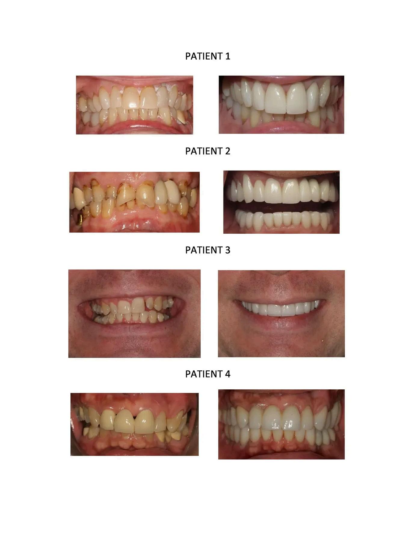
Before-and-after veneer treatment demonstrating how modern cosmetic dentistry can achieve a highly esthetic outcome while enhancing smile symmetry, shade, and overall appearance. Source: Image taken from Dr Swaintek-Lamb’s office website


pidly transformed modern dental practice. Tools such uted tomography (CBCT), and digital treatment grated into clinical workflows, allowing clinicians to er precision than ever before. Beyond improving e technologies are also reshaping how dental lties. Digital dentistry enables orthodontists, ontists, and other specialists to work together using simulations. As a result, treatment planning has d predictable, highlighting the increasingly ry dental care.
One of the most widely adopted technologies in digital dentistry is intraoral scanning. Intraoral scanners allow clinicians to capture highly accurate digital impressions of the dentition and surrounding oral structures without the need for conventional impression materials Digital models can be generated within minutes and easily stored, duplicated, or shared electronically with other providers This capability significantly improves communication between specialists involved in complex treatment plans. For example, an orthodontist can send digital models to a prosthodontist to evaluate spacing requirements for future restorations or implants. Similarly, restorative dentists can collaborate with orthodontists to determine whether minor tooth movement may improve restorative outcomes. By providing a precise and easily transferable representation of the patient’s dentition, intraoral scanning facilitates collaborative decision-making early in the treatment planning process.
Cone beam computed tomography (CBCT) has further expanded the diagnostic capabilities of digital dentistry by providing three-dimensional imaging of craniofacial structures. Unlike traditional two-dimensional radiographs, CBCT allows clinicians to evaluate bone morphology, root positioning, and anatomical structures with greater accuracy. This technology has become particularly valuable in cases requiring interdisciplinary planning. For instance, implant placement often involves collaboration between prosthodontists, periodontists, and oral surgeons CBCT imaging allows clinicians to assess bone volume and proximity to vital structures before surgical intervention, improving both safety and predictability. Similarly, orthodontists frequently use CBCT data when planning complex cases involving impacted teeth, skeletal discrepancies, or orthognathic surgery. By allowing providers to visualize structures in three dimensions, CBCT enhances diagnostic accuracy and provides a shared foundation for interdisciplinary treatment planning.

Digital treatment planning software represents a important advancement in collaborative de Many modern platforms allow clinicians to datasets from intraoral scans, CBCT imaging, an photographs into a single virtual model of the p This integrated digital environment enables pr to simulate treatment outcomes before in clinical procedures. In orthodontics, digital setu predict tooth movement and final occlusion, a orthodontists to coordinate with restorative d regarding ideal tooth positioning prior to pro rehabilitation. Similarly, implant planning s enables clinicians to virtually place implants CBCT data and design surgical guides that precise positioning during surgery. These tool multiple specialists to review and modify tre plans collaboratively, improving communicatio reducing the likelihood of unforeseen complicati

The ability to share digital data across disciplines has fundamentally improved interdisciplinary treatment planning. In many complex cases, successful outcomes depend on careful coordination between different specialties. For example, orthodontic treatment may be required to create appropriate spacing or alignment before implant placement or prosthodontic rehabilitation. In orthognathic surgery cases, orthodontists and oral surgeons must work closely together to determine ideal skeletal and dental relationships before surgical intervention. Digital workflows allow these providers to analyze the same datasets simultaneously, facilitating more informed discussions and collaborative decision-making. This shared digital framework promotes a more integrated approach to patient care, ensuring that each phase of treatment is aligned with the overall therapeutic goals.
In addition to improving provider collaboration, digital technologies also enhance patient communication and education. Three-dimensional visualizations allow clinicians to demonstrate proposed treatment plans in a clear and intuitive manner. Patients can better understand the rationale behind multidisciplinary treatment and visualize potential outcomes before procedures begin This improved communication may increase patient engagement and confidence in the treatment process, particularly in complex cases that require multiple phases of care
As digital technology continues to evolve, the role of digital dentistry in interdisciplinary care will likely expand further Emerging developments such as artificial intelligence, automated treatment simulations, and increasingly integrated software platforms may further streamline communication among dental specialists. These innovations have the potential to enhance diagnostic capabilities, improve treatment efficiency, and increase the predictability of clinical outcomes.
Ultimately, digital dentistry represents more than simply a technological advancement. It reflects a broader shift toward collaborative, patient-centered care within the dental profession. By enabling specialists to share information, visualize treatment outcomes, and coordinate interventions more effectively, digital tools are transforming how multidisciplinary dental teams approach complex cases. As these technologies become increasingly integrated into clinical practice, they will continue to play a central role in shaping the future of collaborative treatment planning in dentistry.
SAMUEL SCHWARTZ
In an era defined by digital design, artificial intelligence, and individualized healthcare, dentistry is undergoing a subtle yet profound transformation, one that is reshaping how we understand growth, function, and long-term wellness. This evolution is especially evident in pediatric dentistry and orthodontics, where emerging technologies are merging with clinical intuition to redefine the experience of care for young patients. What was once a specialty centered on metal brackets and rigid treatment sequences has become a field driven by data, design thinking, and a more nuanced understanding of craniofacial development.
At the forefront of this shift is pediatric aligner therapy, a modality that uses clear aligners not only to guide tooth movement, but also to influence facial growth, airway support, and occlusal development during the years when these structures are most malleable. Leading this movement is Dr. Edo Lavi, founder of the Lavi Institute of Pediatric Aligners and a pioneer of what he terms “timely treatment.” His work represents a broader reorientation within orthodontics: a move from purely mechanical correction to growthcentered, technology-enhanced care.

To explore this changing landscape, I spoke with Dr. Lavi an accomplished orthodontist and Align Technology faculty member whose career traces the profession’s evolution from traditional mechanics to digitally informed, patient-centered innovation. Our conversation illuminates how technology, clinical philosophy, and a deep respect for childhood development are converging to redefine what orthodontic care can achieve. What follows is a discussion not simply about tools, but about timing, intention, and the emerging art of helping children grow into healthier futures.


Dr. Lavi: I should tell you, you’re calling at an interesting time because this is considered cuttingedge now Usually, cutting-edge means experimental you wait to see long-term outcomes But this is different The results are immediately obvious We’ve wanted to do this for years; we just didn’t have the technology Now we do, and people are slowly realizing: maybe we should do things differently than we have for the last 200 years Once that tipping point hits, there’s no going back
Sam: Dr. Lavi, take us back to your early days at UMDNJ and your general practice residency at Long Island College Hospital what first drew you toward orthodontics, and how did those formative experiences shape your philosophy of patient care?
Dr. Lavi: There was a doctor named Dr Erwin Colin, of blessed memory Even though he was older, he looked relaxed and genuinely happy in his work During my residency, I realized that orthodontists seemed to live a different quality of life working with teenagers, which at that stage of my life, felt just right It wasn’t just about enjoying dentistry; it felt like the kind of practice I wanted I did a second year of residency at Interfaith Medical Center in New York, and that experience solidified my path. It gave me a broader perspective on healthcare, exposing me to medically compromised and trauma patients, helping me to understand where dentistry fits in the bigger picture.
Sam: Can you share an experience from that time that shaped your perspective on patient care?
Dr. Lavi: Absolutely One night, I was called to the ER at 3:30 a m for a dental emergency I found a toothbrush in a patient’s large intestine on an X-ray Everyone had a laugh it was meant as a prank but it was humbling It reminded me that we’re part of a larger healthcare ecosystem When patients are fighting for their lives, dentistry is one piece of the puzzle That perspective has influenced how I approach care ever since.
Sam: When you graduated from NYU’s orthodontic program in 2003, digital workflows and clear aligners were in their infancy. How would you describe the state of orthodontics at that time both clinically and culturally within the profession?
Dr. Lavi: Orthodontics hadn’t changed much for over a century The biggest innovation before I started was direct bonding in the 1980s Clear aligners existed, but they were largely for minor adult corrections pediatric aligners didn’t even exist yet At the time, treatment was brackets and wires, often phased: early treatment to guide growth, then a second phase to align teeth once they erupted. The real transformative potential of aligners, I’ve come to realize, is in pediatric dentition guiding bone and teeth development proactively, not just making small cosmetic adjustments for adults.
Sam: You’ve spoken about the holistic potential of aligner therapy particularly in growing children. What does a “holistic” approach mean to you in the context of orthodontics, and how does it differ from the traditional treatment mindset?
Dr. Lavi: Holistic, in this context, isn’t about being “crunchy granola ” It means seeing the entire dentition the teeth that are erupted and those that aren’t and being proactive rather than reactive Traditional treatment is reactive: you see a problem, then address it Holistic treatment means anticipating growth and development, shaping bones and teeth before problems arise. We don’t wait for an impacted canine or a surgical intervention; we develop everything comprehensively from the start.

ou mentioned that relocating to Maryland in 2019 was a turning point. What did yo xed-dentition cases that changed your perspective?

Dr. Lavi: It was almost accidental. I was using aligners for minor anterior crossbites, just very specific objectives. But I started noticing that these limited cases consistently produced significant, or “macro,” movements in the teeth. It was a revelation: this could fundamentally change pediatric orthodontics. I’ve since treated over 3,500 patients using this approach. What started as a small intervention became a transformative method.
Sam: You often distinguish between “early treatment” and what you call “timely treatment ” Can you explain that difference, and why you believe this conceptual shift is critical for the next generation of orthodontists?
Dr. Lavi: Early treatment has always implied something limited or preparatory you fix a little, then wait. I prefer “timely treatment.” It’s comprehensive and happens at the optimal time in development. We’re not waiting for adult dentition to intervene; we’re taking advantage of developmental windows to make meaningful changes now. Missing that window means missing opportunities, and that mindset shift is critical for the next generation of orthodontists.
Sam: How have digital diagnostics, AI-driven treatment planning, and 3D modeling changed the relationship between orthodontist and patient especially in pediatric care?
Dr. Lavi: They’ve transformed precision and visibility For example, with digital scans, we can measure occlusion to 0.1 millimeters and make micro-adjustments that were impossible with traditional brackets and wires. AI helps automate workflows like refinements and scans but it can’t replace the clinician. Every patient is biologically unique; software can’t account for every nuance. AI supports us, but judgment and expertise remain essential.
Sam: So, you’re not concerned about AI replacing orthodontists?
Dr. Lavi: Not at all AI can handle routine tasks, but it can’t replace clinicians who care about quality and patient outcomes There’s a business model for fully automated orthodontics, but for meaningful care where patients truly benefit you need a human guiding it That’s where I focus: teaching doctors to use technology to enhance treatment, not replace it.
Sam: Through the Lavi Institute of Pediatric Aligners, you’ve built a community dedicated to mastering pediatric aligner therapy. What motivated you to create this platform, and what gaps in education or philosophy were you hoping to fill?
Dr. Lavi: Honestly, my wife motivated me Here’s what happened: In June 2020, I’d just come back from Israel and was amazed at how effectively aligners were being used there I assumed everyone in America was doing the same But the more I started using them, the more I realized that wasn’t the case By November 2022, I attended a dental summit in Las Vegas a major orthodontic event I was eager to learn more about pediatric aligners, but no one was really speaking about it. I started giving talks locally, and by November 2024, I was the one presenting on the topic. My wife noticed that top faculty from the fellowship I had taken during the COVID pandemic were in the front row, clearly intrigued. She asked me, “Why aren’t more people doing this?” It clicked: pediatric dentists weren’t learning it because their training focuses on space maintainers, and orthodontists weren’t learning it because they focus on teens and adults. It was a blind spot in the field and that blind spot is what I call the “promised land” for treatment


So, we created an online course under pediatricalliners.com, which, thankfully, was available as a domain. The curriculum is straightforward and digestible: doctors complete self-study online, then join mentorships where we review cases. Today, we see about 200 cases a month, meeting two to three times a week to discuss every case. It’s peer-to-peer learning, practical and impactful, not esoteric. It’s been incredible. Just this morning, my wife and I were reflecting on how far we’ve come less than a year ago, this program didn’t exist And now, as global faculty, I’ve even presented this philosophy in Dubai to CEOs and top teachers in the aligner field It’s clear this approach is here to stay
Sam: Can you outline the benefits of using aligners in pediatric patients? What can aligners do at the stage of mixed dentition that standard treatments cannot?
Dr. Lavi: Traditional braces work best on adult dentition. Pediatric teeth exfoliate, so bonding brackets can be problematic. Aligners, however, work on everything adult teeth, primary teeth, stainless steel crowns, even edentulous areas. The control you get with aligners is unmatched. With braces, one wire controls multiple teeth, exerting force indiscriminately. For example, if you’re moving a canine root in dense bone, a wire may over-force adjacent teeth Aligners allow precise, staged movement controlling both vector and force for every tooth in mixed dentition There are additional benefits: aligners use vertical forces when biting down, which helps open and close bites something braces don’t provide Comparing braces to aligners in pediatric care is like comparing a horse-andbuggy to a Tesla they’re both moving the teeth, but one is fundamentally more precise, predictable, and efficient.
Sam: Why is it important to “get in” at the early stage? What’s the long-term effect of moving primary teeth rather than waiting until permanent dentition?
Dr. Lavi: Timing is critical When premolars and canines erupt, they bring dense alveolar bone If you wait, moving these teeth becomes harder, more prone to relapse, or even risky you might move teeth out of bone Treating early deciduous dentition sets up a favorable bone foundation for permanent teeth In my practice, I’ve seen this firsthand All my children had braces and aligners The younger they started ages six to nine the smoother and faster the results Older children 11 or 12 are much more challenging; it feels like working on adults Early intervention makes a massive difference
Sam: Finally, if you could give one piece of advice to young dentists entering the field today, what mindset or skill do you believe will best prepare them for the future of orthodontics?
Dr. Lavi: Education can make you stupid. Don’t take everything at face value. Use your eyes, your mind, and your heart Be sincere about what’s best for your patients, put yourself in their shoes, and everything else will follow If you do that, you’re golden
Dr Lavi’s work exemplifies how pediatric orthodontics is moving toward a model defined by precision, personalization, and anticipatory care By integrating advanced digital tools with a nuanced understanding of each child’s developmental trajectory, his approach reframes early intervention as an opportunity to support growth rather than merely correct misalignment. This philosophy positions pediatric orthodontics at the leading edge of modern dentistry where technology amplifies clinical judgment, and treatment becomes a guided path toward healthier function and long-term stability.
In this rapidly evolving field, we see the promise of dentistry’s future: data-driven yet human-centered, innovative yet grounded in developmental biology For the next generation of clinicians and patients, this shift represents far more than an update in technique It marks a reimagining of how we nurture ral health by understanding children not simply as patients with teeth to be str owing individuals whose well-being can be shaped from the very start
Oral and maxillofacial radiology (OMFR) is a dental specialty focused on the acquisition, interpretation, and application of imaging to diagnose and manage diseases affecting the teeth, jaws, and surrounding craniofacial structures Using modalities such as digital radiography and cone beam computed tomography (CBCT), oral and maxillofacial radiologists play a critical role in diagnosis, treatment planning, and the evaluation of pathology, trauma, and developmental abnormalities. Without the radiologist, clinicians across all dental specialties would be navigating treatment decisions in the dark.
Dr. Satoko Matsumura’s path into OMFR began in Japan, where the specialty bears little resemblance from its American counterpart. Trained in a system that aligns dental radiology closely with medical practice, she was drawn to the field through an interest in cancer radiology and became board-certified with experience in both diagnosis and radiotherapy Early in her training, she encountered the idea that the radiologist is the “doctor of doctors,” a concept that crystallized her commitment to the field In OMFR, she is not confined to a single discipline but instead contributes across specialties, supporting nearly every area of care
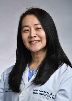
When she transitioned to the United States, the narrower scope of OMFR in the U.S. came as a surprise. In Japan, dental radiologists interpret MRI, MDCT, ultrasound, and fluoroscopy, and may also provide radiotherapy for cancer patients–roles that closely resemble those of medical radiologists or ENT specialists In contrast, OMFR in the U S focuses primarily on periapical radiographs, panoramic imaging, and CBCT Dr Matsumura views this difference as reflective of a broader cultural divide: in Japan, the boundary between dentistry and medicine is less rigid Today, her work includes interpreting approximately ten CBCT scans per day, teaching dental students, and conducting research on CBCT image acquisition
Early in Dr Matsumura’s career, practitioners relied on periapical radiographs, panoramic imaging, and the Waters view, a two-dimensional projection used to evaluate the maxillary sinuses With the introduction of CBCT, clinicians have gained access to true three-dimensional imaging with improved spatial resolution, largely replacing older techniques like the Waters view
Despite this shift, CBCT has not fully replaced traditional radiographs Periapical imaging still offers the highest spatial resolution, and CBCT involves higher radiation exposure Current guidelines from the National Council on Radiation Protection and Measurements recommend a stepwise approach: begin with periapical radiographs, proceed to panoramic imaging if needed, and reserve CBCT for cases requiring three-dimensional assessment. However, as CBCT technology advances and radiation doses decrease, this sequence may evolve. Newer machines already integrate multiple imaging capabilities, suggesting that CBCT could eventually replace conventional modalities altogether.
The greatest clinical advantage of CBCT is the ability to reveal the true three-dimensional extent of pathology Unlike two-dimensional imaging, which compresses structures into a single plane, CBCT allows visualization in axial, coronal, and sagittal sections This capability directly informs surgical planning and risk assessment
In endodontics, CBCT is particularly valuable for identifying the MB2 canal in maxillary first molars, a commonly missed structure that can lead to persistent infection if untreated. CBCT can reveal it before treatment begins, potentially preventing retreatment altogether. In oral surgery, it clarifies the relationship between third molar roots and the inferior alveolar canal, helping assess the risk of nerve injury In implant dentistry, three-dimensional imaging has become essential, replacing earlier reliance on panoramic views that carried a higher risk of complications such as sinus perforation
Looking ahead, Dr Matsumura identifies dental MRI as the most significant development on the horizon
Because MRI does not use ionizing radiation, it could allow for more frequent imaging without the exposure concerns that govern current practice Historically, its use in dentistry has been limited by metal artifacts and insufficient spatial resolution However, advances in dental-specific MRI technology are underway, and she anticipates its integration into clinical practice within the next decade.
Artificial intelligence is beginning to influence aspects of dental radiology, though its role remains supportive rather than diagnostic Current systems can flag caries, identify periapical pathology, and estimate bone loss on radiographs, helping reduce cognitive load and highlight incidental findings
However, detection is not equivalent to diagnosis AI outputs require validation and clinical interpretation, particularly given the limitations of two-dimensional data As Dr Matsumura emphasizes, patients still rely on human expertise for accurate diagnosis She also notes that misconceptions about AI replacing radiologists may discourage students from entering the field
Career paths in OMFR often center on academia, where clinical interpretation, teaching, and research intersect. However, independent practice has become increasingly viable through teleradiology services, allowing radiologists to interpret images remotely without owning imaging equipment. Clinicians can submit digital scans, and radiologists provide detailed reports on a flexible schedule. Some begin with larger case distribution networks before building independent referral systems
Radiology also offers a notable advantage in career longevity Unlike clinical dentistry, which can be physically demanding and lead to musculoskeletal strain, radiology does not require manual procedures As long as cognitive and visual abilities remain intact, practitioners can continue working from virtually any location Dr Matsumura has observed many clinicians transition into radiology later in their careers, often finding it both sustainable and fulfilling
A concern she hears often from dental students is that choosing radiology means abandoning four years of clinical training. Dr. Matsumura challenges this notion, emphasizing that clinical knowledge is essential to radiologic interpretation Understanding endodontics, implantology, periodontics, and surgical principles is what allows a radiologist to produce meaningful, specialty-specific reports Without that foundation, interpretations risk being overly generic or even misleading Without that foundation, interpretations risk being misleading or overly generic
To maintain and refine her diagnostic skills, Dr Matsumura keeps a record of her cases and differential diagnoses, later comparing them with biopsy results This ongoing feedback loop reinforces the impact of her work and sharpens her clinical judgement. Looking forward, she is particularly interested in advancing diagnostic precision. While imaging can often identify abnormalities, it does not always provide definitive answers. Emerging technologies may eventually close this gap, allowing for more precise and confident diagnoses.
Ultimately, Dr Matsumura returns to the principle that first drew her to the field: the radiologist’s role is foundational Every dental specialty depends on accurate imaging and interpretation That sense of being genuinely indispensable across the entire institution and across disciplines is what continues to make her work meaningful
The rapidly growing field of anti-aging medicine has drawn much attention both clinically and culturally Quick buzzwords like “ age gracefully” and “healthy aging” appeal to patients’ self-image and fear of decline. These anti-aging scientific developments and marketing campaigns have gained popularity among medical treatments for a wide breadth of issues such as neurodegenerative diseases, cancers, and metabolic disorders, and they will soon influence the future of periodontics

The hallmark of aging is cellular senescence. Senescent cells have characteristics such as cell-cycle arrest, morphological changes, defective nuclear membranes, and metabolic alterations. The senescent state is a response to chronic physical, chemical, or biological stressors that include persistent inflammation, oxidative stress, and a dysbiotic microbiome
The development of a targeted treatment for senescence in periodontitis was based on previous research from the University of Pennsylvania School of Dental Medicine. The study concluded that the accelerated biological aging of gingival tissues is caused by the deleterious secretions of senescent cells, termed the senescence-associated secretory phenotype (SASP) The secretions disrupt neighboring cells by propagating chronic inflammation and triggering senescence in younger tissues in a process known as “inflammaging.” Neither the SASP nor inflammaging are unique to periodontitis, but rather are shared characteristics of chronic diseases associated with cellular senescence. SASP factors alone do not define a senescent state in vulnerable tissues. For example, elevated levels of gingival p16 gene expression were found in patients with periodontitis compared to their healthy, age-matched counterparts, indicating enhanced activity of cell cycle arrest pathways in periodontitis. As stated in previous research, elevated p16-expressing cells are associated with worse clinical outcomes in pulmonary, neurologic, and musculoskeletal diseases. The findings of this study are significant since they provide the first clinical report to relate elevated senescence and chronic inflammation to the secretome of gingival tissues, allowing future research to target senotherapeutic interventions for periodontitis
In April 2025, the Journal of Dental Research published the most recent findings out of the University of Pennsylvania School of Dental Medicine. Targeted senotherapy using the enzyme inhibitor dasatinib with the natural flavonoid compound quercetin effectively reduced markers of senescence and periodontal inflammation in in vitro and in vivo animal models
Dasatinib and quercetin (DQ) are one of the most promising senotherapies in medicine Preclinical animal research indicates that targeting and reducing senescent cell populations with DQ enhances physiological function in diseases with pathophysiological mechanisms comparable to periodontal disease, including arthritis, obesity, osteoporosis, and pulmonary fibrosis. Current research is exploring the role of DQ therapy in chronic kidney disease and Alzheimer’s disease, in addition to increasing lifespan.
DQ produced hopeful results for the treatment of periodontal disease. DQ reduced markers of inflammatory mediators associated with SASP and alleviated signs of senescence in gingival keratinocytes while also suppressing senescence features in gingival tissues and alveolar bone loss in aged mice. The results of the study present the first systematic evidence that DQ can reduce gingival senescence, which thereby mitigates age-associated periodontal bone loss
The use of quercetin adds to the growing body of knowledge regarding the effectiveness of natural compounds in the treatment of oral and systemic diseases. Researcher S.E. Sahingur hopes to begin an early-stage clinical trial to assess the effectiveness of quercetin on the biological and clinical markers of periodontitis Quercetin is currently labeled and sold as a safe over-the-counter supplement Before we know it, quercetin may be marketed as the newest antiaging “cure-all” for periodontitis that patients and consumers are craving.
Resources
Rattanaprukskul K, Xia X-J, Hysa M, et al. Dasatinib and Quercetin Limit Gingival * Senescence, Inflammation, and Bone Loss Journal of Dental Research 2025;104(4):419-427 doi:10 1177/00220345241299789
Rattanaprukskul, K., Xia, X. J., Jiang, M., Albuquerque-Souza, E., Bandyopadhyay, D., & Sahingur, S. E. (2024). Molecular Signatures of Senescence in Periodontitis: Clinical Insights. Journal of dental research, 103(8), 800–808. https://doi.org/10.1177/00220345241255325

University of Pennsylvania School of Dental Medicine (2025, April 11) New drug therapy with natural compound shows promise against periodontal diseases. Penn Dental Medicine. https://www.dental.upenn.edu/newsevents/2025/04/11/new-drug-therapy-with-natural-compound-shows-promiseagainst-periodontal-diseases/