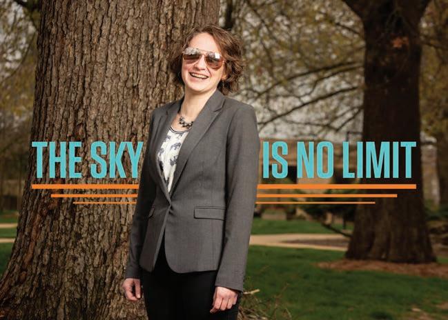
5 minute read
Recipe for organ tissue creation

from Missouri S&T Magazine, Summer 2020
by Missouri S&T Library and Learning Resources | Curtis Laws Wilson Library


Using bioactive glass, stem cells and a 3-D printer, Missouri S&T researchers are creating organ tissue samples in hopes of advancing pharmaceutical testing and providing a better understanding of how diseases affect human cells.
e researchers grow stem cells and add them to hydrogels made of alginate, gelatin or similar substances. en, in a step unique to Missouri S&T, the researchers add bioactive glass to supply needed calcium ions to the hydrogel/ cell mixture and load the mixture as “bio-ink” into a 3-D printer. ey test the samples after fabrication to assess the stem cell function, the material’s tensile strength, degradation and the best glass type to add.
“Dierent cells prefer dierent gels, so we work to find which gel combination suits our research,” says Krishna Kolan, MS ME’11, PhD ME’15, a postdoctoral researcher in mechanical and aerospace engineering. “e challenge is that dissolved glass adds calcium, but it changes the pH, and cells need neutral pH to survive. We figured out which glass and how much to add to maintain neutral pH.”
Kolan says researchers are several years away from making a functioning organ, such as a liver or kidney. e challenge is the vascular system and multiple types of cells in those organs, but S&T researchers are working on ways to develop vascular systems within the bioprinted tissue. Undergraduate students August Bindbeutel in mechanical engineering and Lesa Steen in materials science and engineering are assisting Kolan.
“Endothelial cells form networks in environments they like, such as glass-infused hydrogel,” Kolan says. “As the network grows, it vascularizes the tissue.”
As researchers work toward someday repairing or replacing organs with engineered organs, they create tissue models for pharmaceutical testing, Kolan says. His team is also working on 3-D-printed bone models. Biology graduate student Bradley Bromet is comparing diseased cells with healthy stem cells to see in 3-D how a disease — diabetes, for instance — aects cells.
Kolan is working on the project with Ming Leu, the Keith and Pat Bailey Professor in S&T’s mechanical and aerospace engineering department; Richard Brow, interim deputy provost and Curators’ Distinguished Professor of materials science and engineering; Delbert Day, CerE’58, Curators’ Distinguished Professor emeritus of ceramic engineering, and Julie Semon, assistant professor of biology and director of S&T’s Laboratory of Regenerative Medicine.
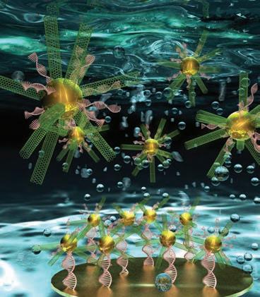
DNA BIOSENSOR COULD SPEED DISEASE DETECTION
Combining nanotechnology and biomedical diagnostics into a process called nanodiagnostics is helping scientists detect diseases at an earlier stage.
S&T chemistry researchers Risheng Wang and Wenyan Liu are creating an ultrasensitive DNA biosensor to detect, transmit and record information about biological substances like DNA and RNA, proteins, antibodies, antigens and other biological components.
Using the sensor to detect biomarkers linked to disease could allow physicians to diagnose cancer and genetic disorders earlier than current testing methods allow. It could also help monitor patient response to therapies.
“Biosensing with nanomaterials has the advantages of greater sensitivity and faster response than traditional analytical methods that require today’s medical devices and time-consuming molecular amplification techniques,” Wang says.
Made from carbon nanotubes and gold nanoparticles, the biosensor has a 3-D radial shape much like that of a sea urchin. e researchers say the biosensor generated a remarkable electrochemical response.
“is biosensor could detect the ultralow-abundance nucleic acids in complex biological media,” says Liu. “It was also highly selective in discriminating single mismatched DNA from fully matched DNA. is type of nanodiagnostic system is a potential candidate for point-of-care medical measurements because of its excellent stability and possibility of miniaturization.” e research, which is funded by the National Science Foundation, was featured on the cover of the April issue of Analytical Chemistry.
DEVELOPING A FASTER, CLEANER, CHEAPER CHEMICAL REACTION


A group of S&T researchers has developed a way for chemists to perform the combined reaction-separation process in chemical reactions without using metals or solvents.
Manufacturers convert substances such as carbon dioxide or biomass into more usable forms through reactive chemistry, often using expensive metal catalysts such as gold or palladium. Reclaiming those catalysts for reuse can be dicult as well as labor- and energy-intensive, especially when the catalyst and reaction agent are homogeneous: both liquids, for example. e expense of precious metals makes catalyst reclamation even more important.
By using hollow composite polymer fibers with organic linkers such as toluene as a binding agent, the researchers immobilized the catalyst on the surface of the fibers while the reaction product flowed through the hollow tube. e process allows chemists to more easily reclaim the catalyst. e researchers used a continuous operation, adding chemicals at dierent points along a system. at allows for quicker heating and a faster reaction rate — seconds or minutes as opposed to the hours needed for batch reactions.
“ink of it as making coee,” says Ali Rownaghi, an assistant teaching professor of chemical and biochemical engineering at Missouri S&T and a member of the research team. “You can brew a big pot of coee, but think how much easier it would be to add together water and coee along a tube and dispense a cup at the end.” e pharmaceutical manufacturing aspect of the research supports the University of Missouri System’s investment in research as part of the NextGen Precision Health Initiative and Institute.
Working with Rownaghi at S&T are assistant professor Fateme Rezaei and graduate students Abdo-Alslam Alwakwak, Yingxin He, Ahmed Almuslem and Matthew Senter, all in chemical and biochemical engineering.