
Director’s Corner
The Cawley Center for Translational Cancer Research at the Helen F. Graham Cancer Center & Research Institute is unique among the 1,500 community cancer centers accredited by the Commission on Cancer in the United States.
The Cawley Center brings innovative solutions to clinical problems and not only bench to bedside research, but bench research to the community. The Center has a workforce diversity and state-of-the-art infrastructure. It is addressing health care disparities and training the next generation of young investigators. To date, the Center has trained over 100 students and physician scientists, many of whom have gone on to successful careers in academia, industry and clinical practice. This year’s report highlights a new program led by Shirin Modarai, Ph.D., and Jennifer Sims Mourtada, Ph.D., focused on the Organoid Core for breast and ovarian cancer. The research aims to speed up the development of new cancer treatments. In 2025, the Cawley Center also welcomed Percy Guzman Montero, M.D., as the first Bacchieri Caspar Wistar Disparities Research Fellow in Cancer Population Health. Montero completed postdoctoral training at the National Cancer Institute.
The Cawley Center is supported by philanthropy and external funding. Over the years the funding has come from the National Institutes of Health, National Cancer Institute, Delaware Ovarian Cancer Foundation, Wistar Institute, Lisa Dean Moseley Foundation, Carpenter Foundation, Longwood Foundation, Friends of Helen F. Graham Cancer Center & Research Institute, Delaware Bioscience Center for Advanced Technology, IDeA Networks of Biomedical Research Excellence (INBRE) Program, ChristianaCare and others. Finally, the Delaware community generously continues its support of this unique Center.
The Graham Cancer Center and the Cawley Center have a 15-year partnership with the Wistar Institute in Philadelphia. In 2025, 16 clinicians at the Graham Center were reappointed as Adjunct Faculty of The Wistar Institute and Affiliate Member of the Caplan Cancer Center for another three years. Collaborative research between the clinicians at the Graham Cancer Center and Wistar scientists involves pancreatic, ovarian, lung, glioblastoma, esophageal and gastric cancers.
The Cawley Center also benefits from the guidance of a dedicated Community Research Advisory Board which ensures that its research efforts align with the needs of the
local community. This focus on community engagement enhances the Center’s impact on public health and cancer care outcomes.
The Cawley Center would not be able to function without the Translational Research Support Team who are responsible for specimen collection and preparation, protocol development, patient consents, data collection and grant preparation.
We thank everyone for their continued support as we continue to work toward improving cancer treatments and impacting the patient disparities in cancer care and prevention.
Nicholas Petrelli, M.D.
Director, Cawley Center for Translational Cancer Research
Helen F. Graham Cancer Center & Research Center
ChristianaCare
Associate Director of Translational Research
The Wistar Institute
Thomas Schwaab, M.D., Ph.D.
Bank of America Endowed Medical Director
Helen F. Graham Cancer Center & Research Institute
ChristianaCare
Adjunct Faculty of The Wistar Institute
Affiliate Member of the Caplan Cancer Center
The Organoid Core
Jennifer Sims Mourtada, Ph.D.
The Cawley Center recently established an Organoid Core, led by Jennifer Sims-Mourtada, Ph.D., to grow tumor organoid models of breast, ovarian and other solid tumors. Organoids are tiny three-dimensional “mini-tumors in a dish,” created from tissue generously donated by patients.
Because they closely replicate the complexity of human tumors, organoids give researchers a way to study cancer in the lab and explore how it responds to different treatments. Unlike animal models and established cancer cell lines, organoids are freshly derived from human tumors and cultured in a supportive framework that mimics the human body. By maintaining the exact genetic and behavioral features of the tumor, organoids are more reliable predictors of which drugs will work, and which types of patients are most likely to benefit.
Dr. Sims-Mourtada and Shirin Modarai, Ph.D., a research scientist at the Cawley Center, are currently using organoid models in preclinical drug screening. The team has developed standard operating procedures for patient consent, tissue procurement and organoid growth, ensuring that each step is carried out ethically and consistently. Using these models, the team is partnering with academic investigators and pharmaceutical companies, including The Wistar Institute and Incyte Pharmaceuticals, to screen early-stage cancer drugs.
By tailoring clinical trials based on data obtained from patient-specific organoids, researchers can better predict how different patients will respond to treatments, optimizing strategies and improving trial outcomes. This approach makes testing more realistic, accelerates drug approvals, reduces costs and increases the likelihood that new therapies will improve patient care.
The organoid core at the Cawley Center is one of only a handful of cancer centers nationwide conducting this work, and the first community cancer center to establish such a program, ensuring that discoveries are grounded in populations that reflect realworld patients. The Cawley Center is actively seeking new academic and industry partners to expand this work and translate discoveries into treatments that truly impact the lives of patients.
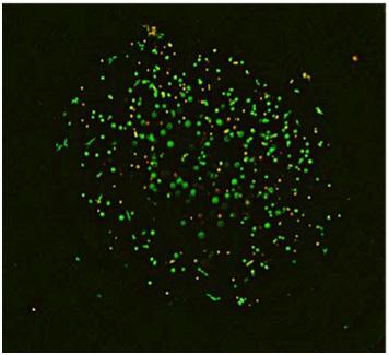
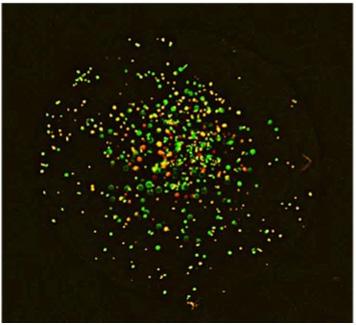
Before Treatment
Lisa Dean Moseley Fellowship in Translational Cancer Research
Alexandria Haraczy, M.D.
The Lisa Dean Moseley Translational Cancer Research Fellowship at the Cawley Center for Translational Cancer Research was created to prepare the next generation of translational scientists in an environment where research and patient care come together.
Fellows gain the opportunity to work side-by-side with scientists on projects designed to improve cancer outcomes.
We are proud to welcome Alex Haraczy, M.D., as our newest Moseley Physician-Scientist Fellow. Dr. Haraczy completed a PGY3 surgical residency at ChristianaCare and is the first ChristianaCare resident to be awarded this fellowship. She received her medical degree from the University of Michigan and previously trained as a post-baccalaureate research fellow at the National Institutes of Health.
As part of her fellowship, Dr. Haraczy is investigating biomarkers of triple-negative breast cancer (TNBC), the most aggressive breast cancer subtype. Unfortunately, Delaware leads the nation in TNBC incidence, with geographic “hot spots” where women, particularly Black women, face higher risk.
By studying the link between inflammatory biomarkers and TNBC in these hotspot regions compared to other areas, Dr. Haraczy’s work will help uncover why these disparities exist and how they can be addressed.
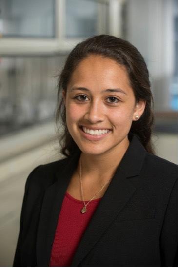
Alexandria Haraczy, M.D.
Lisa Dean Moseley Translational Research Fellow
Triple negative breast cancer update
Triple-negative breast cancer (TNBC) is an aggressive and difficult-to-treat form of breast cancer. Unlike other breast cancers, TNBC does not respond to hormone therapies or HER2targeted drugs, leaving chemotherapy as the main option.
Unfortunately, Delaware has the highest rate of TNBC in the nation, with especially high rates seen in identified hotspot neighborhoods where women face greater exposures to breast cancer risk factors such as obesity, alcohol use and environmental toxins. Researchers at the Cawley Center for Translational Cancer Research are working to understand this challenge and to develop better strategies to predict, prevent and treat the disease.
Through the DESPARIT study, funded by the National Institutes of Health (NIH) and conducted in collaboration with Scott Siegel, Ph.D., Jennifer SimsMourtada, Ph.D., and her team identified epigenetic changes—chemical switches on DNA that turn genes on or off—in genes that control immune function and drive inflammation that can promote tumor growth.
Unlike the healthy inflammation our bodies use to heal injuries or fight infections, tumor-promoting inflammation creates an environment that helps cancer grow and spread.
These changes may help explain the higher incidence of TNBC in these neighborhoods. The team is now working to validate these findings and determine whether they can be used as biomarkers of risk. At the same time, her lab is using organoid models, miniature tumors grown from patient samples, to test how these inflammatory changes can specifically drive TNBC. Preliminary data show that when early-stage tumors are exposed to these changes, they develop features of a TNBC phenotype, suggesting that inflammation may play a direct role in shaping this aggressive disease.
Another area of investigation is the Pan-Immune-Inflammation Value (PIV), a blood-based measure of circulating immune cells. Although PIV has been shown to predict prognosis and outcomes in breast cancer overall, TNBC patients appear to behave differently. As part of his senior thesis in the Sims-Mourtada Lab at the Cawley Center, University of Delaware undergraduate student Atif Bacchus has been analyzing PIV in TNBC patients. His
work so far shows that TNBC patients tend to have lower PIV scores, driven by weaker innate immune responses. The innate immune system is the body’s first line of defense, relying on fast-acting cells such as neutrophils and macrophages to recognize threats and regulate inflammation. When this frontline defense is weakened, tumors may escape immune control and foster chronic, tumor-promoting inflammation that supports cancer growth. This research is providing critical knowledge about the high rates TNBC incidence in the communities served by the Helen F. Graham Cancer Center & Research Institute.
By uncovering these biological drivers and potential biomarkers, Cawley Center scientists are actively developing better methods of screening, preventive strategies and treatments that will directly benefit patients in Delaware and can be applied to communities across the country.
Publications:
Chu T, Wasi M, Guerra RM, Song X, Wang S, Sims-Mourtada J, You L, Skeletal response to Yoda1 and whole-body vibration in mice varied with animal age, bone compartment, treatment duration, and radiation exposure. Wang L. Bone. 2025 Sep; 198:117525. doi: 10.1016/j.bone.2025.117525. Epub 2025 May 17. PMID: 40389188
Ajayi GO, Ma A, Modarai SR, Opdenaker LM, Sims-Mourtada J. CRISPR/Cas9 Targeting of Aldehyde Dehydrogenase 1A1 Reveals Heterogeneous Roles in Radiation Response and Redox Stress Across Clonal Lines in Triple-Negative Breast Cancer. Int J Mol Sci. 2025 Mar 5;26(5):2303. doi: 10.3390/ijms26052303. PMID: 40076923.
Toney NJ, Opdenaker LM, Frerichs L, Modarai SR, Ma A, Archinal H, Ajayi GO, Sims-Mourtada J. B cells enhance IL-1 beta driven invasiveness in triple-negative breast cancer.: Sci Rep. 2025 Jan 16;15(1):2211. doi: 10.1038/s41598-025-86064-1. PMID: 39801513; PMCID: PMC11722549.
Tomiyoshi, K., Wilson, L. J., Mourtada, F., Mourtada, J. S., Namiki, Y., Kamata, W., Yang, D. J., & Inoue, T. (2024). Optimization Processes of Clinical Chelation-Based Radiopharmaceuticals for Pathway-Directed Targeted Radionuclide Therapy in Oncology. Pharmaceutics, 16(11), 1458. https://doi. org/10.3390/pharmaceutics16111458
Mourtada F, Tomiyoshi K, Sims-Mourtada J, Mukai-Sasaki Y, Yagihashi T, Namiki Y, Murai T, Yang DJ, Inoue T. Actinium-225 Targeted Agents: Where Are We Now? Brachytherapy. 2023 Nov-Dec;22(6):697-708. doi: 10.1016/j.brachy.2023.06.228. Epub 2023 Sep 9. PMID: 37690972; PMCID: PMC10840862.
Ajayi GO, Ma A, Modarai SR, Opdenaker LM, Sims-Mourtada: CRISPR/Cas9 knockout of ALDH1A1 increases radiation response in triple-negative breast cancer. Clinical Cancer Research, Vol. 31, Issue 12 Supplement, June 15, 2025.
Ajayi GO, Ma A, Modarai SR, Opdenaker LM, Sims-Mourtada Functional knockout of ALDH1A1 reveals distinct roles in tumor cell heterogeneity. Molecular Biology of the Cell, Vol. 36, No. 1. January 1, 2025.
Presentations:
Jennifer Sims-Mourtada, Ross Budziszewski, Lisa Frerichs, Ashley Stewart, Caitlin Mbuakoto, Madolyn L. MacDonald, Shawn W. Polson, Yuchen Zhang, Scott D. Siegel. An Update on the Delaware INBRE Women’s Health Supplement: Detecting Epigenetic Signatures to Improve the Prevention of Triple-Negative Breast Cancer. NIH Women’s Health IDEA State Symposium, September 16, 2025
Jennifer Sims-Mourtada, Ross Budziszewski, Lisa Frerichs, Ashley Stewart, Caitlin Mbuakoto, Madolyn L. MacDonald, Shawn W. Polson, Yuchen Zhang, Scott D. Siegel Detecting epigenetic signatures to prevent avoidable racial inequities in triple-negative breast cancer (desparit): A pilot study. American Association of Cancer Researchers 18th conference on the Science of Health Disparities, September 20,2025, Baltimore MD.
Better Treatment for the Silent Cancer: Ovarian
Shirin R. Modarai, Ph.D. Research Scientist
Ovarian cancer is a deadly disease that often goes undetected until it has reached an advanced stage. Although many women initially respond to chemotherapy, most patients relapse and develop resistance to treatment.
Unfortunately, new treatment strategies have not been effective for most women with ovarian cancer and even more limited treatment options for those who have relapsed after chemotherapy. The use of patient-derived tumor models makes it possible to design personalized treatment strategies in the future, offering more durable responses to therapy.
Scientists in the Cawley Center are using organoid cultures to better understand how ovarian cancer responds to treatment. Organoid cultures have several advantages over traditional 2D cell culture models. While it takes time to develop and culture the organoids, this 3D culturing technique conserves some of the structural elements of the tissue in a more realistic environment that can be supplemented with growth
factors and additional cell types. In addition, this culturing method serves as an excellent model for drug screening and other downstream applications, including live cell imaging.
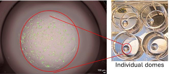
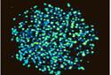
An example of what the organoids look like and how they are grown in 3D is shown in Figure 1.
The image on the left is showing a highly magnified view of ovarian tumor organoids grown in the 3D matrix and imaged with the previously acquired a state-ofthe-art live-cell imaging system, the Tecan Spark Cyto 100, that was purchased with generous donations from the Delaware Ovarian Cancer Foundation. The image on the right shows the broad view of what the matrix drop looks like by the naked eye, and the support for equipment like the imaging system help foster the research and speed along patient relevant research.
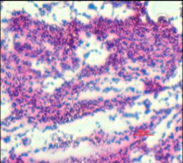
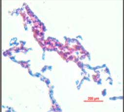
Additionally, organoids maintain the genetic, molecular and histological characteristics of the original tumor, as seen in Figure 2. These images shown in Figure 2 were stained and imaged at the Cawley Center.
for
Figure 1
Figure 2
The H&E of an ovarian tumor tissue (left image) compared to the H&E of an organoid derived from that same ovarian tumor (right image). As the organoid core facility continues to expand, this technology is essential
paving the way for new research avenues to identify and target cancer more effectively.
Tumor tissue Tumor organoid
One project currently underway is the thesis project of one of our University of Delaware undergraduate students, Julia Querey, who is looking at the optimization of different growth media for the growth and expansion of various subtypes of ovarian cancers. Current research in ovarian cancer lacks relevant model systems that can foster reliable drug screenings for initial treatment models, maintenance therapies and acquired resistant tumors. To setup such organoid model systems, it is
crucial to have the best environment for the organoids to grow and this is one avenue Querey is looking at with her research project. Working with Dr. Sims-Mourtada and Dr. Modarai, Querey can utilize the organoid core’s patient repository to perform her optimization of growth media on different patientderived ovarian organoids.
Figure 3 shows some examples of how ovarian organoids grow based on the growth conditions in 3D.
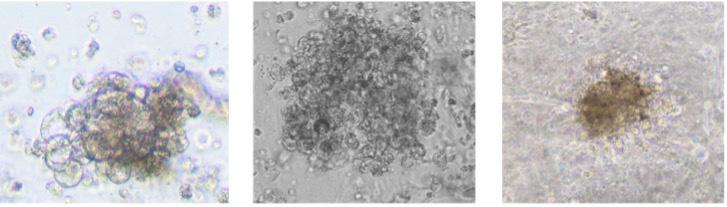
Figure 3
Different types of ovarian cancer organoids that can grow based on the optimal growth conditions.
New Caspar Wistar Bacchieri Disparities Fellowship in Cancer Population Health
Scott Siegel, Ph.D., Director, Cancer Center Population Sciences
In April 2025, Percy Guzman Montero, M.D., joined the Helen F. Graham Cancer Center & Research Institute as the Bacchieri Caspar Wistar Disparities Research Fellow.
In this new role, under the supervision of Scott Siegel, Ph.D., Dr. Montero’s work will support ChristianaCare’s broader mission to reduce the burden of cancer for all members of our community through translational research.
Dr. Montero trained as a physician and breast surgical oncologist in Costa Rica, where he provided specialized care to patients with breast cancer at Hospital Rafael Ángel Calderón Guardia. His clinical practice focused on breast cancer surgery, and he played a key role in national efforts to reduce delays in diagnosis and treatment. This handson experience continues to shape his research approach and his dedication to improving health equity.
Dr. Montero subsequently completed postdoctoral training at the National Cancer Institute (NCI), where he contributed to both global and domestic research initiatives aimed at improving cancer care delivery. At NCI, he worked
extensively with real-world data to generate evidence that informed practice and policy. He also co-founded the Global Health Interest Group to foster interdisciplinary collaboration and support early-career scientists worldwide.
Dr. Montero holds a Master’s in Epidemiology, a specialist degree in Innovation and Management, and a Digital Health certificate from Harvard Business School. These academic and clinical experiences have reinforced his belief in the power of innovation, collaboration and data to transform health systems, particularly for underserved communities.
Thank you to the Bacchieri Family for their generous support of this fellowship.
The Cawley Center is proud to welcome Dr. Montero to ChristianaCare.
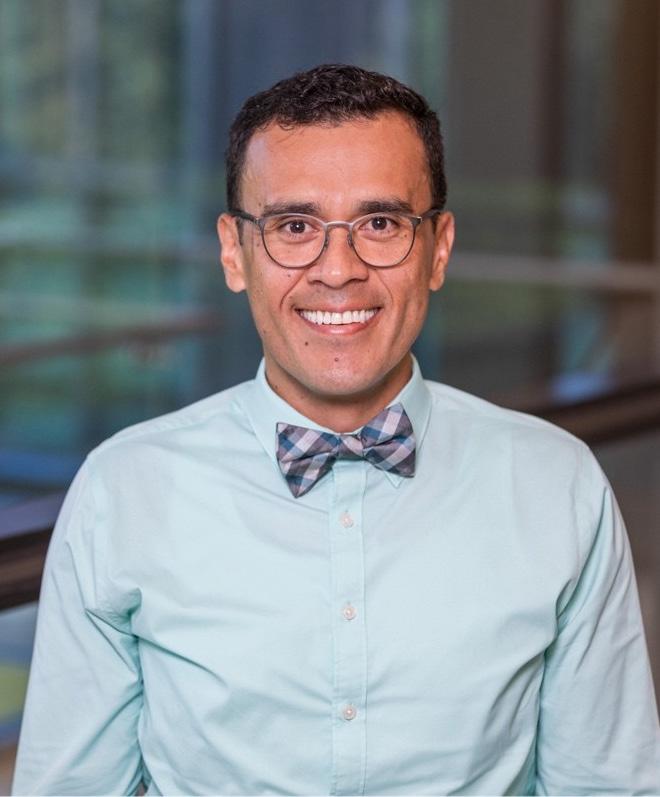
Percy Guzman Montero, M.D. Bacchieri Caspar Wistar Disparities Research Fellow
The Drivers of Delaware’s Advanced Breast Cancer Hotspots
Scott Siegel, Ph.D., Director, Cancer Center Population Sciences
Delaware ranks first in the country for the incidence of late-stage breast cancer among younger women and triple-negative breast cancer (TNBC), an aggressive subtype that does not respond to standard hormonal therapies.
A new study from the Cawley Center for Translational Cancer Research sheds new light on why these advanced forms of breast cancer appear to cluster in certain Delaware communities—and offers a path forward to reduce that burden.
Dr. Siegel and his colleagues’ article was published in JNCI Cancer Spectrum on July 22, 2025 and builds on earlier research that identified two geographic hotspots for advanced breast cancer in Wilmington and Middletown. The new analysis explains what’s driving those elevated rates and how that knowledge is shaping ChristianaCare’s clinical response. The paper represents more than a year of work and thousands of hours of data collection. The research team manually reviewed nearly 3,500 medical records to create a first-of-its-
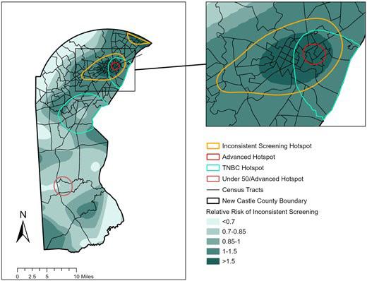

kind dataset that includes method of detection, time since last screening, tumor grade and molecular subtype.
By mapping cancer data down to the level of patient addresses, researchers showed how the Wilmington hotspot sits at the intersection of two other mapped risk zones: one with high rates of TNBC and another where women are less likely to get regular breast cancer screenings.
ChristianaCare has already begun to act. A community health worker is now embedded in the Wilmington hotspot to increase screening and conduct breast
cancer risk assessments. Residents at higher risk will be connected to prevention programs, including those for TNBC. In Middletown, the study found a high number of advanced breast cancer cases among women under 50. Here, too, screening gaps and aggressive tumor characteristics were factors.
The study sends a clear message that community cancer centers can lead the way on improving cancer outcomes by focusing efforts where the risk is highest. With the right data, they can target outreach, expand screening and deliver resources to the neighborhoods that need them most.
Selected Publications:
1. Siegel, S.D., Zhang, Y., Budziszewski, R., Bacchus, A., Rowland, J., Iacocca, M.V., Hall-McBride, R., Curriero, F.C. (2025). Extending catchment area approaches to a community cancer center: a breast cancer hotspot case-study. Journal of the National Cancer Institute Cancer Spectrum, 9(4): pkafo67.
2. Rafalko, N., Siegel, S., Yerkes, P., Eberth, J.M., Burstyn, I., Goldstein, N.D. (2025). Quantifying the primary care workforce in the U.S.: A validation study with and without an imperfectly measured referent standard. Annals of Epidemiology, 108:92-98.
3. Morreale, M., Fenech, A.L., Brownlee, H.A., Siegel, S.D., Laurenceau, J.-P. (2025). A dyadic analysis of optimism, pessimism, and fear of cancer recurrence in couples coping with early-stage breast cancer. Psycho-Oncology, 34(3): e70125.
4. Siegel, S.D. (2024). Reducing breast cancer disparities with precision public health. Delaware Journal of Public Health, 10(3):46-50.
5. Siegel, S.D., Budziszewski, R., Layton, E., Nam, B., & Schnoll, R. (2024). A pilot pragmatic randomized controlled trial of a nicotine metabolite ratio-guided smoking cessation intervention in a lung health and screening program. Drug and Alcohol Dependence Reports, 12:100275.
6. Fenech, A. L., Humphris, G. M., Laurenceau, J.-P., Siegel, S. D., Rogers, S. N., Ozakinci, G., Crawford, J. R., & Pring, M. (2024). Anxiety, depression, and fear of cancer recurrence in head and neck cancer. Health Psychology.
7. Fenech, A.L., Soriano, E.C., Asok, A. Siegel, S.D., Morreale, M., Brownlee, H.A., Laurenceau, J.P. (2024). Fear of cancer recurrence and change in cortisol levels in partners of breast cancer survivors. Journal of Cancer Survivorship.
8. Siegel, S.D., Zhang, Y., Lynch, S.M., Rowland, J., Curriero, F.C. (2024). A novel approach for conducting a catchment area analysis of breast cancer by age and stage for a community cancer center. Cancer Epidemiology, Biomarkers & Prevention, 33(5):646-653.
Discovery of the Code for Tissue Organization
Bruce Boman, M.D., Ph.D., MSPH, FACP Senior Research Scientist
This discovery provides a set of five biological rules that explains how colonic epithelium dynamically maintains its precise organization of cells despite continuous tissue renewal. These rules might even provide a means to understand the mechanisms that underlie the organization of other tissue types, and how tissue disorganization leads to birth defects and tissue pathology like cancer.
Every day, your body replaces billions of cells—and yet, your tissues stay perfectly organized. How is that possible?
A collaboration between Dr. Boman’s research team at the Helen F. Graham Cancer Center & Research Institute and the Mathematics Department at the University of Delaware think they’ve found an answer.
In their study published in Biology of the Cell, they show that just five basic rules may explain how the body maintains the complex structure of tissues like those in the colon, for example, even as its cells are constantly dying and being replaced.
This research challenge took years of preparation and analysis to solve. It was the result of more than 15 years of
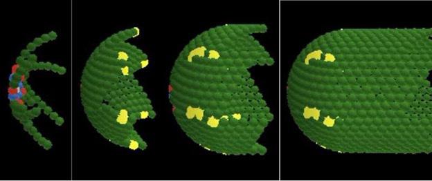
collaboration between cancer biologists and mathematicians at the university, working together to uncover the rules that shape tissue structure and guide how cells behave.
“This may be the biological version of a blueprint,” said Dr. Boman, who is also a faculty member in the departments of Biological Sciences and Mathematical Sciences at the University of Delaware.
“Just like we have a genetic code that explains how our genes work, we may also have a ‘tissue code’ that explains how our bodies stay so precisely organized over time.”
continued on next page
Math Meets Medicine
The researchers used mathematical modeling—essentially, creating a computer simulation of how cells behave—to see if a small number of rules could account for the highly organized structure of the lining of the colon. That’s an ideal place to study: cells in the colon renew every few days, but the overall shape and structure stays remarkably stable.
After running many simulations and refining their models, the team identified five core biological rules that appear to govern the structure and behavior of cells:
Timing of cell division.
The order in which cells divide.
The direction cells divide and move.
How many times cells divide.
How long a cell lives before it dies.
“These rules work together like choreography,” said Gilberto Schleiniger, Ph.D., professor in the University of Delaware’s Department of Mathematical Sciences. “They control where cells go, when they divide and how long they stick around—and that’s what keeps tissues looking and working the way they should.”
Decoding Human Tissue
The researchers believe these rules may apply not just to the colon, but to many different tissues throughout the body— skin, liver, brain and beyond. If true, this “tissue code” could help scientists better understand how tissues heal after injury, how birth defects happen and how diseases like cancer develop when that code gets disrupted.
Dr. Boman explained, “Your tissues don’t just grow and shrink randomly. They know what they’re supposed to look like, and they know how to get back to that state, even after damage. That level of precision needs a set of instructions. What we’ve found is a strong candidate for those instructions.”
This work also has important implications for the Human Cell Atlas, a global scientific collaboration working to map every cell type in the human body. While the Atlas aims to catalog what each cell is and what it’s doing at a given moment, this new research offers a dynamic framework for understanding how those cells stay organized over time.
By identifying simple, universal rules that govern cell behavior and tissue structure, the findings could help guide future efforts to not only describe cells but predict how they behave in health and disease.
Implications for Disease and Discovery
One reason the team turned to mathematical models, rather than traditional biology experiments, is that it’s extremely difficult to observe how every single cell in a tissue behaves in real time. But with computer models, researchers can run simulations that reveal patterns and dynamics hidden from view.
This kind of collaboration between biology and math reflects a broader shift in how scientists approach complex problems. It also aligns with national priorities: the National Science Foundation’s “Rules of Life” initiative challenges researchers to uncover the fundamental principles that govern living systems. This study is a strong step in that direction.
“This is just the beginning,” said Dr. Schleiniger. “Once you can identify the rules, you can begin to ask entirely new questions, and maybe even learn how to fix what’s gone wrong.”
Next steps for the team include testing the model’s predictions experimentally, refining it with additional data and exploring its relevance to cancer biology, especially how disruptions to the tissue code may lead to tumor growth or metastasis.
Dramatic Increase in Colorectal Cancer in Young Adults: Investigation into How Stem Cells Contribute to this Trend
A growing body of scientific evidence reveals that colorectal cancer (CRC) is becoming more prevalent in younger patients. Indeed, an increase in the incidence of CRC in individuals under 50 years of age was reported in over half the countries in a study involving 36 nations. Moreover, younger patients have more advanced cancer at diagnosis compared to older ones. This occurrence of later stage disease is due to a diagnostic delay because cancer is more common in the elderly, so doctors are more inclined to suspect cancer in older patients. And younger patients are more likely than older patients to have consulted with a doctor three or more times prior to referral. Even if referred, younger patients may be referred through a less urgent route compared to older ones. Unfortunately, a delay in diagnosis often results in cancer progressing to a less curable stage. Thus, younger individuals not only have a growing risk for CRC, but young CRC patients present with highly aggressive disease and more advanced stage cancer at diagnosis.
This increase in the incidence of cancer in younger patients indicates that the predictive value of symptoms is much greater than previously estimated. Thus, screening guidelines have been revised to avoid younger people experiencing greater diagnostic delays and consequent later stage at diagnosis and less treatable disease. Indeed, the U.S. Preventive Services Task Force guidelines recommend lowering the screening age from 50 to 45 years old. Disparities also exist in early-onset CRC involving African Americans, Hispanics/ Latinos, and Southern U.S. populations.
To determine the cause of the rise in early onset CRC (EOCRC) cases, patients have been genotyped to see if they may carry cancer-predisposing germline mutations because young age at CRC diagnosis is a cardinal feature of hereditary CRC. However, in the genetics clinic at the Graham Cancer Center, a survey of CRC patients (n=283) shows only a small fraction (15%) of young CRC patients (<50) have cancerpredisposing germline mutations and the spectrum of germline changes did not differ substantially between younger and older patients. Thus, an inherited genetic predisposition does not appear to account for the increasing incidence of EOCRC.
Further study was then done to identify any unique genetic factors that might be acquired during development of EOCRC by genotyping patient tumors. But the spectrum of somatic alterations was largely comparable between EOCRC and adult-onset CRC (AOCRC) with just slight differences in mutation variation and site of mutation. So, at this time, the molecular pathogenesis of EOCRC and AOCRC appears very similar. It turns out that mutation in the adenomatous polyposis coli (APC) gene is the main driver of development of both EOCRC and AOCRC. APC is a tumor suppressor gene that when mutated constitutively activates WNT signaling, which leads to stem cell overpopulation that promotes CRC tumorigenesis.
Given that a difference in genetics between EOCRC and AOCRC was not found that could explain the dramatic rise in CRC among young adults, scientists began to explore whether a
Westernized lifestyle might be to blame. The latest reports by other investigators indicate that the gut microbiome (E. coli and colibactin genotoxin) is the current suspect. However, besides CRC, it doesn’t explain the surge in other early-onset cancer types occurring on a global scale.
Much of the focus has now turned to differences in diet between younger and older adults, particularly a deficit in essential vitamin intake. Vitamin A (retinol, retinoic acid) is a nutritional factor of particular interest because it is not synthesized by the human body, so it is prone to dietary deficiency. We hypothesize that chronic vitamin A deficiency in younger adults is a predisposing condition for cancer development in the colon that occurs upon mutation of the APC gene. At the molecular level, this can be studied by determining if vitamin A deficiency leads to a condition that promotes colon tumor growth by mutant APC. Thus, our overall goal is to determine if reduced vitamin A levels in colon cells increase the ability of mutant APC to drive tumor growth. Vitamin A is also a key nutrient for stem cell function in embryonic development and maintenance of healthy adult tissues. Mechanistically, vitamin A is an activator of the retinoic acid signaling pathway and has anticancer activity by inducing stem cell differentiation.
continued on next page
The Boman research team recently discovered a link between the retinoid signaling pathway and the WNT signaling pathway that is controlled by APC. This discovery occurred as a goal of Dr. Boman’s team to discover how over-population of cancer stem cell drives tumor development and growth. Indeed, his research on stem cells discovered that APC mutations that drive CRC development do so by causing stem cell over-population. The results show that APC mutation leads to dysregulation of two key pathways: 1) Constitutive activation of WNT signaling, 2) Reduction of retinoic acid signaling. Specifically, APC mutations lead to incomplete differentiation of overpopulation of stem cells. These findings provided evidence that the link between WNT and retinoic acid signaling is etiologically involved in CRC development. Namely, when APC is mutant, the imbalance in the WNT:RAlinked mechanism can promote CRC development. Accordingly, to restore normal tissue homeostasis, wt-APC expression was induced in APC-mutant CRC cells. Dr. Boman’s team then found that administering all-trans-retinoic acid (ATRA) induced differentiation and growth arrest of colon cancer stem cells. ln 2024, these findings were reported in the peer-reviewed scientific literature in the journal Cancers (see below). This finding also represents a major step in understanding how vitamin A deficiency may promote the occurrence of APC mutations at a relatively young age in EOCRC patients.
References:
Boman BM, Dinh TN, Decker K, Emerick B, Modarai SR, Opdenaker LM, Fields JZ, Raymond C, Schleiniger G. Dynamic Organization of Cells in Colonic Epithelium is Encoded by Five Biological Rules. Biol Cell. 2025 Jul;117(7): e70017. doi: 10.1111/boc.70017.
Nguyen AL, Facey COB, Boman BM. The Complexity and Significance of Fibroblast Growth Factor (FGF) Signaling for FGF-Targeted Cancer Therapies. Cancers (Basel). 2024 Dec 30;17(1):82. doi: 10.3390/ cancers17010082.
Nguyen AL, Facey COB, Boman BM. The Significance of Aldehyde Dehydrogenase 1 in Cancers. Int J Mol Sci. 2024 Dec 30;26(1):251. doi: 10.3390/ijms26010251.
Facey COB, Hunsu VO, Zhang C, Osmond B, Opdenaker LM, Boman BM. CYP26A1 Links WNT and Retinoic Acid Signaling: A Target to Differentiate ALDH+ Stem Cells in APC-Mutant CRC. Cancers (Basel). 2024 Jan 7;16(2):264. doi: 10.3390/cancers16020264.
Hunsu VO, Facey COB, Fields JZ, Boman BM. Retinoids as ChemoPreventive and Molecular-Targeted Anti-Cancer Therapies. Int J Mol Sci. 2021 Jul 20;22(14):7731. doi: 10.3390/ijms22147731.
Facey COB, Boman BM. Retinoids in Treatment of Colorectal Cancer [Online First], IntechOpen, DOI: 10.5772/intechopen.93699. Available from: https://www.intechopen.com/online-first/retinoids-intreatment-of-colorectal-cancerRetinoids in Treatment of Colorectal Cancer, IntechOpen 2020.
Posters:
Publications 2024-2025:
ASCB Annual Meeting, San Diego California, December 14-18, 2024. Functional knockout of ALDH1A1 reveals distinct roles in tumor cell heterogeneity. Molecular Biology of the Cell, Vol. 36, No. 1. January 1, 2025.
B cells enhance IL-1 beta driven invasiveness in triple negative breast cancer. Toney NJ, Opdenaker LM, Frerichs L, Modarai SR, Ma A, Archinal H, Ajayi GO, Sims-Mourtada J. Sci Rep. 2025 Jan 16;15(1):2211. doi: 10.1038/s41598-025-86064-PMID: 39820772
CRISPR/Cas9 Targeting of Aldehyde Dehydrogenase 1A1 Reveals Heterogeneous Roles in Radiation Response and Redox Stress Across Clonal Lines in Triple-Negative Breast Cancer. Ajayi GO, Ma A, Modarai SR, Opdenaker LM, Sims-Mourtada J. Int J Mol Sci. 2025 Mar 5;26(5):2303. doi: 10.3390/ijms26052303. PMID: 40076923
SABCS Annual breast symposium, San Antonio Texas, December 10 – 14, 2024. CRISPR/Cas9 knockout of ALDH1A1 increases radiation response in triple-negative breast cancer. Clinical Cancer Research, Vol. 31, Issue 12 Supplement, June 15, 2025.
Dynamic Organization of Cells in Colonic Epithelium is Encoded by Five Biological Rules. Boman BM, Dinh TN, Decker K, Emerick B, Modarai SR, Opdenaker LM, Fields JZ, Raymond C, Schleiniger G. Biol Cell. 2025 Jul;117(7): e70017. doi: 10.1111/boc.70017. PMID: 40657955
2024 Radiation Oncology Research Poster Session, Thomas Jefferson University, Sept 11, 2024. Functional knockout of ALDH1A1 increases radiation sensitivity in triple-negative breast cancer. Presenter: Grace Ajayi
3rd Annual Biology Research Day, University of Delaware, January 10, 2025 Targeting ALDH1A1 to Sensitize Triple-Negative Breast Cancer to Radiotherapy. Presenter: Grace Ajayi
Training the Next Generation of Scientists
Lynn Opdenaker, Ph.D.
Part of the mission of the Cawley Center for Translational Cancer Research is to educate budding scientists and provide mentorship and training to undergraduate and graduate students.
Undergraduate students who express interest in laboratory research can be paired with one of our senior scientists and work alongside members of their lab. They will be trained in standard laboratory protocols and participate in weekly lab meetings where they can be involved in data sharing and critiquing. Most of these undergraduate students choose to complete a senior thesis and dedicate a significant amount of time in their junior and senior years to complete an individual project.
Graduate students from the University of Delaware can rotate within the Cawley Center to get a sense of the research and laboratory dynamics. If both the scientist and graduate student feel that they are a good fit, they can decide to complete their master’s thesis or doctoral dissertation with one of the laboratories in the Cawley Center. Students who are working toward their degrees in the
Cawley Center have access to all UD resources but also experience additional and unique opportunities. Students within the Cawley Center can attend tumor conferences where patients and treatment options are discussed among clinicians, genetic counselors, pathologists, radiologists and other specialties. This unique experience allows students to get a glimpse of what goes on in the clinical setting, while being able to take this knowledge back to the laboratory with them and allow it to help shape and guide their projects. Students also participate in a weekly research meeting where they present and discuss their work or “hot topics” in cancer research. During the year, clinicians, genetic counselors, pathologists or any number of specialized of medical personnel are invited to give talks directly to the Cawley Center.
continued on next page
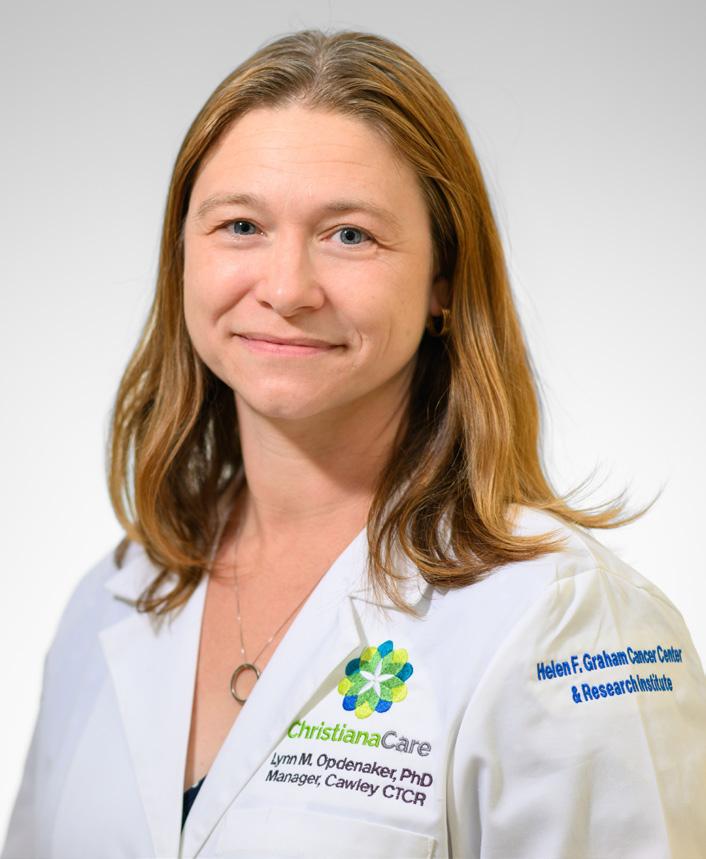
Lynn Opdenaker, Ph.D.
Current graduate students:
Molly Lausten, Ph.D., Student (Bruce Boman Lab)
Project: Investigating the Role of miR-27a-3p in the WNT Signaling Pathway and Chemoresistance in Colorectal Cancer Stem Cells
Summary: Small pieces of RNA (called microRNA) have been shown to affect the expression of specific genes. Some of these are overexpressed in colon cancer and are shown to correlate with treatment resistance. This particular microRNA upregulates signaling pathways that are known to induce resistance to chemotherapy. Ultimately, we seek to uncover how this microRNA is regulated and use this knowledge to reverse the treatments resistance in colorectal cancer.
Grace Ajayi, Ph.D., Student (Jennifer Sims-Mourtada Lab)
Project: Understanding the role of ALDH1A1 in Triple Negative Breast Cancer
Summary: Currently, there are no effective targeted therapies for Triple Negative Breast Cancer (TNBC). Our protein of interest (ALDH1A1) is a potential target for treatment. It plays a role in treatment response but may not function the same in all breast cancer cells. This project seeks to identify the function of ALDH1A1 within the diverse cell populations found in TNBC, which could identify patient populations that would benefit from ALDH1A1 targeted therapies.
Anh Nguyen, Ph.D., Student (Bruce Boman Lab)
Project: FGF19–FGFR4 Axis Promotes Colorectal Cancer Progression via MAPK and WNT/β-catenin Hyperactivation and Drives Therapy Resistance Through RA–WNT/β-Catenin–YAP1-Mediated Oncofetal Reprogramming
Summary: Overexpression of two proteins – FGF19 and FGFR4- is observed in colon cancer and tend to correlate with poor patient survival. We are investigating whether this causes resistance to cancer treatment and the mechanism by which this occurs. The goal of this project is to develop novel FGF-based strategies to improve outcomes for patients with advanced colorectal cancer.
Gene Editing is the Present and the Future
Eric B. Kmiec, Ph.D., Executive Director and Chief Scientific Officer, Gene Editing Institute
Stop Expecting CRISPR Science to Make Big Leaps
Despite the power of this new tool, it’s going to take time to develop its full potential.
Having worked in the precision medicine space for more than three decades, Eric Kmiec, Ph.D., executive director and chief scientific officer at ChristianaCare’s Gene Editing Institute has seen the concept of gene editing morph from merely a concept into clinical reality. Dr. Kmiec believes that the gene editing tool known as CRISPR technology, originally used to help sickle cell anemia patients, has the potential for a much broader application in oncology and even cardiovascular disease.
“All patients are different genetically and we know that cancer functions at the genetic level. We have to target the gene to solve the problem,” explains Dr. Kmiec.
ChristianaCare’s patient-centric approach, addressing health disparities among different ethnic and socioeconomic groups, wants all patients regardless of their background to have access to CRISPR technology. The Institute is particularly focused on using CRISPR technology for head, neck, esophageal and lung cancer. These cancers disproportionately affect those who smoke, use tobacco and
drink alcohol excessively. While cancer treatments such as immunotherapy, radiation and chemotherapy are effective, they are harsh, and some patients even decide to halt treatment.
Dr. Kmiec believes that having the Gene Editing Institute embedded within ChristianaCare is a great advantage for its research. In fact, the National Institutes of Health often include Delaware as a model in population health studies, especially in health disparities, because its population diversity and health care-access issues are reflective of broader national concerns. It also gives Dr. Kmiec and the Gene Editing Institute clinical input from the oncologists of all disciplines at the Helen F. Graham Cancer Center & Research Institute where the Institute started in July 2015.
The impact that the Cawley Center is making in disparities is evident in the reports by the scientists in this Stewardship report.
“We don’t only want to be using CRISPR technology on people who can afford it. We want everyone to have access to it,” Dr. Kmiec said.
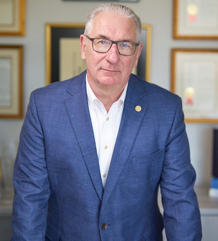
“We’ve gone through the hype of when CRISPR was first introduced, but now with the emergence of CRISPR being used in vivo, directly delivering it into tumors, it is an exciting time to be researching it.”
The medical promise of CRISPR gene editing can be seen most easily in current research on oral cancer, which affects more than 50,000 Americans every year. An increasing challenge in the treatment of oral cancer is that tumors become resistant to chemotherapy. By injecting CRISPR gene editing directly in the tumors and targeting the genetic resistance to common agents, the Gene Editing Institute could potentially enable that standard of care for cancer to work once again for patients, overcoming one of the toughest hurdles in treatment today. In doing so, we may improve outcomes for a disease whose demographics skew toward low-income communities.
CRISPR is not a miracle product that can magically cure cancer. It is, however, an exceptionally powerful tool that is creating entirely new possibilities in cancer treatment.
Fighting Breast Cancer Disparities Through Partnership
A 16 Year Partnership Between the Wistar Institute
and the Helen F. Graham Cancer Center & Research Institute at ChristianaCare
A decade-long collaboration between Wistar and ChristianaCare is leading to new discoveries and better treatment for triple-negative breast cancer patients. Over a decade ago, Dario C. Altieri, M.D., president, CEO and director of the Ellen and Ronald Cancer Center at The Wistar Institute, and Nicholas J. Petrelli, M.D., FACS, then medical director of the Helen F. Graham Cancer Center and Research Institute, formed a partnership between the two institutions that would expedite the pipeline of cancer interventions from bench to bedside.
“We had the patients, and they had the world-class science,” said Dr. Petrelli. “So, we felt, this is a great partnership between an NCI basic science laboratory research center and an NCI community cancer center. This partnership is unique in cancer research.”
This partnership has yielded more than 40 translational cancer research papers to date and advanced research discoveries made in Wistar labs into early clinical trials at ChristianaCare. One of the newest projects to come from this collaboration is a population health study on treating triple-negative breast cancer that is briefly reported on in this Stewardship Report. Jennifer Sims Mourtada, Ph.D., and Scott Siegel, Ph.D., share two intersecting research interests. The first is breast cancer. The second is health disparities. At a community level, identifying where rates of risk for triple-negative cancer are highest—as Dr. Siegel hopes to do— could mean more targeted outreach. There are only so many public health resources available. To be able to use those resources in the areas where they could make the biggest impact could impact many lives.
As this project demonstrates, Wistar and ChristianaCare’s partnership has repeatedly yielded a two-way benefit: basic scientists learn directly from clinicians about the issues those clinicians are seeing at the bedside, and the clinicians learn from the scientists about the challenges the scientists face as they’re trying to solve problems that will ultimately lead to clinical solutions.
“We will be publishing in basic science journals, but we will also be changing how we practice in the cancer center, potentially how we do our community outreach and we may actually prevent loss of life as a result of this research,” said Dr. Siegel. “I’ve never, ever been involved with any kind of project that could actually have impacts that widespread.”
The following Graham Cancer Center individuals are collaborators and have appointments at the Wistar Institute:
Arvind Sabesan, M.D., Surgical Oncology
Brendan Hagerty, M.D., Surgical Oncology
Jamal Misleh, M.D., Medical /Hematologic Oncology
Scott Hall, M.D., Ph.D., Hematologic Oncology
Greg Masters, M.D., Medical Oncology
Ariel Katcher, M.D., Gynecologic Oncology
Mark Cadungog, M.D., Gynecologic Oncology
Scott Siegel, Ph.D., Psychology, Population Health
Thomas Schwaab, MD, Ph.D., Urologic Surgery
Pulak Ray, M.D., Neurosurgery
Charles Mulligan, M.D., Thoracic Surgery
Brian Nam, M.D., Thoracic Surgery
Neil Hockstein, M.D., Head & Neck Surgery
Percy Montero, M.D., Population Health
Eric Kmiec, Ph.D., Gene Editing
Nick Petrelli, M.D., Surgical Oncology
Mary Iacocca, M.D., Pathology
A New Partnership Model for Collaborative Cancer Research and Care
THE WISTAR INSTITUTE - HELEN F. GRAHAM CANCER CENTER & RESEARCH INSTITUTE EXPERIENCE
“In summary, this represents a unique, highly innovative, non-overlapping partnership with near-perfect complimentary skills. The Helen F. Graham Cancer Center & Research Institute presents as a perfect testing ground for preclinical models developed at Wistar (there is no such opportunity at Wistar itself); likewise, the Wistar provides a great source of basic science discovery at Graham Cancer Center. This includes putting to use thousands of clinical samples (with annotated clinical data) into the hands of capable scientists for discovery, clinical correlation and hypothesis testing. Indeed, there is translational research already ongoing between these two distinct institutions.” April 2015, NCI External Advisory Board Chair
Michael A. Caligiuri, M.D.
Director, Comprehensive Cancer Center CEO, James Cancer Hospital and Solove Research Institute
Cawley Center for Translational Cancer Research
Translational Research Support Team
Innovation Solutions to Clinical Problems
Fostering collaboration
Addressing Health Disparities
Bench to Bedside Research Workforce Diversity
State of the art research infrastructure
Training the next generation of scientists
ChristianaCare’s Center of Translational Cancer Research Excellence
Helen F Graham Cancer Center and Research Institute
Essie Layton Translational Cancer Research Supervisor
Allan Bernabe Research Coordinator
Ross Budziszewski Research Manager
Lee Anne Cassidy Program Manager, Cancer Center Research Administration
Lisa Frerichs Research Coordinator
Latoya Johnson Research Administrator
Raymond Richardson Research Coordinator
Andrea Ward Histology Technician