7 minute read
A Breakthrough Discovery Into How Cancers Grow

Gerta Hoxhaj, Ph.D., and her lab have challenged a long-held belief about how tumors acquire purine nucleotides.
When looking for a new place to live, you could use an empty lot and build a structure from the ground up, or you might take an existing house and renovate some rooms or add on. In biology, cells do a similar accounting. To make a particular molecule, they can build it efficiently by assembling with salvaged components – if the parts are lying around. If not, cells have to spend a lot of energy creating the molecule from scratch.
For a class of molecules called purine nucleotides, a collaborative group of UT Southwestern researchers has uncovered how cells create and maintain their supplies of purines, vital components of DNA, RNA, and other cellular systems.
Chemotherapy drugs that target purine nucleotide synthesis have been a cornerstone of cancer treatments for more than 70 years. Recent findings from UT Southwestern scientists have upended the traditional view of how healthy and cancerous cells acquire purines, showing how some chemotherapy resistance occurs and connecting metabolic dysfunction in other diseases.
Exploring Purine Nucleotides
Gerta Hoxhaj, Ph.D., Assistant Professor in Children’s Medical Center Research Institute at UT Southwestern (CRI), began looking in 2020 at how cells generate their purine nucleotides. Despite scientists knowing these molecules were important to many metabolic processes, “we still didn’t know the fundamentals of how normal or cancer cells acquire their purines,” Dr. Hoxhaj says.
Purine nucleotides are composed of a purine base attached to a sugar molecule – ribose or deoxyribose – and one or more phosphate groups. These nucleotides are essential for cell growth and function because they serve as nucleic acid building blocks, signaling molecules, and energy carriers.
Building purine nucleotides from scratch – called de novo synthesis – costs the cell a lot of energy. A more efficient process of recycling, the salvage pathway, takes much less energy but requires a supply of pre-assembled subunits.
Historically, scientists thought normal adult tissues relied on the salvage pathway, while proliferating cells, such as those in cancerous tissues, produced most of their purines through de novo synthesis.
Dr. Hoxhaj, also a member of UT Southwestern’s Harold C. Simmons Comprehensive Cancer Center, wanted to look at both pathways broadly in an unbiased way – not just in a specific organ, tissue, or tumor type – to determine how much each pathway contributed to purine pools.
An Interdisciplinary Approach
Taking an interdisciplinary approach, Dr. Hoxhaj started by conferring with Ralph J. DeBerardinis, M.D., Ph.D., Professor and Director of the Eugene McDermott Center for Human Growth and Development, as well as in CRI, whose lab was also on to its own purine metabolism discovery.
As part of his research, Dr. DeBerardinis studies babies born with genetic disorders called inborn errors of metabolism. In a recent study, his team cultured cells from 10 patients with various genetic mitochondrial diseases and measured hundreds of metabolites to find abnormalities common to all the diseases.
“We were specifically asking: When the mitochondria don’t work properly and energy production is slowed, which other metabolic pathways also don’t work?” says Dr. DeBerardinis, a Howard Hughes Medical Institute Investigator since 2018, as well as Director of CRI’s Genetic and Metabolic Disease Program. “It turned out purine metabolism was the most consistently altered pathway, even more than pathways classically associated with mitochondria.”
The result surprised them because purine metabolism takes place in the cell’s cytoplasm, but the mutations affected the interior workings of the mitochondria. His lab eventually found that mitochondrial dysfunction causes cells to activate purine salvage, presumably allowing them to conserve energy due to impaired energy production.
The research brought in other physician-scientists, such as Kemp Kernstine, M.D., Ph.D., Professor of Cardiovascular & Thoracic Surgery, with whom Dr. DeBerardinis had built a database of altered metabolism in lung cancer.
“Why is a pediatrician working on lung cancer?” Dr. DeBerardinis asks of himself. “Lung cancer also involves altered metabolism, and we thought insights from metabolic diseases in children might help us understand metabolism in these tumors.”
Drs. DeBerardinis and Kernstine quickly discovered lung tumors with low mitochondrial activity activated the purine salvage pathway, both in mouse models and patients.
Collaboration and Discovery
Meanwhile, Dr. Hoxhaj’s team was tracing purine synthesis by infusing mice with a variety of nutrients, including amino acids and purine bases such as adenine and guanine, all tagged with heavy isotopes in order to track how purines were metabolized in both healthy and cancerous mouse tissues.
“We noticed that different organs have unique preferences for using the de novo and salvage pathways,” Dr. Hoxhaj says. “The small intestine, for instance, depended on de novo purine synthesis, while kidneys relied on salvage.”
“Our findings about the kidneys led us to Jim Brugarolas, who was so open to sharing his research and expertise,” she adds.
James Brugarolas, M.D., Ph.D., Professor of Internal Medicine and Director of UTSW’s Kidney Cancer Program, provided mouse models of tumors, allowing Dr. Hoxhaj’s team to study purine metabolism in kidney cancer. Additionally, other CRI labs contributed to the purine research: Hao Zhu, M.D., provided a liver cancer model, and Sean Morrison, Ph.D., offered specialized expertise in experimental design and data analysis.
“The magic often happens at the interface between fields or traditional disciplines,” says Dr. Morrison, Director and Professor in CRI, and Professor of Pediatrics. “One of our main goals is to create an environment in which people can make discoveries together that they wouldn’t have been able to make on their own.”
Dr. Hoxhaj’s study ultimately showed tumors are just as effective at using the salvage pathway as they are using the de novo pathway, challenging the traditional view that cancer cells primarily rely on the de novo pathway.
The Hoxhaj lab also discovered that blocking the salvage pathway reduced tumor growth, as well as that feeding mice a nucleotide-rich diet accelerated tumor growth.
“Although nucleotides are present in all foods, they are especially abundant in animal products like meat,” Dr. Hoxhaj says. “Our research suggests nucleotides act as nutrient sources for tumors. These insights could prompt doctors to consider dietary interventions, such as reducing nucleotide intake, especially for patients undergoing chemotherapy or as part of broader cancer treatment strategies.”
Gaining Recognition
Since she joined UTSW, Dr. Hoxhaj’s research has been widely recognized. Already a Cancer Prevention and Research Institute of Texas (CPRIT) Scholar, she was the recipient of the Texas Academy of Medicine, Engineering, Science and Technology’s 2025 Mary Beth Maddox Award and Lectureship. She was awarded the Vilcek Prize for “Creative Promise in Biomedical Science” in 2024, named a Pew-Stewart Scholar for Cancer Research in 2023, an American Cancer Society Scholar in 2022, and a V Foundation Scholar in 2021.
“We were this new lab in town, and we knew we were on to something important,” Dr. Hoxhaj says. “Having the support of the CRI and UTSW communities made it all possible. I’m grateful to be at an institution where people are so open and generous.”
James Brugarolas, M.D., Ph.D., Professor of Internal Medicine and Director of the Kidney Cancer Program at the Harold C. Simmons Comprehensive Cancer Center
Ralph J. DeBerardinis, M.D., Ph.D., Professor and Director of the Eugene McDermott Center for Human Growth and Development
Gerta Hoxhaj, Ph.D., Assistant Professor in Children’s Medical Center Research Institute at UT Southwestern
Sean J. Morrison, Ph.D., Professor and Director of Children’s Medical Center Research Institute at UT Southwestern
Hao Zhu, M.D., Professor in Children’s Medical Center Research Institute at UT Southwestern
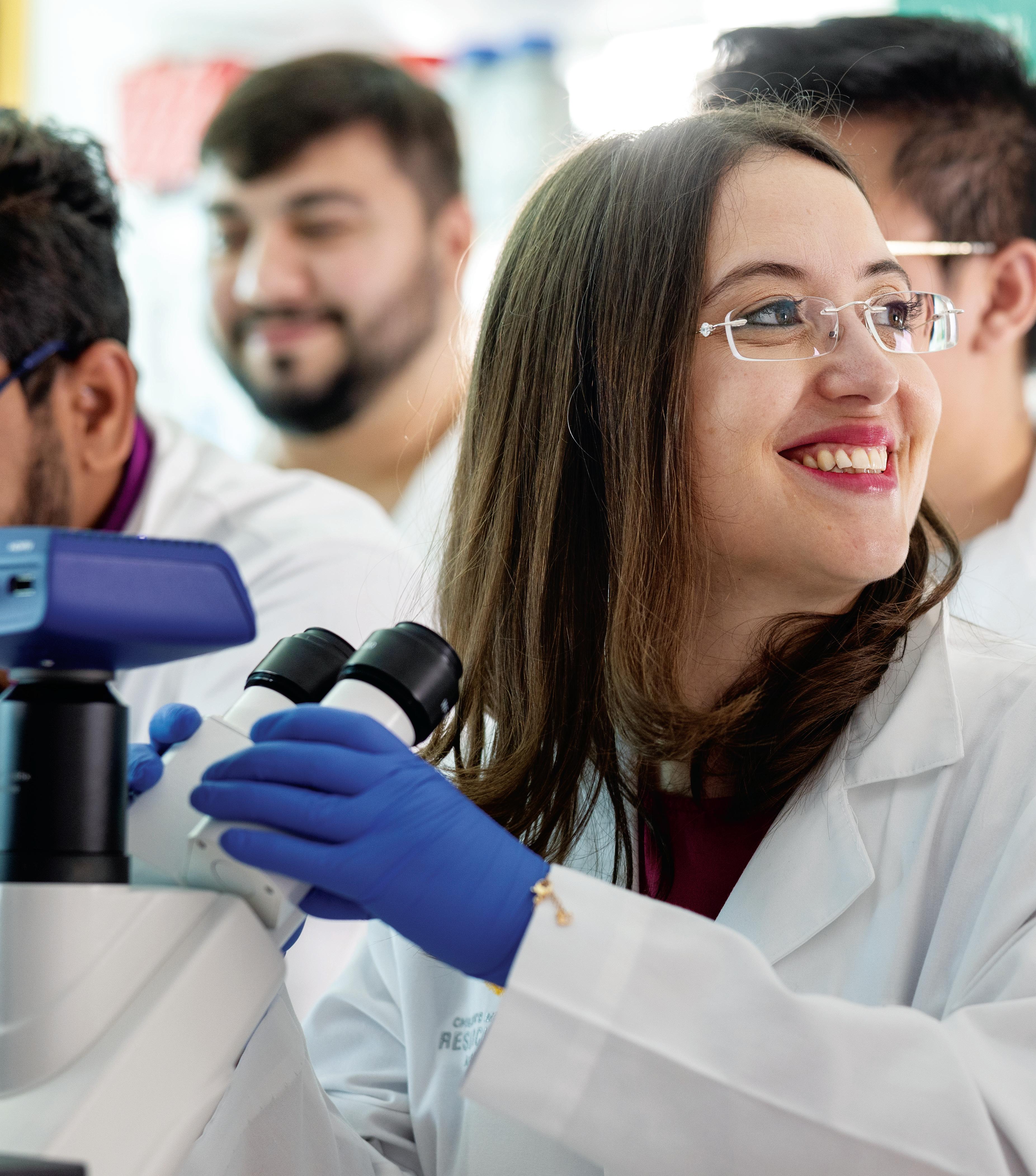
Photo credit: Gerta Hoxhaj, Ph.D., uses a microscope alongside her laboratory members. A multidisciplinary approach has helped her team uncover targets for chemo-resistant cancers. Photo courtesy of the Vilcek Foundation (Photographer Stewart Cohen )
