Immunocytochemical Identification of LayerSpecific Markers in iPSCDerived Cortical Neurons
Zihan Zhou
Senior Thesis | 2026

Title:ImmunocytochemicalIdentificationofLayer-Specific
MarkersiniPSC-DerivedCorticalNeurons
Author:ZihanZhou
ImmunocytochemicalIdentificationofLayer-SpecificMarkers in
iPSC-DerivedCorticalNeurons
Abstract:
Stemcellshavetheuniqueabilitytoreplicateindefinitely andtransformintodifferentcelltypes,makingthemcrucialfor applicationsliketissuerepairanddrugdevelopment.Traditional methods,suchasembryonicstemcellsorbonemarrow,raise ethical,political,andscientificproblems.In2006,Japanese scientistShinyaYamanakaintroducedinducedpluripotentstem cells(iPSCs),whichenablessomaticcellsintoapluripotentstate (theabilityofanimmaturecelltogrowintoseveraldifferentcell types),allowingthemtobecomenearlyanycelltypeexcept reproductivecells.Unlikeconventionalstemcells,iPSCsbypass ethicalcontroversiesandholdpromisefortreatingawiderangeof diseasesaffectingvariousorgans.Inducedpluripotentstemcells (iPSCs)provideaversatileplatformforgeneratingpatient-specific cellsforregenerativemedicine.Inthisstudy,iPSCswere differentiatedintocorticalneuronsandcharacterizedtoidentify theircorticallayeridentity,whichmeansthestemcellswere turnedintobraincellsandthencheckedtoseewhichtypeofbrain cellandwhichlayerofbraintheybecame.Immunocytochemical staining(awaytotagpartsofacellsotheycanbeseenundera microscope)wasperformedusingantibodiesagainstneuronaland progenitormarkers,includingCtip2,MAP2,PSD95,Pax6and Tuj1.Secondaryantibodiesconjugatedtofluorescentdyesenabled visualizationwithaKeyenceBZ-X810fluorescencemicroscope. TheresultsshowedthatlayerVneuronalmarkersweremost commonlyexpressedinthetestsamples,combinedwithpositive stainingforimmatureneuronalmarkers,indicatingthepresenceof aheterogeneouspopulationthatincludedimmaturecellsaswellas alreadydifferentiatedlevelVcorticalneurons.Thisstudy demonstratestheabilityofiPSC-derivedneuronstopartially
mimiccorticallayerspecification,indicatingtheirpotential relevanceinsimulatingcorticaldevelopmentandillness.
Introduction:
Stemcellsarecellsthathavetheabilitytoself-renew indefinitelyasundifferentiatedcellsandundercertainconditions differentiateintodifferentcelltypesofatissueororgan.Extensive researchandclinicaltrialshaveexploredtheirpotentialintissue regeneration,drugtesting,andsoon.However,traditionalsources ofstemcellssuchasembryonicstemcellsorbonemarrowoften raiseethical,political,andscientificchallengeslikedebateover usageofhumanembryos,governmentinterference,andimmune rejection(Poulos,2018).
Thediscoveryofinducedpluripotentstemcells,oriPSCs, byJapanesescientistShinyaYamanakain2006openedanewdoor forregenerativemedicine.HewasawardedtheNobelPrizeof PhysiologyorMedicineonlysixyearsafterhisbreakthrough becauseofthesignificanceofiPSCs.iPSCsaresomaticcells, meaninganycellsinthehumanbodyotherthaneggorsperm cells,thathavebeenreprogrammedintoapluripotentstate (Stadtfeld&Hochedlinger,2010).Inthisstate,theyregainthe capacitytodevelopintonearlyanycelltypeinthebodyexceptfor reproductivecells.Unliketraditionalstemcells,iPSCscanbe generatedfromalreadydifferentiatedcells,eliminatingtheneedto relyonembryonicsources.Thistechnologyhassignificant potentialintreatingdiseasesthataffectnearlyeveryorgansystem.
OneofthecellsthatiPSCscandifferentiateintoisthe corticalneuron,whichistheprimaryinformation-processingcell inchargeofenablingcomplexhumancognition.Becausehuman corticalneuronshavesignificantlylargerandmorecomplex dendritictreesthanotherspecies,humanscanintegrateawider rangeofsynapticinputslikereasoning,language,andabstract thinking(Galakhovaetal.,2022).Becauseoftheirkeyrolein
intelligenceandbrainfunction,studyingcorticalneuronsis essentialforunderstandingbothhowthebrainworks.
Thisresearchfocusesonidentifyingwhichregionofthe brainthespecificgroupofregeneratedcorticalneuronsderived fromiPSCsmostcloselyresembles.Corticalneuronsarelocatedin thecerebralcortexofthebrain.Thecerebralcortexformstheouter layerofthebrainandisorganizedintosixdistinctlayers,labeledI throughVIfromthesurfaceinward.Eachlayercontainsdifferent typesofneuronsandhasuniquefunctionsandconnectionswithin thebrain.Toidentifywhichcorticallayertheregeneratedneurons belongto,weuseantibodystaining.Eachantibodybindstoa singletargetprotein.Bytaggingtheseproteins,wecanlocateand identifyspecifictypesofcells.Sinceaproteinmaybepresentin differentlayersofthebrain,panelscontainingmultipleantibodies areusedtoincreaseaccuracy.Becauseeachcorticallayer contributesdifferentlytobrainfunction,itiscrucialtoknowwhich layertheregeneratedneuronsbelongto.Thisinformationis essentialforunderstandingbraindevelopmentandforassessing whetherthesecellsaresuitableforfurtherresearchorclinical applications.Therefore,theabilitytoaccuratelyidentifycortical neuronsderivedfromiPSCsisofgreatclinicalandscientific importance.
PrinciplesBehindtheFunctionofKeyProcedures ObtainingCorticalNeurons
Corticalneuronsamplesusedinthisstudywerederived frombloodcellscollectedfrompatientsdiagnosedwith Creutzfeldt–Jakoddisease(CJD).Thesesomaticbloodcellswere firstreprogrammedintoiPSCsusingestablishedtranscription factorprotocols.Thedisease-carryingiPSClineswerethen geneticallycorrectedtoremovetheCJD-associatedmutation, resultinginahealthycontrollinereferredtoasCJD13JN2.To inducedifferentiationintocorticalneurons,doxycycline(DOX)
wasaddedtotheculturemediumstartingonDay1.Doxycycline activatedatranscriptionalprogramdesignedtorapidlyexpresskey neuronallineagegenes,triggeringdirectdifferentiationofthe iPSCsintoneurons.Thisprocesspushesthestemcellstowarda corticalneuronalfatebyupregulatingneurogenicfactors.Overthe courseofdifferentiation,cellsweremonitoredformorphologyand markerexpressiontoconfirmsuccessfulneuronalconversionfor 14days.
PFAFixation,andBSABlocking,andPBSWashing
Tostarttheexperiment,cellswerefixedusing paraformaldehyde,PFA,throughouttheexperiment.PFAisa commonlyusedcross-linkingfixativeinimmunocytochemistry.It worksbydepolymerizingintoformaldehydeinsolution,which thencovalentlycross-linksproteins,formingthemethylenebridges betweenprimaryaminegroups(Kimetal.,2017).Thebridges effectively“freezes”cellularandsubcellulararchitecture, preventingdegradationandpreservingantigenicityforantibody binding.Afterthecellsarefixed,weareabletopreservethecell andstudythemattheidealstate,preventingthemfromdegrading orfurtherdeveloping.However,excessivePFAconcentrationor prolongedfixationcanleadtoover-crosslinkingwhichmayalter theconfirmationandreducethebindingefficiencythatleadstoa weakerstaining.
Afterfixationandbeforeanyadditionofantibody,blocking isconductedusinga5%bovineserumalbumin(BSA)solutionin thephosphate-bufferedsaline(PBS).Atthemolecularlevel,BSA bindstohydrophobicandchargedsitesonbothcellularandplastic surfaces,occupyingnonspecificbindingareasthatcouldotherwise bindantibodiesandproducebackgroundsignals(ThermoFisher Scientific,2020).Thissaturationstepensuresthatprimary antibodiesbindselectivelytotheirintendedantigensratherthanto
unoccupiedproteinorglasssurfaces.ProperBSAblockingis importantforaccurateandefficientantibodystaining.
Throughouttheprocessofblockingandstaining,another solutionphosphate-bufferedsaline(PBS)alsoplaysacriticalrole ingeneratingthebeststainingresult.PBSisawashingbuffer typicallycomposedofsodiumchloride,sodiumphosphate,andin someformulations,potassiumchlorideandpotassiumphosphate (Tomaszewski,2024).Thisisotonicandnon-toxicsolution preservescellularosmoticbalanceandstructure(Stradleigh& Ishida,2015).PBSalsoprovidespHstability,between7.2to7.4, whichisessentialformaintainingantigen-antibodybinding conditions.PBSisappliedafterfixation,betweenantibody incubations,andpost-stainingtoremoveexcessPFA,unbounded antibodies,andBSAwithoutdisruptingthecells.Throughwashing preventsongoingbackgroundnoise,reducedsignalclarity,and chemicalinterferenceinthelaterprocess.
Antibodies
Inthisexperiment,arangeofantibodiesandstainswere usedtolabeldifferentneuronalstructuresandcorticallayers.In well1,whichservedasthestandardforcorticalneurons,DAPI stainedthenucleiofallcellsinblue,providingageneralreference forcellnumberandlocation.MAP2isamicrotubule-associated proteinfoundindendriticandneuronalcellbodies.Itisusedto identifymatureneuronsandvisualizetheiroverallmorphology. MAP2wasdetectedusingananti-chicken488secondaryantibody, producinggreenfluorescence.PSD95isapostsynapticdensity proteinlocatedatexcitatorysynapses.Itwasusedtoassess synapticmaturityandconnectivity,andwasvisualizedwithananti rabbit647antibodyemittingredfluorescence.Togetherthese markersconfirmedthestructureandmaturityofthedifferentiated corticalneurons.
Inwell2,thegoalwastovisualizecytoskeletalarchitecture andidentifyupper-layercorticalneurons.WeusedDAPIagainto labelthenucleiinblue.Phalloidinisatoxinsecondaryantibody thatbindsfilamentousactin(F-actin)tohighlightthecytoskeletal fiberssuchasaxonsandgrowthcones,revealingtheshapeand structureofextendingneuronalprocesses.SATB2isanuclear transcriptionfactor,whichwasusedtolabelupper-layerneurons foundincorticallayersII-IV.SATB2wasdetectedusingananti mouse647antibody,resultinginredfluorescence.
Inwell3,weaimtocharacterizedeep-layercortical neurons.DAPIcontinuedtomarknucleiinblue.CTIP2,anuclear markerspecifictolayerVcorticalneurons,wasusedtoidentify projectionneuronsthatsendacornstosubcorticaltargets.CTIP2 waslabeledusinganantiratFit-cantibody,whichfluoresced green.Inaddition,weusedTuj1,apan-neuronalmarkerfoundin themicrotubulesofaxonsanddendritestohighlightearly-stage neuronalstructureanddifferentiation.Tuj1wasdetectedusingan antimouse647antibody,emittingaredsignal.
Lastly,well4servedasacontrolforcorticalidentification staining.DAPIstainednucleiblue,whilePAX6,atranscription factorexpressedinradialglialcellsandneuralprogenitors,was usedtoidentifyearly-stagecorticalneurons.PAX6reflectsthe developmentalidentityofneuronsduringcorticalformation.Itwas visualizedusinganantirabbit647antibody,whichfluorescedred. Thiswellservedtoconfirmthepresenceofthecellandtheir immaturitylevelintheexperimentalsetup.
Primaryvs.SecondaryAntibodies
Inimmunocytochemistry,primaryantibodiesare responsibleforbindingdirectlytothetargetproteinorantigen withinthecell.Theseantibodiesarehighlyspecificandarechosen
basedonthehostspeciesinwhichtheywereraisedandtheknown affinityforaparticularcellularmarker.
Becausemostprimaryantibodiesarenotdirectly fluorescentlylabeled,secondaryantibodiesareusedtodetectand visualizethem.Secondaryantibodiesareraisedagainstthespecies inwhichtheprimaryantibodywasproduced(e.g.,anti-mouse, anti-rabbit,anti-chicken)andareconjugatedtofluorescentdyes suchasAlexaFluor488(green),AlexaFluor594(red),orFITC. Thesecondaryantibodymustcomefromadifferentspeciesthan theprimarysoitbindsonlytotheprimaryantibodyandnotto otherproteinsinthesample(SouthernBiotech,2021).For example,iftheprimaryantibodyismadeinamouse,the secondaryantibodyshouldbeanti-mouseandraisedinadifferent specieslikeadonkeyorrabbit.
Thesefluorophoresemitlightatspecificwavelengthswhen excitedbyafluorescencemicroscope,allowingthelocalizationof targetproteinstobevisualizedanddocumented. Usingsecondaryantibodiesprovidesseveraladvantages.It amplifiesthefluorescentsignal,asmultiplesecondaryantibodies canbindtoasingleprimaryantibody,increasingbrightness.Italso offersexperimentalflexibility,allowingthesamesecondary antibodytobeusedformanyprimaryantibodiesofthesamehost species.Inthisstudy,inwell1,weusedantichicken488for primaryantibodyMap2andantirabbit647forprimaryantibody PSD95.Forwell2,weusedantimouse647forprimaryantibody SATB2andPhalloidin.Forwell3,weusedantiratfir-cfor primaryantibodyCtip2andantimouse647forprimaryantibody Tuj1.Forwell4,weusedantirabbit647forprimaryantibody Pax6.SecondaryantibodyDAPIwasaddedintoallfourwellslater intheprocedurefora20minutesHoechstincubationtostainthe nucleus.Theoverallvolumeofthesolutionforeachwellis 1000µL,andtheexactamountofantibodytobeaddedcanbe
calculatedusingthedilutionformula:
theconcentrationofthestockantibody, isthevolumetoadd, �� 1 �� 2 isthedesiredfinalconcentration,and isthefinalvolume �� 2 (1000µL).
FixationandPermeabilization
Topreservecellularstructureandimmobilizeproteinsfor immunocytochemicalanalysis,cellswerefixedusing paraformaldehyde(PFA).Anequalvolumeof8%PFAsolution wasaddeddirectlytotheculturemediumineachwelltoachievea finalconcentrationof4%PFA.Thisfixationstepwasperformedat roomtemperaturefor15minutes.Fixationcross-linksproteinsand stabilizescellulararchitecture,whichisessentialforaccurate stainingandimaging.
Followingfixation,thesolutionwasgentlyaspiratedtoavoid disruptingthemonolayer,asthecellsaredelicateandhighly susceptibletomechanicaldamage.Wellswerethenwashedtwice with1mLofPBSperwash,witheachwashlasting5minutes.All washingandsolutionexchangesthroughouttheprocedurewere performedslowlyandcarefullyusingapipetteatthesideofthe welltominimizeshearstressandpreservecellintegrity.
Topermeabilizethefixedcellsandallowantibodiesto accessintracellulartargets,0.1%TritonX-100inPBSwasadded toeachwellfor5minutesatroomtemperature.TritonX-100isa nonionicsurfactantthatselectivelypermeabilizestheplasma membranewhilepreservingtheoverallstructureofthecells.After permeabilization,cellswerewashedtwicemorewithPBS,each timeallowingthesolutiontositundisturbedfor5minutes,totaling 10minutesofpassive washingtogentlyremoveresidualdetergent.
Materials:
Allreagentsusedinthisstudywerepreparedandhandled accordingtostandardimmunocytochemistryprotocols.Cellswere fixedusingan8%paraformaldehyde(PFA)solution,whichwas addeddirectlytotheculturemediumata1:1ratiotoreachafinal concentrationof4%.Phosphate-bufferedsaline(PBS)wasused throughouttheprotocolforwashinganddilutingreagents.For permeabilizationofcellularmembranes,a0.1%solutionofTriton X-100(Sigma-Aldrich)inPBSwasapplied.Toblocknonspecific antibodybinding,1%bovineserumalbumin(BSA)wasprepared bydiluting7.5%BSAstocksolutioninPBS,withTritonX-100 addedtomaintainpermeability.
Forantibodystaining,bothprimaryantibodiesand secondaryantibodieswereused.Antibodiesusedincludes:Map2, PSD95,Phalloidin,SATB2,CTIP2,Tuj1,Pax6,DAPI,Anti Chicken488,AntiRabbit647,AntiMouse647,andAntiRat Fit-C.Antibodiesweredilutedtodifferentconcentrations dependingonthewellofcellstheywereaddedinto.
Last,allfluorescenceimagingwasconductedusinga KeyenceBZ-X810fluorescencemicroscope,equippedwiththree differentcolorsoflaser:red,green,andblue.Imageswere acquiredunder4x,10xand,20xobjectivelenses.
Methods:
Tominimizenonspecificantibodybinding,cellswere incubatedinablockingbufferconsistingof1%BSAinPBS containing0.1%TritonX-100.Theblockingstepwasconductedat roomtemperaturefor30minutes.BSAactsasaproteinblocker thatoccupiesnonspecificbindingsites,whileTritonX-100 maintainsmembranepermeabilityduringsubsequentantibody incubations.
Primaryantibodiestargetingneuronalandprogenitor markerswerepreparedinthesameblockingbufferandappliedto
thecellsin200μLdropletsperwell.Antibodydilutionswere preparedasfollows:
● Well1:
Map2(Dilution1:5000(1µLMap2to99µLsolventand then20µLofthatsolutionto980µLsolvent))andPSD95 (Dilution1:100(10µLPSD95into990µLsolvent))
● Well2:
SATB2(Dilution1:50(20µLSATB2into980µLsolvent))
● Well3:
Ctip2(Dilution1:100(10µLCtip2into90µLsolvent)) andTuj1(Dilution1:100(10µLTuj1into90µLsolvent))
● Well4:
Pax6(Dilution1:100(10µLPax6into90µLsolvent))
Primaryantibodiesweregentlypipettedintoeachwell,and theplateswereincubatedovernightinthecoldroomonagentle shakertoensureevenantibodydistributionandbinding.Allwells werekeptcoveredduringincubationtoprotectfluorophoresfrom lightexposure.
Thenextday,cellswerewashedthreetimeswithPBSfor5 minuteseachtoremoveunboundprimaryantibodies.Secondary antibodiesweredilutedinasolutionof1%BSAinPBSwith0.1% TritonX-100.Thissolutionwaspreparedbyadding1.3mLof 7.5%BSAstocksolutionand10μLofTritonX-100to10mLof PBS.
Fluorescentlyconjugatedsecondaryantibodieswereselectedbased onthehostspeciesoftheprimaryantibodiesandwereappliedin thefollowingcombinations:
● Well1: antichicken488(Dilution1:400(2.5µLantichicken488
into997.5µLsolvent))andantirabbit647(Dilution1:250 (4µLantirabbit647into996µLsolvent))
● Well2:
antimouse594(Dilution1:250(4µLantimouse594into 996µL))andphalloidin(Dilution1:40(25µLphalloidin into975µLsolvent))
● Well3:
antimouse594(Dilution1:250(4µLantimouse594into 996µLsolvent))andantiratfit-c(Dilution1:400(2.5µL antiratfit-cinto997.5µLsolvent))
● Well4: antirabbit647(Dilution1:400(2.5µLantirabbit647into 997.5µLsolvent))
Eachantibodycocktailwasincubatedwithcellsfor1hour atroomtemperatureonashaker.Wellswereprotectedfromlight usingaluminumfoilthroughouttheincubationtopreserve fluorescence.Followingincubation,cellswerewashedthree additionaltimeswithPBSfor5minuteseachtoremoveunbound secondaryantibodies.
Tovisualizenuclei,cellswerecounterstainedwithDAPIat afinaldilutionof1:2000,preparedbyadding0.5μLofDAPIto 999.5μLPBS.Thestainwasaddedtoeachwellandincubatedfor 5minutesatroomtemperatureinthedark.Afterstaining,cells werewashedthreefinaltimeswithPBSfor15minuteseachto ensurecompleteremovalofbackgroundfluorescenceandunbound dye.Lastly,cellswereblockedby1volumeofBSAbefore imagingandanalysing.
Followingthefinalblock,cellswereimageddirectlyinthe 12-wellplatesusingaKeyenceBZ-X810fluorescencemicroscope. Eachwellwasvisualizedunderappropriatefiltersetstodetectthe emissionspectraofDAPI,AlexaFluor488,AlexaFluor594, FITC,andphalloidinconjugates.Fluorescenceimageswere
capturedusinga4x,10x,20xobjectivelensdependingonthe resolutionrequiredforthemarkersofinterest.Focusandlight settingswereoptimizedperchanneltominimizebackground signalandavoidoversaturation.Representativeimagesfromeach wellweresavedfordownstreamanalysisforfluorescenceintensity quantificationandcellmorphologyassessment.
Results:
ToevaluatethedifferentiationstatusofiPSC-derived neurons,immunostainingwasperformedonfixedcellcultures. WhileantibodystainingwasperformedonWells1through4,only imagesfromWells3and4areincludedinthissectionduetoclear signalqualityandmorerepresentativestainingpatternsobservedin thesewalls.Thesewellsweredesignedtodetectarangeof neuronalidentityandcorticallayerspecification.
Well3:Ctip2andTuj1
Well3wasstainedwithCtip2(amarkerofdeep-layercortical neurons)andTuj1(anearlyneuronalmarker),alongwithDAPI nuclearcounterstain.Theredfluorescencerepresents Ctip2-positivecells,whilethegreensignalcorrespondstoTuj1 expression.AsshowninFigure1below,acentralclusterof red-stainedcellsissurroundedbyafaintbutnoticeablehaloof greensignal.Thisspatialdistributionsuggeststhatmanyofthe cellsinthisregionexpressbothdeep-layerandearlyneuronal characteristics,consistentwithcellstransitioningthroughneuronal differentiation.
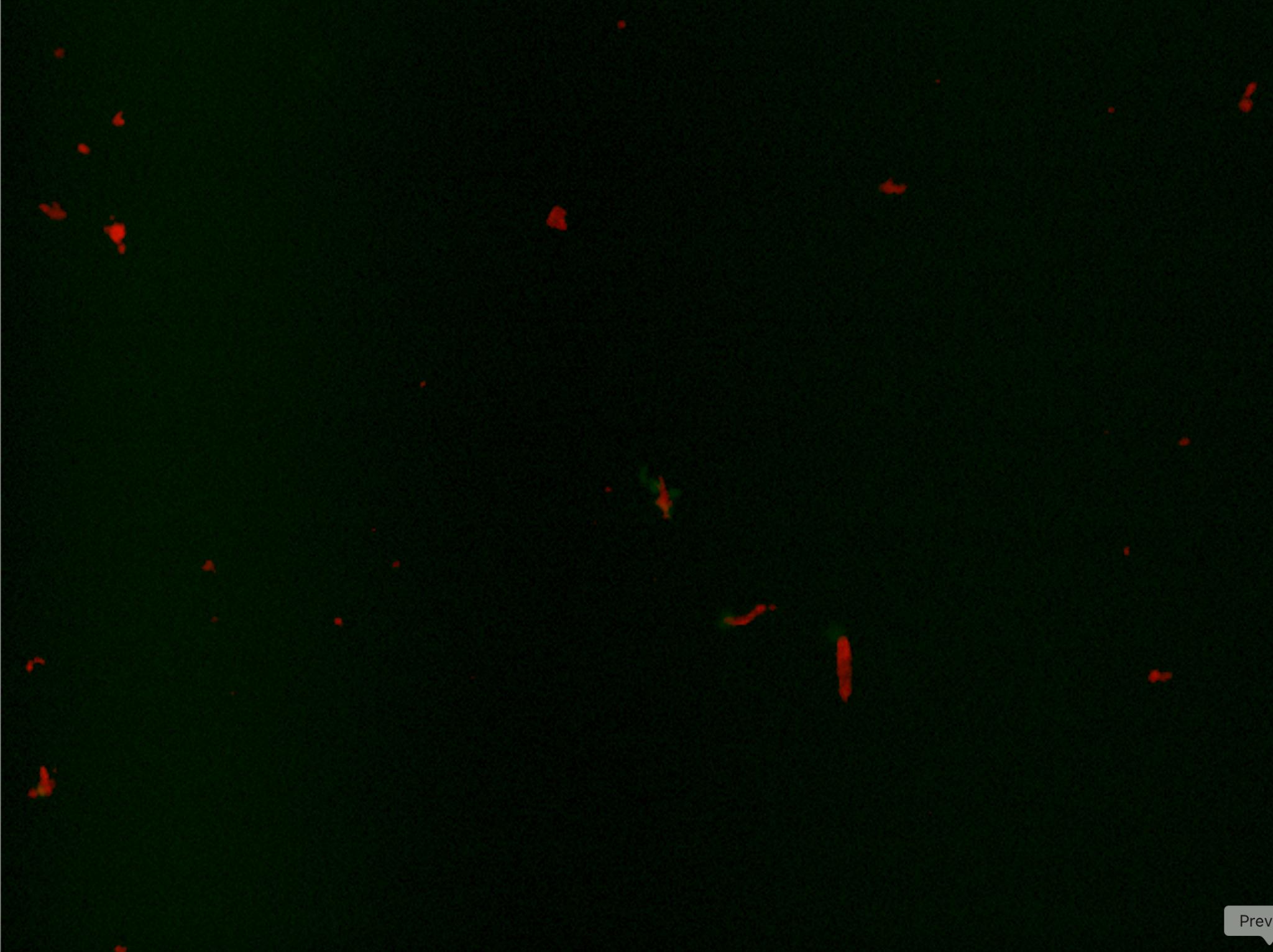
Figure1:ImmunofluorescentimageofWell3stainedwithCtip2 (red),Tuj1(green),DAPI(blue)atday14cells.Theredsignalis concentratedinthecenter,withfaintgreensurroundingthearea, suggestingoverlappingexpression.
Well4:Pax6
CellsinWell4werestainedwithprimaryantibodiesagainstPax6, amarkerforneuralprogenitorcells,followedbysecondary antibodyanti-rabbit647,whichhasaredfluorescence.Thered fluorescenceindicatesPax6-positivecells.AsseeninFigure2,red signalsarepresentinasubsetofcells,suggestingthepresenceof immatureorprogenitor-likeneuronsinthisculture.
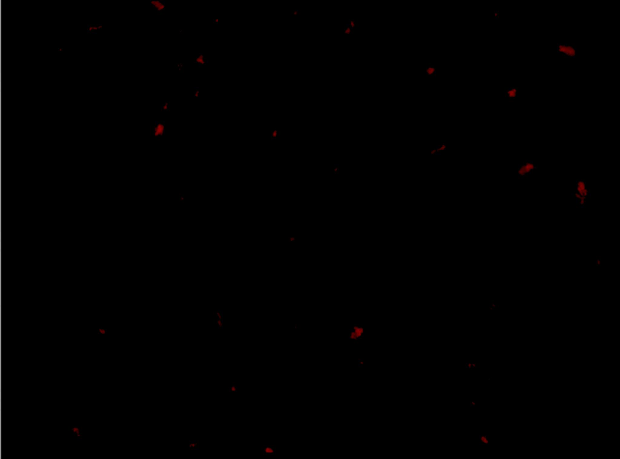
Figure2:ImmunofluorescentimageofWell4showingPax6(red) andDAPI(blue)ofday14cells.Strongrednuclearsignals indicatePax6-positivecells.
DiscussionandFurtherStudy:
Whilethisexperimentdemonstratedtheuseof immunocytochemistrytoidentifystructuralanddevelopmental markersincorticalneurons,severallimitationsmayhaveaffected theoutcome.Amajorsourceofpotentialhumanerrorstemsfrom humanhandlingduringthemultiplewashingsteps.Although extensivepipettingandrepeatedPBSwashesareessentialtothe samplequality,thosewashescanphysicallydislodgeordamage delicatecells,especiallyimmatureneuronsatearlydevelopmental stages.What’smore,ifthewasheswereincomplete,unbound antibodiescanreducesignalintensity.
Anotherconcernisthelightsensitivityof fluorescent-conjugatedantibodies.Duringimagingorevenjust handlingunderlaboratorybrightlightconditions,theseantibodies mayphotobleach,leadingtothestainingunclear.Consistentuseof
darkincubationandfoilpapertoachieveminimallightexposureis criticaltopreservingfluorescencequality.
What’smore,DAPIstainingdidnotappearinWells3and 4,limitingvisualizationofnuclei.WethinkitisbecausetheDapi wasusedintheexperiment.WeusedHoechst,whichisdetectedin thesamechannelasDAPI,insteadofpureDAPI,whichmay explaintheweakorundetectablenuclearsignal.However,other markers(Ctip2,Tuj1,andPax6)showingstrongandlocalized fluorescence,allowsustointerpretcelltypeidentitydespitethe missingnuclearsignal.
Biologically,anotherimportantfactortoconsideristhe developmentalstageoftheneurons.Thecorticalneuronscells werefixedandstainedonDay14ofdifferentiation,arelatively earlystateforcorticalneurons,ascanbeseenfromthestaining results.Someneuronalmarkersmaynotyetbefullyexpressedor properlylocalizedatthistimepoint.Assuch,thestainingis overallweakerduetotheprematuredevelopmentalstateofthe cells.Figure3and4showthematureantibodystainingresultfrom day28cellsafterdifferentiation.Thishighlightstheimportanceof therighttimingoftheantibodystaining,asdifferentmarkers becomedetectableatdifferentstagesofmaturation.
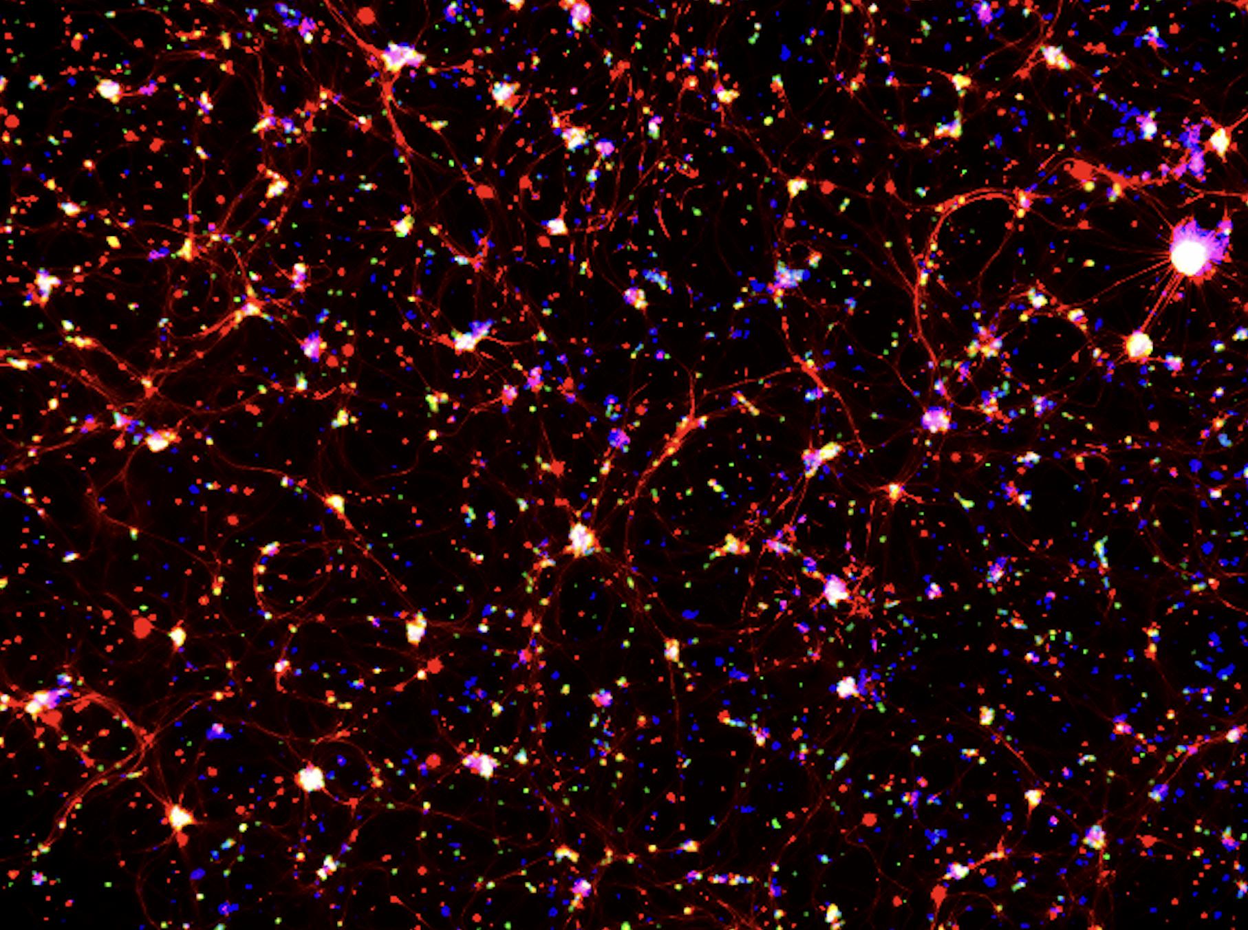
Figure3:ImmunofluorescentimageofWell3stainedwith Ctip2(red),Tuj1(green),DAPI(blue)whenthecellisatday28 differentiation.Strongred,green,andbluenuclearsignals.
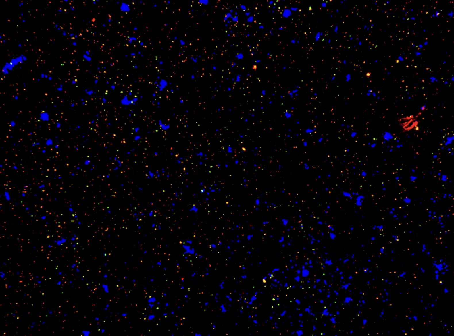
Figure4: ImmunofluorescentimageofWell4showing Pax6(red),Nestin(green)andDAPI(blue)ofday28cells.Strong red,green,andbluenuclearsignals.
Despitetheselimitations,thedatafromthisexperiment havepromisingimplications.WeareabletoidentifyCtip2-positive
layerVneuronsalongsidePax6-positiveprogenitors,underscoring theearlyemergenceofdistinctcorticalidentitiesinour differentiatedcells.LayerVneuronsarecriticaloutputcellsof cortex,sendingaxonstosubcorticalstructuressuchasthe brainstem,spinalcord,basalganglia,andthalamus,whichare functionalvitalforvoluntarymotorcontrolandsensorimotor integration(Kingetal.,2024).Thesuccessfuldifferentiationof theseneuronsholdspromiseformodelingneurologicaldisorders thataffectthisneuronalsubtype,includingamyotrophiclateral sclerosis(ALS)andcorticospinaltractinjuries(Leoneetal.,2009). Toconfirmandfurtheridentifythespecifictypeofneuronsinthe internalpyramidallayer,furtherresearchshouldincorporate additionallayerVmarkersviaantibodyco-stainingor transcript-leveltechniqueslikeRT-qPCRthatofferhigher specificity.Additionally,longitudinalstudiesextendingbeyond14 days,combinedwithcarefuladherencetoprotocolandconsistent monitoringofcellhealthandantibodyquality,willhelpdetermine whetherearlymarkerexpressionleadstofullneuronalmaturation andfunctionalspecializationovertime.
References
Galakhova,A.A.,Hunt,S.,Wilbers,R.,Heyer,D.B.,de Kock,C.P.J.,Mansvelder,H.D.,&Goriounova,N.A. (2022,September15). Evolution of Cortical Neurons Supporting Human Cognition.U.S.NationalLibraryof Medicine.
https://pmc.ncbi.nlm.nih.gov/articles/PMC9561064/
Kim,S.-O.,Kim,J.,Okajima,T.,&Cho,N.-J.(2017,March 13). Mechanical Properties of Paraformaldehyde-Treated Individual Cells Investigated by Atomic Force Microscopy and Scanning Ion Conductance Microscopy.U.S.National LibraryofMedicine.
https://pmc.ncbi.nlm.nih.gov/articles/PMC5359366/#:~:text= PFA%20causes%20covalent%20cross%2Dlinks,treatment% 20%5B7%E2%80%9310%5D
King,S.G.,Maire,P.,Mergenthal,A.,Walker,S.,&Hires, S.A.(2024,March17). Cell-Type Specialization of Layer 5 Excitatory Neurons in Tactile Behavior.Biorxiv.
https://www.biorxiv.org/content/10.1101/2024.03.15.585205 v1.full
Leone,D.P.,Srinivasan,K.,Chen,B.,Alcamo,E.,& McConnell,S.K.(2009,February1). The Determination of Projection Neuron Identity in the Developing Cerebral Cortex.U.S.NationalLibraryofMedicine.
https://pmc.ncbi.nlm.nih.gov/articles/PMC2483251/ Poulos,J.(2018,January2). The Limited Application of Stem Cells in Medicine: A Review - Stem Cell Research & Therapy.BioMedCentral.
https://stemcellres.biomedcentral.com/articles/10.1186/s1328 7-017-0735-7
SouthernBiotech.(2021,December2). Secondary Antibody Selection Guide.
https://www.southernbiotech.com/secondary-antibody-selecti on-guide/#:~:text=the%20selection%20process.-,Your%20se condary%20antibody%20should%20be%20directed%20at% 20the%20host%20species,mouse%20secondary%2C%20and %20so%20on
Stadtfeld,M.,&Hochedlinger,K.(2010,October15). Induced Pluripotency: History, Mechanisms, and Applications.U.S.NationalLibraryofMedicine.
https://pmc.ncbi.nlm.nih.gov/articles/PMC2956203/
Stradleigh,T.W.,&Ishida,A.T.(2015,April17). Fixation Strategies for Retinal Immunohistochemistry.U.S.National LibraryofMedicine.
https://pmc.ncbi.nlm.nih.gov/articles/PMC4543575/
ThermoFisherScientific.(2020). Bovine Serum Albumin: Thermo Fisher Scientific - US.BovineSerumAlbumin. https://www.thermofisher.com/us/en/home/life-science/cell-c ulture/mammalian-cell-culture/media-supplements/bovine-se rum-albumin.html
Tomaszewski,C.(2024,January4). TBS vs PBS in Immunohistochemistry: Navigating the Buffers.Visikol. https://visikol.com/blog/2024/01/04/tbs-vs-pbs-in-immunohi stochemistry-navigating-the-buffers/