Mapping Tumor-Induced Neuronal Hyperactivity in Glioblastoma Mouse Models
Mason Chan
Senior Thesis | 2026

MasonChan
Song/MingLab,PerelmanSchoolofMedicineatTheUniversityofPennsylvania
Glioblastoma(GBM)isthemostcommonandaggressiveprimarybrain tumorinhumanadults,withamediansurvivalofapproximately15 months.Recentstudieshavedemonstratedthatgliomacellsformfunctional synapseswithneuronsandthatneuronalactivitypromotestumor progression.Sunetal.(2025)usedviraltracingtomaptheanatomical connectivitybetweenneuronsandpatient-derivedglioblastomacells, identifyingbrainregionsthatsendsynapticinputtothetumor.Thisstudy extendsthisworkbymappingneuronalhyperactivityacrossthebrainin tumor-bearingmiceusingc-Fosimmunohistochemistry. Immunocompromisedmicereceivedstereotacticinjectionsofpatientderivedglioblastomaorganoids(UPO-10072)intothesomatosensory cortex(n=10)orshamsurgerywithHibernateAvehicleatthesame coordinates(n=10).Openfieldtestingwasconductedatweeks2and4 post-injection.Micewereperfusedimmediatelyfollowingtheweek4 behavioralsession,andbrainsectionswereprocessedforc-Fosandhuman nestinimmunostaining.Automatedcellquantificationwasperformedusing QuPathfollowingalignmenttotheAllenMouseBrainAtlas.Tumorbearingmiceshowednobehavioraldifferencesatweek2butexhibited significantlyreducedcentertimeatweek4,indicatingananxiety-like phenotype.Brain-widec-Fosmappingrevealedregion-specificchanges, withtheamygdalashowingsignificantlyincreasedc-Fosexpressionin tumor-bearingmicecomparedtocontrols.Mostotherquantifiedregions showednosignificantdifferencesbetweengroups.Ourfindingsindicate thatGBMinducesincreasedneuronalactivityintheamygdala,which providesapotentialneuralcorrelatefortumor-associatedanxiety-like behavior.
Glioblastoma(GBM)isthemostaggressiveprimarybraintumorinadults.GBM alsorepresentsthemostcommonmalignantbraintumor,havinganoccurrence rateof3.2casesper100,000peopleannually.Despitedecadesofresearchand therapeuticdevelopment,theprognosisforGBMpatientshashadlimited improvement.Thecurrentmediansurvivaltimeis15monthspost-diagnosis,even withthecurrenttherapeuticintervention.Thiscurrentstandardofcareconsistsof surgicalresectionwithconcurrentradiotherapyandtemozolomidechemotherapy. Thisapproach,however,onlyprovidesmodestsurvivalbenefitsofanadditional2.5 months.GBMcellsmigratealongwhitemattertractsandinvadenormalbrain parenchymabeyondthevisibletumormargins,whichmakescompletesurgical resectiondifficult.Becauseofitsinfiltrationmethod,combinedwiththetumor's therapeuticresistance,GBMisknownasoneofthemostintractabletumorsin oncology.
Evidencebeginningtoaccumulatein2019indicatesthattherelationship betweenglioblastomaandsurroundingbraintissueismorethanthehistoricalmass effectmodel,whichattributedthepatientdamagetothetumorbeingaforeign masscompressingthesurroundingtissue.Gliomacellsintegrateintotheneural microenvironmentandcommunicatebidirectionallywiththenervoussystemin additiontothephysicaldamageandpressureofthetumor.Venkateshetal. demonstratedin2019thatgliomacellsformglutamatergicsynapseswithneurons. Usingelectrophysiologicalrecordingsfromgliomacellsinacutebrainslices, combinedwithimmunoelectronmicroscopyandoptogeneticstimulationin tumor-bearingmice,theyestablishedthatneuronsformfunctionalAMPA receptor-mediatedsynapsesontogliomacells.Theseneuron-to-gliomasynapses exhibitconventionalsynapticarchitecture,includingpresynapticvesicleclustering insynapticcleftsandpostsynapticreceptoraggregatesongliomacellmembranes. Neuronalstimulation,eitherelectricaloroptogenetic,evokespostsynapticcurrents ingliomacells.Thesynapticinputdepolarizesgliomacellmembranesandinduces calciuminfluxthroughAMPAreceptors.Theirinvivooptogeneticexperiments demonstratethatneuronalactivitypromotesgliomacellproliferation,while pharmacologicalblockadeofAMPAreceptorswithperampanelsuppressestumor growthandprolongssurvivalinxenograftmodels.Venkateshetal.identifiednonsynapticcommunicationmechanismsaswell.Gliomacellsextendcellularprocesses termedtumormicrotubesthatformgapjunction-couplednetworks,propagating depolarizingcurrentsbetweencells.Neuronalactivityevokedpotassiumcurrentsin gliomacellsareamplifiedthroughthisnetwork,extendingmembrane depolarizationbeyondcellsreceivingdirectsynapticinput.Theirworkestablished neuronalactivityasamicroenvironmentaldriverofgliomaprogressionandshowed thatintrinsicelectricalpropertiesofneuraltissuecontributetotumorbiology.
TamimiAF,JuweidM.EpidemiologyandOutcomeofGlioblastoma.In:DeVleeschouwerS,editor.Glioblastoma[Internet]. Brisbane(AU):CodonPublications;2017Sep27.Chapter8.Availablefrom:https://www.ncbi.nlm.nih.gov/books/NBK470003/ doi:10.15586/codon.glioblastoma.2017.ch8
1 StuppR,MasonWP,vandenBentMJ,WellerM,FisherB,TaphoornMJ,BelangerK,BrandesAA,MarosiC,BogdahnU, CurschmannJ,JanzerRC,LudwinSK,GorliaT,AllgeierA,LacombeD,CairncrossJG,EisenhauerE,MirimanoffRO; EuropeanOrganisationforResearchandTreatmentofCancerBrainTumorandRadiotherapyGroups;NationalCancerInstitute ofCanadaClinicalTrialsGroup.Radiotherapyplusconcomitantandadjuvanttemozolomideforglioblastoma.NEnglJMed.
2 2005Mar10;352(10):987-96.doi:10.1056/NEJMoa043330.PMID:15758009. Stuppetal.(2005)
3 TamimiAF,JuweidM.(2017)
4 Venkatesh,H.S.,Morishita,W.,Geraghty,A.C.,Silverbush,D.,Gillespie,S.M.,Arzt,M.,Tam,L.T.,Espenel,C., Ponnuswami,A.,Ni,L.,Woo,P.J.,Taylor,K.R.,Agarwal,A.,Regev,A.,Brang,D.,Vogel,H.,Hervey-Jumper,S.,Bergles,D. E.,Suvà,M.L.,…Monje,M.(2019).Electricalandsynapticintegrationofgliomaintoneuralcircuits.Nature,573(7775),539–545.https://doi.org/10.1038/s41586-019-1563-y
6
5 Venkateshetal.(2019)
ThedemonstrationthatgliomacellsformfunctionalsynapsespromptedHuangHobbsetal.’sinvestigationintowhetherneuronalactivitycouldinfluencetumor behavioracrosslongdistanceswithinthebrain.Huang-Hobbsetal.testedthis possibilityusingamousemodelofglioblastomawheretumorswereimplantedin onehemisphere.Theythenusedchemogenetictoolstoselectivelyactivatecallosal neuronslocatedintheopposite,tumor-freehemisphere.Contralateralneuronssend axonalprojectionsthroughthecorpuscallosumtoreachthehemispherecontaining thetumor.Activationofthedistantneuronsquickenedgliomaproliferationand invasion,whichdemonstratedthatthetumorrespondstoremoteneuralactivity. Toidentifythemediatorsoftheobservedremoteinfluence,theyperformedhighthroughputscreeningofgenesenrichedinthepopulationofgliomacellsfoundat theleadingedgeoftumorinvasion.Thisscreenidentifiedseveralaxonguidance molecules.SEMA4Fwasspecifiedasakeyregulator.Contrarytotheconventional viewofaxonguidancemoleculesasneuronalproducts,theyfoundthatgliomacells themselvesexpressSEMA4F,andthatthisexpressionincreasesinresponseto neuronalactivity.SEMA4Fsecretedbytumorcellsactslocallytoremodelsynapses inthesurroundingbraintissuewhichshiftthebalancetowardincreasedexcitatory transmissionandelevatedactivity.Thiscreatesaself-reinforcingcyclewhere remoteneuronalactivitystimulatesSEMA4Fproductioningliomacells,which thenamplifieslocalneuronalactivitythroughsynapticrestructuring.Supporting thefunctionalimportanceofthismechanism,aknockoutofSEMA4Finglioma cellsreducedbothtumorgrowthandinfiltration.Surgicaltransectionofthecorpus callosumeliminatedthegrowth-promotingeffectsofcontralateralneuronal activation,alsoconfirmingthatphysicalconnectivitybetweenhemispheresis requiredforremoteneuronstoinfluencetumorprogression.Theirexperiments establishedthatgliomasintegrateintobrain-wideneuralnetworks,respondingto neuronsintheirimmediatevicinityandalsotoactivitypatternsdistributedacross moredistantbrainregions.
Huang-Hobbs,E.,Cheng,Y.T.,Ko,Y.,Luna-Figueroa,E.,Lozzi,B.,Taylor,K.R.,McDonald,M.,He,P.,Chen,H.C., Yang,Y.,Maleki,E.,Lee,Z.F.,Murali,S.,Williamson,M.R.,Choi,D.,Curry,R.,Bayley,J.,Woo,J.,Jalali,A.,…Deneen, B.(2023).RemoteneuronalactivitydrivesgliomaprogressionthroughSEMA4F.Nature,619(7971),844–850. https://doi.org/10.1038/s41586-023-06267-2
Sun et al.
7 Whilethefunctionalconsequencesofneuronalactivityongliomacellshadbeen establishedthroughsynapticandparacrinemechanisms,thecomprehensive anatomicalarchitectureunderlyingtheseinteractionsremainedincompletely characterized.Sunetal.mappedtheconnectivitybetweenpatient-derived glioblastomacellsandneuronsthroughoutthemousebrainusingviraltracing techniques.Theytransplantedpatient-derivedglioblastomaorganoidsthathad beengeneticallyengineeredtoexpresstheTVAreceptorandrabiesglycoprotein intoimmunocompromisedmice.Followingtumorengraftment,theyinjected EnvA-pseudotyped,replication-incompetentrabiesvirusengineeredtoexpress fluorescentreportersintothetumormass.Thevirusselectivelyinfectedcells expressingtheTVAreceptor,ensuringinitialinfectionremainedrestrictedto tumorcells.Thebrainthenunderwenttrans-monosynapticretrogradetracing, crossingbackacrosssynapsestolabelneuronsthatformdirectpresynaptic connectionsontothegliomacells.Thisapproachenabledcomprehensive identificationofallbrainregionssendingsynapticinputtotheglioma.Their analysisconcernedpatternsspanningbothlocalandlong-rangeconnections, includingcorticalareasandneuromodulatorycenters,suchasthebasalforebrain cholinergicsystemanddorsalrapheserotonergicneurons.Theconnectivityprofile variedwithtumorlocation,indicatingthatgliomacellsintegrateintopre-existing neuronalnetworksinamannerdependentonlocation,withdifferenttumor positionsexposingcellstodifferentactivity.Theirresearchprovidedtheanatomical foundationforunderstandinghowbrain-wideneuralactivityconvergesontothe tumormicroenvironmentandestablishedthatgliomacellsbecomeembedded withinstructuredcircuitarchitecture.
Krisnha et al.
Krishnaetal.directlytestedwhetherneuron-gliomaconnectivityinfluences patientoutcomesbymeasuringfunctionalconnectivityandcorrelatingitwith survivalinbothmousemodelsandhumanglioblastomapatients.Theyemployed magnetoencephalographywithacoherenceanalysis,aneuroimagingtechnique thatquantifiesconnectivitybetweenbrainregionswhilereducingthecommon artifactsfromvolumeconduction.Theirapproachallowedforanon-invasive measurementofintegrationbetweenthetumorandtherestofthebrain.Usingthis method,theywereabletoidentifyregionswithintumorsthatexhibitedhigh functionalconnectivity(HFC)versuslowfunctionalconnectivity(LFC)withthe broaderneuronalnetwork.Theythenperformedsite-directedbiopsiesduring
surgicalresection,samplingtissuespecificallyfromHFCandLFCtumorregionsto LFCxenografts.Extendingthesefindingstohumanpatients,theyanalyzed preoperativemagnetoencephalographydatafromglioblastomapatientsand quantifiedthefunctionalconnectivitybetweeneachpatient'stumoranddistributed brainnetworks.Patientswhosetumorsexhibitedhigherfunctionalconnectivity showedsignificantlyshortersurvivaltimes,evenaftercontrollingforother prognosticfactors,includingage,tumorsize,extentofresection,andmolecular markers.Theconcordancebetweenmousemodelsandhumanpatientsestablished thatneuron-gliomafunctionalconnectivityhasdirectclinicalrelevanceforpatient outcomes.Theirworkstrengthenedthebiologicalandclinicalsignificanceof neuron-gliomainteractionsandsuggestedthatdisruptingneuron-glioma communicationmayrepresentapromisingtargetfornoveltherapeuticstrategies.
Thestudiesdescribedpriorestablishedthatneuronsformfunctionalsynapseswith gliomacellsandthatneuronalhyperactivitypromotestumorprogressionthrough synapticandparacrinemechanisms.Whilethesestudieshaveestablishedneurongliomasynapticintegrationanddemonstratedelevatedneuronalexcitabilitywithin aGBM-infiltratedcortexspecifically,acomprehensivespatialmappingofneuronal hyperactivityhasnotbeendone.Toaddressthisgap,ourstudyemployed immunohistochemicaldetectionofc-Fos,animmediateearlygenewhose expressionactsasaproxyforrecentneuronalactivity.Thec-Fosproteinisa transcriptionfactorthatdimerizeswithJunfamilyproteinstoformtheAP-1 transcriptionfactorcomplex,anditsexpressionisrapidlyinducedfollowing neuronaldepolarizationandcalciuminfluxthroughNMDAreceptorsandvoltagesensitivecalciumchannels.Thec-FosmRNAisdetectablewithinminutesof neuronalactivationandpeaksapproximately30to60minutesafterstimulation, whilec-Fosproteinlevelsreachtheirmaximumbetweenoneandthreehourspoststimulationbeforegraduallydeclining. Thismakesc-Fosimmunostaininga snapshotofneuralactivitypriortocollectionofthetissue.Thisallowsfordetection atsingle-cellresolutionforquantificationofactiveneuronsaccuratelywithin specificareas.Theprimarylimitationofc-Fosmappingisthesamerestrictiontoa singletemporalwindow,whichprecludesanalysisofactivityovertime. Additionally,c-Fosexpressiondoesnotdistinguishbetweenexcitatoryand inhibitoryneuronpopulationsandonlyallowsforcompositemeasuring. 8 9 10 11
8 Chung(2015)
ChungL.ABriefIntroductiontotheTransductionofNeuralActivityintoFosSignal.DevReprod.2015Jun;19(2):61-7.doi: 10.12717/DR.2015.19.2.061.PMID:27004262;PMCID:PMC4801051.
9 Kovács,K.J.(2008),MeasurementofImmediate-EarlyGeneActivation-c-fosandBeyond.JournalofNeuroendocrinology, 20:665-672.https://doi.org/10.1111/j.1365-2826.2008.01734.x
10 Kovács(2008) 11
Sunetal.,inmappingtheanatomicalconnectivitybetweenneuronsand glioblastomacells,promptedtheobjectiveofthisstudy.Thehypothesiswasthat brainregionsidentifiedbySunetal.assendingsynapticinputtogliomacells wouldexhibitincreasedneuronalactivity.Thiswastestedbycomparingc-Fos expressionbetweentumor-bearingandsham-injectedcontrolmiceacrossmultiple brainregions.Openfieldtestingrevealedananxiety-likephenotypeintumorbearingmiceatfourweekspost-injection,characterizedbyreducedcentertime. Centertimeisdefinedastheextentthemousespendswithinadefinedregionin thecenterofthefield.Thisbehavioralfindingdirectedadditionalattentiontobrain structuresinvolvedinanxietyprocessing,includingtheamygdala,duringc-Fos quantification.
Immunocompromisedalbinojuvenilemicewereusedforallexperiments.Mice wererandomlyassignedtoeitherthetumorgroup(n=10)orshamcontrolgroup (n=10).Followingsurgery,themicerecoveredfortwoweeksbeforebehavioral testingbegan.(Fig1)
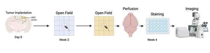
[Fig.1:Timelineschematicshowingday0asthetumorimplantationtime,followedby openfieldtestinginweeks2&4,andthesubsequentperfusion,staining,andimagingin week4.]
Themicewereanesthetizedwithketamine(50μL).Onceunresponsive,themice weresecuredinastereotacticframeandmaintainedonisoflurane.Theireyeswere lubricatedtopreventdryingduringtheprocedure.Thesurgicalsitewassterilized withalternatingbetadineandethanol.Averticalincisionwasthenmadealongthe midlineofthescalp,and10%hydrogenperoxidewasappliedtodissolve connectivetissueandvisualizeskulllandmarks.Thebasiccoordinateswereset usingbregmaandlambdaaslandmarks.ANanojectsystemwasprefilledwith mineraloil,thenloadedwithtumorcellsuspension.Asmallholewasdrilledat coordinatestargetingthesomatosensorycortex.TheNanojectwasloweredtothe injectionsiteandtumorcellsofline10072weredeliveredinfoursequential injectionstotaling100μL.Controlmiceunderwentidenticalprocedureswith HibernateAvehiclesubstitutedfortumorcells.Theincisionwasclosedwith suturesandmiceweremonitoredduringrecovery.
Anxiety-likebehaviorwasassessedusingtheopenfieldtestattwoandfourweeks post-surgery.Theopenfieldparadigmmeasuresananimal'swillingnesstoexplore anexposedcentralareaversusremainingnearthewallsofanarena,abehavior knownasthigmotaxis. Micewithheightenedanxietytendtoavoidthecenterof thearenaandspendmoretimealongtheperiphery.Foreachsession,amousewas placedinthearenaandallowedtoexplorefreelyfor10minuteswhileanoverhead camerarecordeditsmovement.Testingwaslimitedtotwosessionspermouseto preventhabituationtothearena.Mousepositioncoordinateswereextractedfrom thevideorecordingsusingDeepLabCut,amachinelearning-basedposeestimation toolthatidentifiesandtracksanatomicallandmarksacrossframes.Centertime, definedasthecumulativedurationspentinthecentralzoneofthearena,servedas theprimarymeasureofanxiety-likebehavior.
Themicewereperfusedtranscardiallyat4weekswithphosphatebufferedsaline (PBS)followedby4%paraformaldehyde.Brainswerepost-fixedin4% paraformaldehydeonarotator,thentransferredtosucroseuntilsaturated.Brains weresectionedcoronallyat40μmthicknessmanually.Everysixthsectionwas collectedintoantifreezemediumandstoredinsix-wellplates.
Free-floatingsectionswereincubatedin0.3%TritonX-100inPBSfor1-2 hoursatroomtemperature,thenblockedfor1houratroomtemperaturein blockingbuffercontaining1%mouse-on-mouseblockingreagent.Thesections werethenincubatedovernightat4°Cwithprimaryantibodiesagainstc-Fos (rabbit,1:500)andhumannestin(mouse,1:500).FollowingthreewashesinTBST, thesectionswereincubatedfor1-2hoursatroomtemperaturewithAlexaFluor 488-conjugateddonkeyanti-rabbitandAlexaFluor555-conjugateddonkeyantimousesecondaryantibodies(1:500),withDAPIincludedfornuclear counterstaining.AfterthreeadditionalwashcyclesinTBST,thesectionswere mountedonslideswithFlouromount.
FluorescentimagingwasdoneonaZeissLSM800confocalmicroscopewitha DefiniteFocus2.Whole-sectiontilescanswereimagedat10×magnificationusing the488nmlaserlinetovisualizec-Fosimmunoreactivity.Thelow-magnification imagesprovidedbrain-widecoverageforregionalquantificationandatlas alignment.Forcloserexaminationoftheamygdala,highermagnificationimages weredoneat20×usingboththe405nmchannelforDAPInuclearstainingandthe 488nmchannelforc-Fos,allowingquantificationofc-Fosexpressionatcellular resolutionwithintheregion.
Rawimagefiles(.czi)acquiredfromtheZeissLSM800confocalmicroscopewere processedusingFiji(ImageJ).Theywerethencompressedviaaz-stackand
maximumintensityprojection.Brightnessandcontrastwereadjustedforeasier alignment.Acustomscriptthenstandardizedimagedimensionsandconvertedfiles to.tifformat.ProcessedimageswereimportedintoQuPath(v0.5.1)andloaded intotheABBAplugin(v0.10.4)viaFijiforregistrationtotheAllenMouseBrain CommonCoordinateFramework(CCFv3).Eachtissuesectionunderwentaffine transformationandwasmanuallyassignedtoitscorrespondinganterior-posterior positionbasedonanatomicallandmarks.Thesectionswerethenwarpedtoits matchedatlasreferenceusingmulti-pointdeformationtoalignitsbiological structurestoatlas-definedregionalboundaries.Registeredsectionswerethen
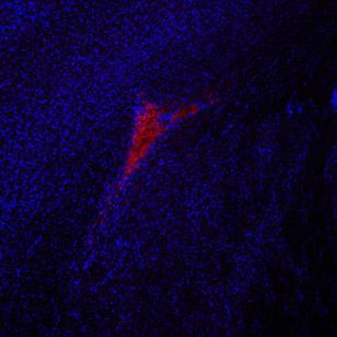
Humannestinimmunostainingwasperformedtoverifysuccessfulengraftmentof patient-derivedglioblastomaorganoidsintumor-injectedmice.Allmiceinthe tumorgroupdisplayednestin-positivestainingattheinjectionsite,confirmingthe presenceofhumantumorcellsatfourweekspost-implantation.Tumorswere centeredinthesomatosensorycortexatthetargetedcoordinates,withvariable degreesoflocalinfiltrationintothesurroundingbrainparenchymaobservedacross animals.Theextentoftumorspreadvariedbetweensubjects,althoughitwas functionallyconsistent.ControlmicethatreceivedshaminjectionswithHibernate Avehicleshowedcompleteabsenceofnestinimmunoreactivity,confirmingthat nohumancellswerepresentandthatanydifferencesobservedbetweengroups couldbeattributedtotumorpresenceratherthansurgicalprocedurealone(Fig.2).
Toassesswhethertumorpresenceaffectedanxiety-relatedbehavior,micewere evaluatedusingtheopenfieldtestattwoandfourweekspost-surgery.This paradigmquantifiesananimal'stendencytoavoidtheexposedcenterofanarenain favoroftheperiphery,abehaviorindicativeofanxiety-likestatesinrodents. Centertimeservedastheprimarydependentmeasure,withreducedcentertime reflectingincreasedanxiety.
Atweek2post-injection,tumor-bearingandcontrolmiceshowedno significantdifferenceincentertime(p>0.05).Bothgroupsspentcomparable amountsoftimeinthecenterzoneofthearena,suggestingthatearly-stagetumor developmentdidnotproducedetectablechangesinanxiety-likebehavior.Byweek 4,however,tumor-bearingmiceexhibitedamarkedreductionincentertime comparedtocontrols,andthisdifferencereachedstatisticalsignificance(p<0.05). Tumormicedisplayedpronouncedthigmotaxis,spendingmuchofthesessionin theperipheralzoneadjacenttothearenawallswhileavoidingthecenter.
Todeterminewhetherthisbehavioralchangereflectedaspecificanxiety phenotyperatherthangeneralmotorimpairmentorsicknessbehavior,total distancetraveledwasanalyzedatbothtimepoints.Therewasnosignificant differenceinactivitybetweentumorandcontrolgroupsateitherweek2orweek4 (p>0.05),indicatingthattumor-bearingmiceretainednormalambulatorycapacity. Theselectivereductionincentertimewithoutcorrespondingchangesinoverall movementsupportstheinterpretationthattumor-bearingmicedevelopedan i lik h h f k id i idi ih h
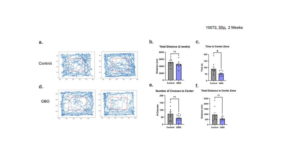
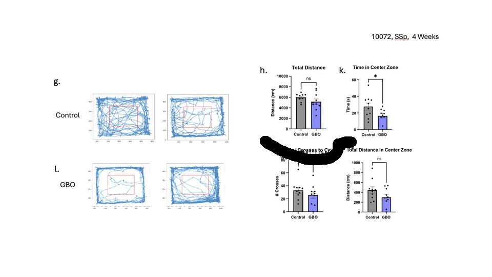
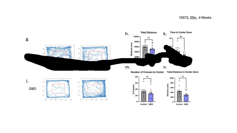
[Fig.3:Openfieldbehavioralanalysis.(a)Representativetrackingmapsshowingmouse movementtrajectoriesintheopenfieldarenaforcontrolmiceatweek2.(b)Total distancetraveledatweek2,control(right)andtumor(left).(c)Timeincenterzoneat2 weeks,control(right)andtumor(left).(d)Representativetrackingmapsshowingmouse movementtrajectoriesintheopenfieldarenafortumormiceatweek2.(e)Numberof crossestocenteratweek2,control(right)andtumor(left).(f)Totaldistancecoveredin centerzone,control(right)andtumor(left).(g)Representativetrackingmapsshowing mousemovementtrajectoriesintheopenfieldarenaforcontrolmiceatweek4.(h)Total distancetraveledatweek4,control(right)andtumor(left).(j)Representativetracking mapsshowingmousemovementtrajectoriesintheopenfieldarenafortumormiceat week4.(k)Numberofcrossestocenteratweek4,control(right)andtumor(left).(l) Totaldistancecoveredincenterzone,control(right)andtumor(left).(m)Representative trackingmapsshowingmousemovementtrajectoriesintheopenfieldarenaforcontrol miceatweek4.ns=notsignificant;p<0.05.]
Mappingofc-Fosexpressionshoweddistinctdifferencesinactivitybetweentumor andcontrolgroups.Controlmicedisplayedc-Fosexpressionconsistentwith patternsexpectedfollowingopenfieldexposureandcageactivity.Thedistribution ofc-Fos+cellsincontrolanimalswasconsistentacrosssubjectsandcorrespondedto regionsknowntobeengagedduringexploratorybehaviorandspatialnavigation.
Tumor-bearingmiceexhibitedanaltereddistributionofc-Fosexpression comparedtocontrols.Importantly,thesechangeswerenotuniformacrossthe brain.Someregionsshowedelevatedc-Fosexpressionintumormice,whileother areasappearedcomparablebetweengroups.Theregion-specificpatterning suggestedthattumorpresenceselectivelymodulatedneuronalactivityinparticular braincircuitsratherthanproducingaglobalincreaseordecreaseinneural activation(Fig.4).
Seibenhener,M.L.,Wooten,M.C.UseoftheOpenFieldMazetoMeasureLocomotorandAnxiety-likeBehaviorinMice. J.Vis.Exp.(96),e52434,doi:10.3791/52434(2015).
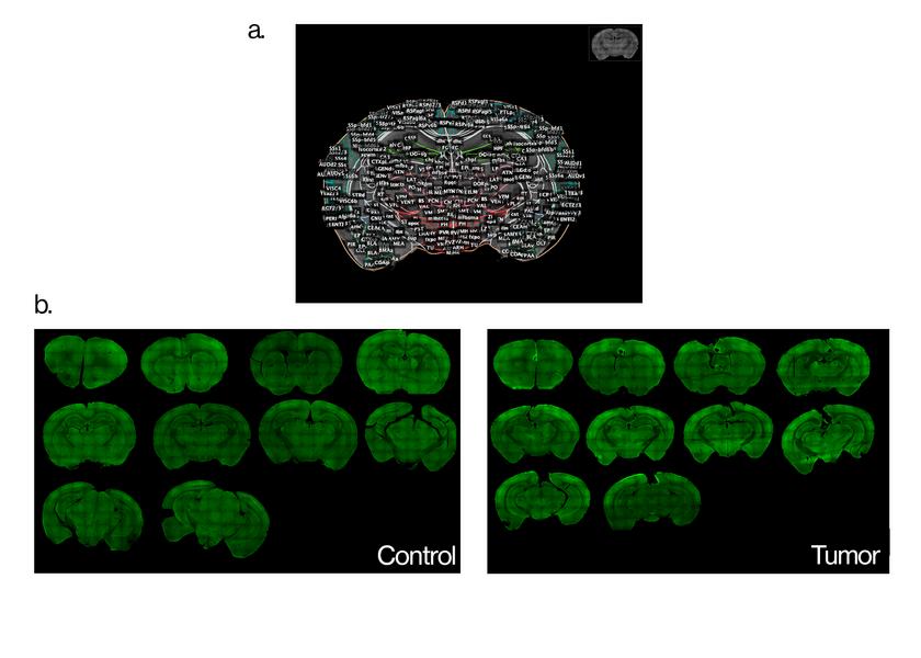
[Fig.4:Brain-widec-Fosdistribution.(a)Singletumorsamplewithatlasregion boundariesoverlaid.(b)Representativecoronalsectionsatmatchedanterior-posterior levelsfromcontrol(left)andtumor-bearing(right)miceshowingc-Fosimmunoreactivity density.]
Toquantifyregionaldifferencesinneuronalactivitybetweengroups,automated celldetectionwasperformedinQuPathusingauniformintensitythresholdapplied acrossallsamples.Thenormalizedpositivepercentageofc-Fossignalwas calculatedforeachatlas-definedbrainregion,andvalueswerecomparedbetween tumorandcontrolgroups.
Quantitativeanalysisacrossmultiplebrainregionsrevealedthattheamygdala showedthelargestandmostconsistentelevationinc-Fosexpressionintumorbearingmice.Thenormalizedc-Fos+percentageintheamygdalawassignificantly greaterinthetumorgroupcomparedtocontrols(p<0.05).Thisfindingwas consistentacrossmiceandwasobservedbilaterally,thoughthedatawasanalyzed withthebrainhemispheresparsedseparatelytoassesspotentiallateralizationeffects.
Incontrasttotheamygdala,mostotherbrainregionsexamineddidnotshow statisticallysignificantdifferencesbetweentumorandcontrolgroups.Theregions wequantifiedincludedhippocampalsubfields,somatosensoryandmotorcortices, thalamicnuclei,andvariousposteriorbrainstructures.Whilesomeregionsshowed trendstowardincreasedordecreasedc-Fosexpressionintumormice,specifically thesomatosensorycortexinthecontrol,thesedidnotreachstatisticalsignificance
atthep<0.05threshold.Thespecificityoftheamygdalafindingsuggeststhat tumor-inducedchangesinneuronalactivityareconcentratedincircuits,potentially thosemostrelevanttothebehavioralphenotypeobserved(Fig.5).
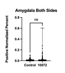
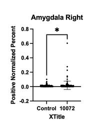
[Fig.5:Regionalc-Fosquantification.Graphshowingnormalizedc-Fos+percentagein theamygdalaforcontrol(left)versustumor(right)groups.GraphshowingnormalizedcFos+percentageintherightlateralizedamygdalaforcontrol(left)versustumor(right). p<0.05,unpairedt-test.Datapresentedasmean±SEM.]
Brain-widevisualizationofc-Fosexpressionpatternswasgeneratedusing BrainrendertomapthenormalizedpositivepercentagesontotheAllenCCFv3 atlas.Heatmaprepresentationrevealedtheregionalspecificityoftumor-induced changes,withtheamygdalaprominentlyhighlightedasasiteofelevatedactivityin tumor-bearinganimals.Thisvisualizationprovidesacomprehensiveoverviewof thespatialdistributionofneuronalactivitychangesassociatedwithglioblastoma presence(Fig.6).
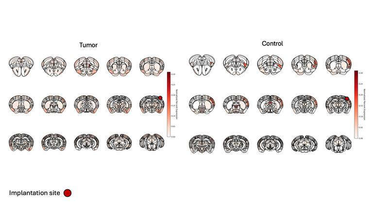
[Fig.6:Heatmapvisualizationofbrain-widec-Fosexpression.Three-dimensional renderingoftheAllenCCFv3atlaswithregionscoloredbynormalizedc-Fos+percentage forcontrol(left)andtumor(right)groups.Colorscaleindicatesexpressionlevel. GeneratedusingBrainrender.]
Highermagnificationconfocalimagingoftheamygdalawasperformedtoexamine c-Fosexpressionatcellularresolution.Imagesacquiredat20×magnificationwith bothDAPInuclearcounterstainandc-Fosimmunofluorescenceconfirmedthe presenceofelevatedc-Fos+celldensityintumor-bearingmicecomparedto controls.Individualc-Fos+nucleiwereclearlyidentifiable,andtheincreasedsignal intumoranimalscanbeseeninrepresentativeexamplesections(Fig.7).
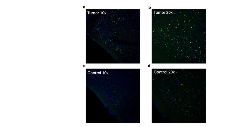
[Fig.7:20x(b,d)and10x(a,c)fluorescenceimagesoftheamygdalafromcoronalbrain sectionsshowingc-Fosactivity(green)withDAPInuclearcounterstaining.Control(c,d) andtumor(a,b).]
Theamygdalaisthecentralnodeinmediatingfearandanxiety-relatedbehaviors, andhyperactivityspecifictotheareahasbeenlinkedtostatesofanxietyacross species.Theco-occurrenceofamygdalahyperactivityandincreasedthigmotaxisin tumor-bearingmicesuggeststhatglioblastomamaydriveanxiety,asitmaybeable todowithotherregions,throughaberrantactivationoflimbiccircuitry.Whether thisrepresentsadirecteffectofthetumor-causedsignalsoranindirect
14
consequenceofalteredsignalingfromotherbrainregions,orevenaresponseto othersystemicfactorsassociatedwiththetumorremainstobedetermined. Thesefindingsextendthepriorworkcharacterizingneuron-glioma interactions.Venkateshetal.demonstratedthatneuronsformfunctional glutamatergicsynapsesontogliomacellsandthatneuronalactivitypromotestumor proliferation. WhereasSunetal.identifiedwhichneuronsprojecttothetumor, ourcurrentworkaddresseswhichregionsexhibitalteredactivity. The combinationofanatomicalconnectivityandfunctionalhyperactivitywould supportthehypothesisthatneuron-gliomasynapticintegrationproduces measurablechangesinbrainactivitypatternsandpotentiallyanimalbehavior.
AnxietyandaffectivedisturbancesareprevalentinGBMpatients,though theiretiologyinthispopulationisconsideredmultifactorialandsoincompletely understood.Boeleetal.reportedaprevalenceofanxietyrangingfrom24to48% ingliomapatientsassessedbyself-reporting,withthesesymptomscarrying significantimpactonqualityoflifeandoverallsurvival. Thecontributingfactors aregenerallyunderstoodtoincludeacombinationofpsychologicalresponsesto diagnosisandtreatmentaswellasneurophysiologicalchangesassociatedwith tumorpresenceandprogression.Theopenfieldtestweusedmeasuresthigmotaxis, whichisawell-establishedindexofanxiety-likebehaviorinrodents,andthe behavioraldifferencebetweentumor-bearingandcontrolmiceemerged specificallyatweekfourwithoutcorrespondingchangesintotallocomotion, supportinginterpretationasananxiety-likephenotyperatherthangeneralsickness behaviorormotorimpairment.Whethertheamygdalahyperactivityobservedhere contributestothatbehavioralphenotype,andwhetheranalogouscircuit-level changesarepresentinhumanGBMpatientswithanxietysymptoms,arequestions ourdataraisebutcannotresolve.Thetranslationalrelevanceofopenfield thigmotaxistoclinicalanxietyinhumanpatientsislimited,andanyconnection betweenthemousefindingsandthehumanliteraturewouldrequiredirect investigationinpatientpopulations.
Severallimitationsconstraintheinterpretationofourfindings.Ourstudy usedc-Fos,which,first,onlycapturesasinglewindowofneuronalhyperactivity andcannotresolvechangesacrosstime,andsecond,makesdistinguishing inhibitoryandexcitatoryneuronsimpossible.Observingthecorrelationbetween amygdalaandhyperactivityandanxiety-likebehaviordoesnotestablishcausality, asthehyperactivitymaydrivethebehavioralphenotype,resultfromit,orarise fromaseparatefactor.Thestudyutilizedimmunocompromisedmiceforless omplicationswithtumorintroduction,howeverthechoicemayinfluencetumor
15
Venkateshetal.(2019) 13 Sunetal.(2025) 14 Boele,F.W.,Klein,M.,Reijneveld,J.C.,&Heimans,J.J.(2023).Depressionandanxietyingliomapatients.Neuro-Oncology Practice,10(4),335–344.https://doi.org/10.1093/nop/npad013
biologyandneuralfunctioncomparedtoimmunocompetentmice.Thesample size,whileadequatefordetectingtheamygdalaeffect,mayhavebeentoosmallto detectsmallerdifferenceinotherregions.Additionally,GBMisinfiltrative,and tumorsspreadvariablyfromtheinjectionsiteacrossanimals.Differencesinthe eventualtumorlocationandextentofinfiltrationcouldinfluencewhichbrain regionsareaffectedandmaycontributetovariabilityinc-Fosexpressionacross mice.Storingthebrainsectionsfree-floatingincryoprotectantmakes distinguishingtheorientationofhemispheresimpossiblebeforemounting, meaningthelateralaccuracyoftheheatmapcouldsufferifaspecificsectionwas hardtodefineasflippedoraccuratelyalignedwhenimaging. Futureresearchshouldtestwhetheramygdalahyperactivitydrivestumor progressionorreflectsthedisruptioncausedbythetumor.Specifically, chemogeneticsilencingofamygdalaneuronswouldaddressthisdirectly.If silencingtheamygdalareducesbothanxietybehaviorandtumorgrowth,this wouldsuggesttheamygdalatobeofevenmoreinterest.GiventhatSunetal. identifiedwidespreadconnectivityfromdiversebrainregionstoglioblastomacells, theamygdalamaycontributetoalargercircuitarchitecturethatcollectively regulatestumorbehavior.
Thetimingofamygdalahyperactivityrelativetoanxietysymptomsandtumor growthisalsounresolved.Ourstudycapturedactivityatfourweekswhen behavioralphenotypeshadappeared,butwedonotknowwhetheramygdala activationprecedesanxietyordevelopsalongsideit.Longitudinalc-Fosmappingat earliertimepointswouldshowthetemporalsequenceofevents.Weekoneortwo mappingmaypotentiallyshowwhichregionsactivatefirstduringtumor developmentandwhetheramygdalahyperactivitypredictssubsequentbehavioral changes.Beyondtheamygdala,usinganothermethodofneuronalactivity monitoringwithalargersamplesizemaybeabletodeducethefunctioneffectsof glioma-brainsynapsesmoreconfidentlyandconsistently.
1.Ahmed,M.H.,Canney,M.,Carpentier,A.,&Idbaih,A.(2023).Overcoming thebloodbrainbarrieringlioblastoma:Statusandfutureperspective.InRevue Neurologique(Vol.179,Issue5,pp.430–436).ElsevierMassons.r.l. https://doi.org/10.1016/j.neurol.2023.03.013
2.Boele,F.W.,Klein,M.,Reijneveld,J.C.,&Heimans,J.J.(2023).Depressionand anxietyingliomapatients.Neuro-OncologyPractice,10(4),335–344. https://doi.org/10.1093/nop/npad013
3.ChungL.ABriefIntroductiontotheTransductionofNeuralActivityintoFos Signal.DevReprod.2015;19(2):61-67. https://doi.org/10.12717/DR.2015.19.2.061
4.Huang-Hobbs,E.,Cheng,Y.T.,Ko,Y.,Luna-Figueroa,E.,Lozzi,B.,Taylor, K.R.,McDonald,M.,He,P.,Chen,H.C.,Yang,Y.,Maleki,E.,Lee,Z.F., Murali,S.,Williamson,M.R.,Choi,D.,Curry,R.,Bayley,J.,Woo,J.,Jalali,A., …Deneen,B.(2023).Remoteneuronalactivitydrivesgliomaprogression throughSEMA4F.Nature,619(7971),844–850.https://doi.org/10.1038/s41586023-06267-2
5.Jacob,F.,Salinas,R.D.,Zhang,D.Y.,Nguyen,P.T.T.,Schnoll,J.G.,Wong, S.Z.H.,Thokala,R.,Sheikh,S.,Saxena,D.,Prokop,S.,Liu,D.,Qian,X., Petrov,D.,Lucas,T.,Chen,H.I.,Dorsey,J.F.,Christian,K.M.,Binder,Z.A., Nasrallah,M.,…Song,H.(2020).APatient-DerivedGlioblastomaOrganoid ModelandBiobankRecapitulatesInter-andIntra-tumoralHeterogeneity.Cell, 180(1),188-204.e22.https://doi.org/10.1016/j.cell.2019.11.036
6.Kovács,K.J.(2008).MeasurementofImmediate-EarlyGeneActivation:c-fos andBeyond.JournalofNeuroendocrinology,20,665–672. https://doi.org/10.1111/j.1365-2826.2008.01734.x
7.Krishna,S.,Choudhury,A.,Keough,M.B.,Seo,K.,Ni,L.,Kakaizada,S.,Lee, A.,Aabedi,A.,Popova,G.,Lipkin,B.,Cao,C.,NavaGonzales,C.,Sudharshan, R.,Egladyous,A.,Almeida,N.,Zhang,Y.,Molinaro,A.M.,Venkatesh,H.S., Daniel,A.G.S.,…Hervey-Jumper,S.L.(2023).Glioblastomaremodellingof humanneuralcircuitsdecreasessurvival.Nature,617(7961),599–607. https://doi.org/10.1038/s41586-023-06036-1
8.LeDoux,J.E.(2000).Emotioncircuitsinthebrain.AnnualReviewof Neuroscience,23,155–184.https://doi.org/10.1146/annurev.neuro.23.1.155
9.Montgomery,M.K.,Kim,S.H.,Dovas,A.,etal.(2020).Glioma-induced alterationsinneuronalactivityandneurovascularcouplingduringdisease progression.CellReports,31(2),107500. https://doi.org/10.1016/j.celrep.2020.107500
10.Nørøxe,D.S.,Yde,C.W.,Østrup,O.,Michaelsen,S.R.,Schmidt,A.Y., Kinalis,S.,Torp,M.H.,Skjøth-Rasmussen,J.,Brennum,J.,Hamerlik,P., Poulsen,H.S.,Nielsen,F.C.,&Lassen,U.(2020).Genomicprofilingofnewly diagnosedglioblastomapatientsanditspotentialforclinicalutility–a prospective,translationalstudy.MolecularOncology,14(11),2727–2743. https://doi.org/10.1002/1878-0261.12790
11.Seibenhener,M.L.,&Wooten,M.C.(2015).UseoftheOpenFieldMazeto MeasureLocomotorandAnxiety-likeBehaviorinMice.JournalofVisualized Experiments,(96),e52434.https://doi.org/10.3791/52434
12.Stupp,R.,Mason,W.P.,vandenBent,M.J.,Weller,M.,Fisher,B.,Taphoorn, M.J.B.,Belanger,K.,Brandes,A.A.,Marosi,C.,Bogdahn,U.,Curschmann,J., Janzer,R.C.,Ludwin,S.K.,Gorlia,T.,Allgeier,A.,Lacombe,D.,Cairncross,J. G.,Eisenhauer,E.,&Mirimanoff,R.O.(2005).RadiotherapyplusConcomitant andAdjuvantTemozolomideforGlioblastoma.NewEnglandJournalof Medicine,352(10),987–996.https://doi.org/10.1056/NEJMoa043330
13.Sun,Y.,Wang,X.,Zhang,D.Y.,Zhang,Z.,Bhattarai,J.P.,Wang,Y.,Park,K. H.,Dong,W.,Hung,Y.F.,Yang,Q.,Zhang,F.,Rajamani,K.,Mu,S., Kennedy,B.C.,Hong,Y.,Galanaugh,J.,Sambangi,A.,Kim,S.H.,Wheeler, G.,…Song,H.(2025).Brain-wideneuronalcircuitconnectomeofhuman glioblastoma.Nature,641(8061),222–231.https://doi.org/10.1038/s41586-02508634-7
14.Tamimi,A.F.,&Juweid,M.(2017).EpidemiologyandOutcomeof Glioblastoma.InS.DeVleeschouwer(Ed.),Glioblastoma.CodonPublications. https://doi.org/10.15586/codon.glioblastoma.2017.ch8
15.Taylor,T.E.,Furnari,F.B.,&Cavenee,W.K.TargetingEGFRforTreatment ofGlioblastoma:MolecularBasistoOvercomeResistance.CurrentCancerDrug Targets.
16.Venkatesh,H.S.,Morishita,W.,Geraghty,A.C.,Silverbush,D.,Gillespie,S. M.,Arzt,M.,Tam,L.T.,Espenel,C.,Ponnuswami,A.,Ni,L.,Woo,P.J., Taylor,K.R.,Agarwal,A.,Regev,A.,Brang,D.,Vogel,H.,Hervey-Jumper,S., Bergles,D.E.,Suvà,M.L.,…Monje,M.(2019).Electricalandsynaptic integrationofgliomaintoneuralcircuits.Nature,573(7775),539–545. https://doi.org/10.1038/s41586-019-1563-y
17.Vleeschouwer,S.(Ed.).(2017).Glioblastoma.ExonPublications. https://doi.org/10.15586/codon.glioblastoma.2017