
6 minute read
Squamous Odontogenic Tumor


Squamous Odontogenic Tumor
A Case Report
Sara Endo, D.D.S.; Timothy Chang, D.D.S.; Daria Vasilyeva, D.D.S.; Scott M. Peters, D.D.S.
ABSTRACT
The squamous odontogenic tumor (SOT) is a slowgrowing, benign, rare intraosseous neoplasm. SOT is most commonly found in the anterior region of the maxilla and the premolar-molar region of the mandible. These lesions have no sex predilection and show peak occurrence in the third decade of life.
A 25-year-old female, in apparent good health, presented to an oral surgeon with referral from a general practitioner for evaluation of an incidentally discovered lesion of the maxilla. A review of her medical history was noncontributory, and the extraoral and intraoral examination was unremarkable.
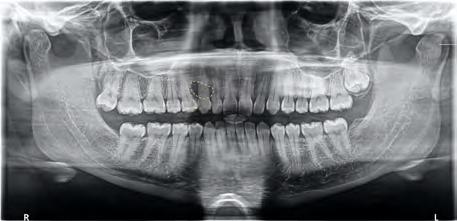
Panoramic radiograph revealed a single, unilocular radiolucency between her right maxillary canine and premolar (Figure 1). A biopsy of the radiolucent lesion was performed to establish a diagnosis. Microscopic examination revealed islands of bland squamous epithelium (Figure 2a) with occasional microcyst formation (Figure 2b). Calcified bodies were noted in some of the islands. Based on these findings, a diagnosis of squamous odontogenic tumor was rendered.
Discussion
The squamous odontogenic tumor (SOT) is a slow-growing, benign, rare intraosseous neoplasm that was first described by Pullon et al. in 1975 and recognized by the World Health Organization in 2005.[1-3] According to more recent literature, SOT is most commonly found in the anterior region of the maxilla and the premolar-molar region of the mandible.[4] Tumors in the maxilla tend to show more locally aggressive behavior compared to those found in the mandible.
SOT shows no sex predilection, can occur at a wide range of ages, from 9 to 67 years old, and has a peak occurrence in the third decade of life.[4] This tumor has been suggested to originate from gingival epithelium or gingival rests of Serres, but origination from the rests of Malassez has the most supporting data. This is due to the observations that SOT is commonly associated with erupted vital teeth, has tight adherence to the roots of teeth, and is easily separated from the soft tissue.[5]
Clinically, patients with SOT present with asymptomatic swelling, mobility of the adjacent teeth and localized periodontal bone loss. The tumor is often associated with erupted, vital teeth and is commonly discovered incidentally on routine radiographic examination. Radiographically, the tumor has a characteristic appearance of a triangular unilocular radiolucency between the roots of two teeth, with the base of the triangle toward the root apices; rarely, does SOT appear as large, multilocular lesions.[4] When considering a unilocular radiolucency in the maxilla, multiple differential diagnoses must be considered.
Histologically, SOT contains islands of squamous epithelium of varying size and shape that are well-demarcated from the dense fibromyxoid connective tissue stroma by a layer of flattened cells.[3] Some neighboring islands are connected via epithelial cords. Intracellular keratin, focal microcystic formation and dystrophic calcifications can also occur.[2]


The clinical differential for SOT may include ameloblastoma, odontogenic keratocyst (OKC), lateral periodontal cyst (LPC), squamous cell carcinoma (SCC), among others.[4] The correct diagnosis is made only after histologic analysis of the lesion. While some cases of SOT can display aggressive clinical behavior, the cells in the epithelial nests do not exhibit atypia, allowing clinicians to distinguish it from squamous cell carcinoma.[6]
While SOT does not usually cause pain or discomfort to patients, it can mimic periodontitis, with features such as gingival inflammation, deep probing depths and alveolar bone loss.[5] Therefore, the management of SOT includes local surgical excision and, less commonly, en bloc resection.[4,7] Based on 46 cases of SOT with follow-up data, there is an estimated recurrence rate of approximately 19.4% following surgical excision.[4]
Although radiolucent lesions are not an infrequent finding on radiographic examination, care must be taken to perform a thorough clinical examination and biopsy, as there is significant overlap in features with a number of odontogenic cysts and tumors. Clinical and radiographic features should inform the differential diagnoses of this type of lesion.
This case report serves as a reminder for oral healthcare providers to remain vigilant during examination and obtain a biopsy as a means for establishing a definitive diagnosis.
The authors declare no conflicts of interest. Nor did their study receive commercial funding. Queries about this article can be sent to Dr. Endo at endos1@unlv.nevada.edu.
REFERENCES
1. Pullon PA, Shafer WG, Elzay RP, Kerr DA, Corio RL. Squamous odontogenic tumor: report of six cases of a previously undescribed lesion. Oral Surgery, Oral Medicine, Oral Pathology 1975;40(5):616-630. doi:10.1016/0030-4220(75)90372-2.
2. Jones BE, Sarathy AP, Belinda Ramos M, Foss RD. Squamous odontogenic tumor. Head and Neck Pathology 2010;5(1):17-19. doi:10.1007/s12105-010-0198-y.
3. Philipsen HP, Reichart PA. Squamous odontogenic tumor (Sot): a benign neoplasm of the periodontium. Journal Clinical Periodontology 1996;23(10):922-926. doi:10.1111/j.1600-051x.1996. tb00512.x.
4. Upadhyaya JD, Banasser A, Cohen DM, Kashtwari D, Bhattacharyya I, Islam MN. Squamous odontogenic tumor: review of the literature and report of a new case. Journal Oral and Maxillofacial Surgery 2021;79(1):164-176. doi:10.1016/j.joms.2020.06.031.
5. Haghighat K, Kalmar JR, Mariotti AJ. Squamous odontogenic tumor: diagnosis and management. Journal Periodontology 2002;73(6):653-656. doi:10.1902/jop.2002.73.6.653.
6. Ide F, Shimoyama T, Horie N, Shimizu S. Intraosseous squamous cell carcinoma arising in association with a squamous odontogenic tumour of the mandible. Oral Oncology 1999;35(4):431-434. doi:10.1016/s1368-8375(99)00010-x.
7. Baden E, Doyle J, Mesa M, Fabié M, Lederman D, Eichen M. Squamous odontogenic tumor. Oral Surgery, Oral Medicine, Oral Pathology 1993;75(6):733-738. doi:10.1016/0030-4220(93)90432-4.


Sara Endo, D.D.S., was a member of the Class of 2023, Columbia University College of Dental Medicine, New York, NY. She is a resident, Division of Orthodontic and Dentofacial Orthopedics, University of Nevada Las Vegas, Las Vegas, NV.
Timothy Chang, D.D.S., was a member of the Class of 2023, Columbia University College of Dental Medicine, New York, NY. He is a resident, Section of Oral and Maxillofacial Surgery, University of California Los Angeles, Los Angeles, CA.
Daria Vasilyeva, D.D.S., is a resident, Division of Oral and Maxillofacial Pathology, Columbia University Irving Medical Center, New York, NY.
Scott M. Peters, D.D.S., is former assistant professor, Division of Oral and Maxillofacial Pathology, Columbia University Irving Medical Center, New York, NY. He now holds title of associate professor, Oral and Maxillofacial Pathology, Geisinger Health System, Danville, PA.


