
29 minute read
Platelet-derived Materials for the Treatment of Infrabony Periodontal Defects


Platelet-derived Materials for the Treatment of Infrabony Periodontal Defects
A Systematic Review and Meta-analysis
Armando Uribe-Rivera, D.D.S, M.S.; Daniel Heffes, D.D.S.; Hans Malmström, D.D.S.; Dongmei Li, Ph.D.; Yan-Fang Ren, D.D.S, Ph.D, M.P.H.
ABSTRACT
Aim: To compare the clinical outcomes of plateletderived biomaterials, including recombinant human platelet-derived growth factors receptor BB (rhPDGFBB), platelet-rich plasma (PRP) and platelet-rich fibrin (PRF), for treatment of periodontal infrabony defects.
Materials and Methods: A meta-analysis was conducted to compare pocket depth reduction (PRD) and clinical attachment gain (CAG) with and without rhPDGF-BB, PRP or PRF for surgical treatments of periodontal infrabony pockets. Eligible studies were randomized controlled clinical trials in adults with periodontal infrabony defects.
Results: Thirty-four randomized controlled trials involving 939 patients with a mean follow-up of 9.29(±2.49) months were included in this meta-analysis. All three platelet-derived materials achieved better outcomes in both PDR and CAG (p <0.00001) in comparison to controls. The effect size of the plateletderived biomaterials was on average 0.81(95%CI 0.62 to 0.99)mm [(0.96mm for PRF, 0.80mm for rhPDGFBB and 0.63mm for PRP)] for PDR, and 0.82 (95%CI 0.60 to 1.03)mm [( 0.92mm for PRF, 1.01mm for rhPDGF-BB and 0.60mm for PRP)] for CAG.
Conclusion: Periodontal therapies using plateletderived biomaterials improved PDR and CAG in the short term, compared to controls without these materials. However, there is not clinically significant PDR and CAG between the platelet materials.
Periodontitis is the most common inflammatory condition of bacterial origin. It causes destruction of the periodontal apparatus, including the alveolar bone, periodontal ligament and cementum, in addition to the gingival tissues.[1] Management of periodontal diseases is aimed at eliminating bacterial infections, reconstructing the periodontal apparatus, and restoring the masticatory function and general health of the individual.[2,3]
Fundamental periodontal treatments include two phases:
1. A disease-control phase that involves mechanical debridement and root scaling and planing to eliminate the bacterial pathogens, thus, modulating the host response to control or slow down periodontal disease progression.[4]
2. A reconstructive phase that aims at restoring periodontal anatomical structures destroyed by disease progression and involves periodontal therapies, including flap surgery, in addition to connective tissue and bone grafts (e.g., autografts, allografts, synthetic materials), for the purpose of tissue regeneration.
Based on evidence from histological studies,[5,6] these therapies resulted in tissue repair rather than tissue regeneration and, thus, failed to accomplish the goals of regenerative periodontal treatments.[7-9] Today, periodontal therapies for regeneration of periodontal tissues often incorporate molecular or cellular constructs, such as extracellular matrix proteins, growth factors, morphogens, biological mediators and/or precursor cells onto the surgical sites.[10] However, clinical outcomes of these novel therapeutic modalities remain to be thoroughly investigated.
It has been shown that an array of products containing platelet-derived growth factors led to regeneration of alveolar bone, vascular structures and soft tissues of the periodontium.[11,12] These include PRP, PRF and rhPDGF-BB. [13,14] PRP is a concentrate that contains from 2- to 8.5-fold increase of platelets obtained from autologous blood. When delivered locally, PRP has the capability of increasing the proliferation of fibroblasts, osteoblasts and angiogenesis, aiding the formation of new periodontal tissues.[15]
PRF is considered a second-generation platelet concentrate that enables the operator to obtain fibrin membranes enriched with platelets, growth factors, leucocytes, cytokines and circulating stem cells from autologous blood. This natural scaffold was found to play a key role in hard- and soft-tissue repair processes.[16]
On the other hand, rhPDGF-BB is often used with various scaffolds as carriers to enhance the molecular and cellular functions involved in regeneration of the target periodontal structures. [17-20] Application of rhPDGF-BB may enhance mitogenic and chemotactic activities that facilitate tissue regeneration in the targeted areas.[10,21] Carrier scaffolds that emulate the characteristics of native jaw bone have shown to increase the inductive capacity of cells and facilitate blood vessel distributions, which are conducive to new tissue formation and vascular regeneration in the affected site.[22] For instance, Beta-tricalcium phosphate (β-TCP), a synthetic material commonly used in conventional periodontal therapies, led to the improvement of clinical outcomes due to its biocompatibility with human tissues in the periodontium.
[10] Combination of rhPDGF-BB with such carrier scaffolds may further enhance the clinical outcomes.
Though these platelet-derived biomaterials are purported to act through a similar mechanism to enhance tissue regeneration, they vary in preparation, application and cost. Numerous studies involving these products have claimed varying degrees of success in clinical applications for periodontal tissue regeneration. But, it is not known if one product is superior to another in terms of clinical outcomes. The objectives of this study were, therefore, to perform a systematic review and meta-analysis using infrabony periodontal defects to estimate clinical outcomes measured (by pocket depth reduction [PDR] and clinical attachment gain [CAG]), following applications of PRP, PRF or rhPDGF-BB in comparison to conventional periodontal techniques. We aim to answer the following two questions: 1. Is PRP, PRF or rhPDGF-BB more effective than controls in achieving PDR and CAG in patients with periodontal intrabony pockets? 2. Is one of these products superior to another in improving treatment outcomes measured in PDR and CAG?
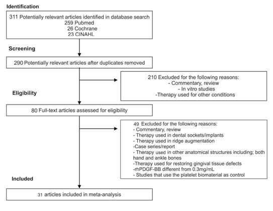
Figure 1. Flowchart of Inclusion Criteria or Articles in this Study.
Materials and Methods
This systematic review and meta-analysis were designed and conducted in accord with the Preferred Reporting Items for Systematic Reviews and Meta-Analysis (PRISMA) statement.
A. Focused PICO Question
The following statements were used to conduct the systematic search:
• Population (P) = systemically healthy humans with periodontal intrabony defects.
• Intervention (I) = use of a platelet-derived material, either rhPDGF-BB or PRF or PRP, in periodontal surgery for treatments of infrabony pockets.
• Comparison (C) = periodontal surgery without the use of the platelet-derived materials for treatments of infrabony pockets. The protocols for periodontal surgery are the same in the comparison group as in the intervention group, with the sole difference being with or without the platelet-derived materials.
• Outcome (O) = PDR and CAG at a minimum of 6 months and maximum of 12 months.
A PICO question was created to carry out the literature search: “Among clinical trials that combine either rhPDGF-BB, PRF or PRP with periodontal surgery for the management of periodontal intrabony defects, do adults with periodontal diseases exhibit improved PDR and CAG compared to periodontal surgeries without these materials?”
B. Literature Search
We searched the literature from January 1997 through January 2021 utilizing four electronic databases (PubMed [http://www.ncbi.nlm. nih.gov/oubmed]; Cochrane [http://www.cochrane.org]; CINAHL [https://www.ebscohost.com/nursing/products/cinahl-databases]; and Web of Science [htpp://apps.webofknowledge.com]).
We used specific headings and keywords as follows:
For PubMed—(Platelet Derived Growth Factor or PGDF) (Platelet-Rich Fibrin or PRF) and (Platelet-Rich Plasma or PRP or Platelet Concentrate) and (Bone Regeneration or Bone Regenerat* or Osteoconduction) and (Adult or Adult* or Elder or Elder* or Middle Aged or Middle Age or Young Adult or Young Adult*) and (Randomized Controlled Trial[ptyp])[(Platelet Derived Growth Factor or PDGF) and (Bone Regeneration or Bone Regenerat* or Osteoconduction) and (Adult or Adult* or Elder or Elder* or Middle Aged or Middle Age or Young Adult or Young Adult*) and (Randomized Controlled Trial[ptyp]).
For Cochrane—(Platelet Derived Growth Factor or PGDF) and (Bone Regeneration or Bone Regenerat* or Osteoconduction) and (Adult or Adult* or Elder or Elder* or Middle Aged or Middle Age or Young Adult or Young Adult*).
For Web of Science—(Platelet Derived Growth Factor or PGDF) (Platelet-Rich Fibrin or PRF) and (Platelet-Rich Plasma or PRP or Platelet Concentrate) and ((Bone Regeneration or Bone Regenerat*) or Osteoconduction) and (Adult or Adult* or Elder or Elder* or Middle Aged or Middle Age or Young Adult or Young Adult*) and (clinical trial* or research design or comparative stud* or evaluation stud* or controlled trial* or follow-up stud* or prospective stud* or random* or placebo* or single blind* or double blind*)).
For CINHAL—(MH “Platelet-Derived Growth Factor”) or “Platelet Derived Growth Factor” or PGDF) and ((MH “Platelet-Rich Fibrin”) or “Platelet Rich Fibrin” or “PRF”) and (MH “PlateletRich Plasma”) or “Platelet Rich Plasma” or “PRP” or “Platelet Concentrate”)(MH “Bone Regeneration+”) or “Bone Regeneration” or Osteoconduction) - Narrow by Subject Age: - all adult.
C. Screening and Eligibility
Studies were included only if all of the following eligibility criteria were met: 1) published in English; 2) reported the use of rhPDGFBB, PRF or PRP; 3) randomized controlled clinical trials; 4) reported descriptive statistics on clinical periodontal outcomes, including PDR and CAG (sample size, mean, standard deviation for two experimental arms, including both baseline and postoperative assessments). Studies were excluded if they showed: 1) different modalities other than the use of rhPDGF-BB, PRF and PRP; 2) case reports or 3) case series; and 4) animal studies. Of note, the titles and abstracts obtained from the first search were screened independently by two reviewers (M.L., F.A.M.). When publications did not meet the inclusion criteria, they were excluded upon reviewers’ agreement.
A second screening of articles was performed by Drs. H.D. and U.R.A., which included more recent publications to be included in the study. Any disagreement between the reviewers was resolved by reaching consensus through discussion. All full texts of the potential articles were obtained and examined by two reviewers (U.R.A. and Y.F.R). Data from the articles were reviewed twice and agreement between the two reviewers was accomplished, so that final eligibility of the articles was assured. All studies that fulfilled all selection criteria were processed for data extraction.
D. Quality Assessment of Clinical Trials
The Oxford quality scoring system (JADAD scoring) was utilized to appraise the quality of the clinical trial. [23] For purposes of this meta-analysis, those studies that received a JADAD score between four and five points were considered for analysis (Table 1).
E. Data Management and Statistical Analysis [23]
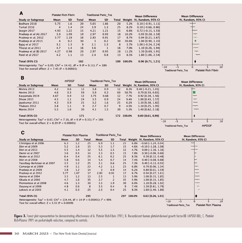
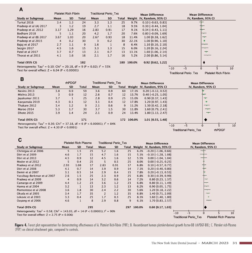
The outcome variables were PDR and CAG in millimeters measured at a minimum of six months of follow-up after the periodontal surgery. Mean values and standard deviations (SD) were extracted for each group (Figures 2,3). All data were arranged in groups for the inter-group comparison (experimental versus control groups between rhPDGF-BB, PRF and PRP therapies). A meta-analysis was performed using the random model for all three platelet biomaterials, when heterogeneity (I2 proportion) was high and the fixed model when heterogeneity was low. The mean difference was calculated and a 95% confidence interval was computed. Forest plots were created to display the analysis using Review Manager (RevMan Version 5.3. Copenhagen: The Nordic Cochrane Centre, The Cochrane Collaboration, 2014). All statistical tests were two-sided, with alpha <0.05 being considered significant.

F. Clinical Significant PDR and CAG
For purposes of this meta-analytic study, the mean difference of >0.7mm for both PDR and CAG was considered clinically significant based on the study by Zamet JS, et al. with similar outcomes, which most of the studies included in this study used for power analysis.[24]
Results
A. Literature Search and Study Selection
The database-search identified 311 studies, of which 21 were duplicates (Figure 1). A total of 290 articles were included for title and abstract screening. From those, 80 articles were included for full-text review. Forty-nine articles were excluded after full-text review, which was conducted independently by two reviewers (A.U.R., D.H.). After reviewing the included articles, there was a consensus of these studies between Y.F.R. and A.U.R. for verifying values, clinical outcomes and quality of studies. Thirty-one randomized control trials fulfilled the inclusion criteria and were included for analysis (Table 1). Of note, JADAD scores of all the included studies reached a score of four out of five points due to lack of placebo treatments in their designs.
B. Description of the Studies
In the 31 studies that underwent data extraction, 939 individuals underwent periodontal open-flap surgeries combined with either tissue debridement or guided-tissue regeneration that included the use of bone grafting materials or a combination of both techniques and were followed up at 9.29+2.49 (range of 6 to 12 months). For the only study that had a length of follow-up greater than 12 months,[25] only the measurements at 12 months were included for the purpose of this meta-analysis.
Articles included for this study (n=31) were grouped into traditional periodontal surgery in combination with platelet-derived materials (experimental group) and traditional periodontal methods without platelet-derived materials (control group). The platelet-derived materials were broken down into three subgroups: rhPDGF-BB, PRP and PRF (Table 1).
B.1. rhPDGF-BB group
Seven studies used rhPDGF-BB (n=331 subjects) in combination with periodontal surgery. Of the seven articles, five studies reported the use of β-TCP as grafting material.[6,22,25-27] One study used 1% sodium hyaluronate gel,[28] and one study used stem cells cultured in β-TCP in the experimental group.[29] The same grafting materials were used in the control group, without the incorporation of rhPDGF-BB in them for all the seven studies.
B.2. PRP group
Fourteen studies used PRP (n=374 subjects) in combination with periodontal surgery.[9,13, 29-42] Of the 15 articles, 5 used a combination of bovine porous bone mineral;[13,30-33] three studies used a combination of PRP and β-TCP.[34-36] Two studies used a combination of PRP with enamel matrix derivatives and natural bone mineral.[37,38] Two studies used a combination of PRP with alloplastic grafts, including either bioactive glass[39] or hydroxyapatite.[40] One study utilized a combination of PRP with demineralized freeze-dried bone allograft.[41] Two studies used PRP alone in the experimental group.[9,42]
B.3. PRF group
Ten studies were analyzed for PRF (n=234 subjects) in combination with periodontal surgery.[9,43-52] Of the nine studies, one used a combination of PRF with enamel matrix derivatives,[50] and one used a combination of PRF with anorganic bovine bone mineral.[52] One study used a combination of PRF with alloplastic (bioactive glass) graft,[53,54] and one used the combination of PRF and hydroxyapatite graft in the experimental groups.[46] Six studies used PRF alone as membrane for periodontal regeneration. [9,45-47,49,51] It is important to highlight that one study included two combinations of PRF (PRF alone and PRF + hydroxyapatite). [46] The same products, but without the PRF, were used in the control groups.
C. Assessment of heterogeneity
Heterogeneity was evaluated among the studies for the meta-analysis comparison of PDR and CAG. In the present study, a high heterogeneity was found among studies for both PDR (I2 = 82%) and CAG (I2 = 85%). However, a low heterogeneity was found between studies which evaluated the use of rhPDGF-BB (I2 = 16%) and PRF (I2 = 38%) for PDR. The other subgroups and clinical outcomes showed high heterogeneity, including; rhPDGF-BB (I2 = 84%) and PRF (I2 = 55%) for CAG and PRP for both the PDR and the CAG (I2 = 90%).
C.1. Study design
All studies were randomized clinical trials and frequently presented a split-mouth design. The articles and study characteristics are presented in Table 1.
C.2. Surgical protocol
Surgical protocols used in the included studies involved traditional periodontal flaps and open-flap debridement as presented in Table 1.
C.3. Clinical periodontal measurements
A standardized assessment was used for pocket-depth defect and clinical attachment level. Anatomical landmarks for both baseline and postoperative periodontal tissues assessment included cemento-enamel junction, alveolar crest and base of the defect. The clinical parameters were recorded to the nearest millimeter with the help of a periodontal probe. This assessment method was
described in 30 studies. Therefore, only one study did not describe the technique used for periodontal tissues assessment.[25]
D. Meta-analyses
Overall, the results of the random effect meta-analysis favored the use of platelet-derived materials over controls. Experimental groups achieved greater PDR (P = <0.00001) and CAG (P = <0.00001) than the control groups. The random-effect mean difference between control and experimental therapies was 0.81mm (95%CI 0.62 to 0.99mm) in PDR (Figure 2A) and 0.82mm (95%CI 0.60 to 1.03mm) in CAG (Figure 2B).
D.1: PRF vs. controls
In this subgroup, PRF in combination with periodontal surgery achieved a mean difference of 0.96mm (95% CI 0.71 to 1.21mm) higher in PDR (P = < 0.00001) and a mean difference of 0.92mm (95% CI 0.62 to 1.22mm) larger in CAG (P = < 0.00001), when compared to controls. (Figures 3A, 4A).
D.2: rhPDGF-BB vs. controls
In this subgroup, rhPDGF-BB in combination with traditional periodontal surgery achieved a mean difference of 0.80mm (95% CI 0.61 to 0.99mm) higher in PDR (P = <0.00001) and 1.01mm (95%CI 0.55 to 1.48mm) larger in CAG (P = <0.0001) than controls (Figures 3B, 4B).
D.3: PRP vs. controls
In this subgroup, PRP in combination with periodontal surgery achieved a mean difference of 0.63mm (95% CI 0.26 to 1.01mm) higher in PDR (P = 0.0009) and 0.60mm (95%CI 0.17 to 1.03mm) larger in CAG (P = 0.006) than controls (Figures 3C, 4C).

TABLE 1 Characteristics of Included Studies for Meta-analysis
Discussion
The purpose of this study was to evaluate the effects of plateletderived biomaterials on PDR and CAG when used for treating infrabony defects in periodontal patients. We found that the use of
rhPDGF-BB, PRP and PRF with periodontal surgery improved the clinical outcomes when compared to the controls without these biological materials. Additionally, compared to rhPDGF-BB and PRP, PRF moderately enhanced both PDR and CAG.
In the field of dentistry, in the last decade, platelet biomaterials have been used extensively in periodontal tissues.[55,56]
The meta-analysis results of our study showed that incorporating platelet-derived biomaterials in traditional periodontal methods, displayed an improvement in both the PDR and the CAG when compared with controls. Other reports showed similar clinical outcomes and suggested that the advantage of using platelet biomaterials with other tissue-regenerative methods in the periodontum is linked with the continuing release of the autologous growth factors from platelets onto periodontal tissues.[57-59] Furthermore, it has been shown that these autologous growth factors stimulate biologic processes, including chemotaxis, angiogenesis, cell proliferation and differentiation for enhancing tissue regeneration.[60-62] Hence, the application of the platelet biomaterials
in periodontal surgery is gaining momentum with promising clinical outcomes.
A major periodontal surgery goal is to improve the formation of tooth-supporting structures affected by periodontal disease.[63] Platelet biomaterials are assumed to increase the effectiveness of periodontal techniques.[64] However, little is known about the
comparison of the effectiveness of each of the platelet biomaterials, including rhPDGF, PRP and PRF, on infrabony periodontal defects. This study demonstrated that PRF was superior to rhPDGF and PRP for enhancing PDR and CAG.
Other studies reported similar outcomes using PRF. They suggested that PRF is a more standardized, slow-type polymerization technique which may enhance platelet capability for releasing growth factors for longer periods.[16,25,62,65-67] In addition, the findings in our study are also similar, when compared to other systematic reviews on PRF and periodontal outcomes.[55,68] For instance, Verma et al. showed that PRF combined with other biomaterials for grafting methods displayed superior periodontal outcomes to those demonstrated when the same grafting materials were used alone.[55] It is noteworthy that other reports suggested that PRF effectiveness is questionable when it is the only material used in periodontal methods.[68]
In contrast with the study by Najeeb et al. the PDR and CAG of >1.00mm in our meta-analysis included those studies which utilized PRF alone as a membrane. Hence, we suggest that the systematic review by Najeeb et al. combined studies that included both case reports and randomized clinical trials with low methodological quality. This factor is important for proper analysis and interpretation of the data and for minimizing publication bias.[23] Another important factor in our study, different from Najeeb’s report, is that all of the included papers in our study were considered of high methodological quality.
Limitations for this study included the high heterogeneity between some of the comparisons in this meta-analysis. This potentially biased some of the assumptions taken into account in the meta-analysis results of this study and the findings should be viewed with caution. A second limitation in this study is found on the follow-up period among studies, which ranges from 6 to 12 months. It is paramount that more studies with longer followup evaluations, using platelet-derived biomaterials, be designed to determine the platelet biomaterial’s efficacy in the long term.
The strengths of this study are found in the appraisal and thorough search and analysis of the randomized clinical trials using the three different platelet biomaterials in a specific periodontal condition. In our opinion, there was a dearth of information in the dental literature when comparing the three platelet-derived therapies and their efficacy in periodontal outcomes. This question is properly addressed in this meta-analysis.
It is important to mention that the findings in this study showed not only a high degree of statistical significance in the comparison of clinical outcomes between the experimental and control groups, it also demonstrated that these findings were clinically significant to enhance periodontal tissues affected in periodontal disease.
This study opened other questions for future studies, including: 1) what is the cost-effectiveness of each platelet biomaterial, when compared to another; and 2) what is the long-term effect of platelet biomaterials in the periodontum and what is their benefit in incorporating them in periodontal surgery methods?
In conclusion, our results provide dental professionals with a scientific foundation for making appropriate decisions in the short-term with respect to the use of platelet-derived biomaterials in conjunction with periodontal surgery for the management of infrabony periodontal defects. Based on our meta-analysis and literature search, we determined that there is clinically significant PDR and CAG, when incorporating platelet biomaterials in surgical periodontal therapies. However, there is not clinically significant PDR and CAG between the platelet materials.
The authors thank the Clinical and Translational Institute at the University of Rochester for services on statistical methods, as well as the following: Librarian Lorraine Porcello, Eastman Institute for Oral Health, and Librarian Daniel Castillo, School of Medicine and Dentistry, University of Rochester, for collaboration and literature search; Drs. Edith Gonzalez-James, D.D.S.., Lakshmidevi Ranjan Mandava R.L, B.D.S., and Marcela Flores-Amador, D.D.S., former residents at EIOH, for assistance with initial literature search; and Dr. John H. Campbell, D.D.S., M.S., FACS, Department of Oral and Maxillofacial Surgery, University at Buffalo, and OMS Dr. Gilbert H. Schulenberg, D.D.S., of Buffalo, for assistance in editing of this manuscript. Queries about this article can be sent to Dr. Uribe-Rivera at aur@bu.edu.
REFERENCES
1. Williams RC. Periodontal disease. N Engl J Med 1990;322(6):373-82.
2. Costa FO, et al. Surgical and nonsurgical procedures associated with recurrence of periodontitis in periodontal maintenance therapy: 5-year prospective study. PLoS One 2015;10(10):e0140847.
3. Pajnigara N, et al. Diagnostic accuracy of cone beam computed tomography in identification and postoperative evaluation of furcation defects. J Indian Soc Periodontol 2016;20(4):386-390.
4. Howell TH, et al. A phase I/II clinical trial to evaluate a combination of recombinant human platelet-derived growth factor-BB and recombinant human insulin-like growth factorI in patients with periodontal disease. J Periodontol 1997;68(12):1186-93.
5. Caton J, Nyman S. Histometric evaluation of periodontal surgery. I. The modified Widman flap procedure. J Clin Periodontol 1980;7(3):212-23.
6. Maroo S, Murthy KR. Clinical and radiographic evaluation of recombinant human platelet derived growth factor with beta tricalcium phosphate in the treatment of a periodontal intrabony defect. J Indian Soc Periodontol 2014;18(6): 789-93.
7. Wang HL,Greenwell H. Surgical periodontal therapy. Periodontol 2000 2001;25:89-99.
8. Polimeni G, Xiropaidis AV, Wikesjo UM. Biology and principles of periodontal wound healing/regeneration. Periodontol 2000 2006;41:30-47.
9. Pradeep AR, et al. Comparative evaluation of autologous platelet-rich fibrin and plateletrich plasma in the treatment of 3-wall intrabony defects in chronic periodontitis: a randomized controlled clinical trial. J Periodontol 2012;83(12):1499-507.
10. Nevins M, et al. Platelet-derived growth factor stimulates bone fill and rate of attachment level gain: results of a large multicenter randomized controlled trial. J Periodontol 2005;76(12):2205-15.
11. Dereka XE, Markopoulou CE, Vrotsos LA. Role of growth factors on periodontal repair. Growth Factors 2006;24(4):260-7.
12. Babo PSR, RL, Gomes ME. Periodontal tissue engineering: current strategies and the role of platelet rich hemoderivatives. Journal of Materials Chemistry B 2017;5:3617-3628.
13. Ouyang XY, Qiao J. Effect of platelet-rich plasma in the treatment of periodontal intrabony defects in humans. Chin Med J (Engl) 2006;119(18):1511-21.
14. Chang YCZ, JH. Effects of platelet-rich fibrin on human periodontal ligament fibroblasts and application for periodontal infrabony defects. Australian Dental Journal 2011;56(4):365-71.
15. Marx RE, et al. Platelet-rich plasma: Growth factor enhancement for bone grafts. Oral Surg Oral Med Oral Pathol Oral Radiol Endod 1998;85(6): 638-46.
16. Kumar YR, et al. Platelet-rich fibrin: the benefits. Br J Oral Maxillofac Surg 2016;54(1):57-61.
17. Andrew JG, et al. Platelet-derived growth factor expression in normally healing human fractures. Bone 1995;16(4):455-60.
18. Heldin CH, Ostman A, Ronnstrand L. Signal transduction via platelet-derived growth factor receptors. Biochem Biophys Acta 1998;1378(1):F79-113.
19. Rosenkranz S, Kazlauskas A. Evidence for distinct signaling properties and biological responses induced by the PDGF receptor alpha and beta subtypes. Growth Factors 1999;16(3):201-16.
20. Furuhashi M, et al., Platelet-derived growth factor production by B16 melanoma cells leads to increased pericyte abundance in tumors and an associated increase in tumor growth rate. Cancer Res 2004;64(8):2725-33.
21. Lynch SE, et al., The effects of short-term application of a combination of platelet-derived and insulin-like growth factors on periodontal wound healing. J Periodontol 1991;62(7): 458-67.
22. Thakare K, Deo V. Randomized controlled clinical study of rhPDGF-BB + beta-TCP versus HA + beta-TCP for the treatment of infrabony periodontal defects: clinical and radiographic results. Int J Periodontics Restorative Dent 2012; 32(6):689-96.
23. Berger VW, Alperson SY. A general framework for the evaluation of clinical trial quality. Rev Recent Clin Trials 2009;4(2):79-88.
24. Zamet JS, et al. Particulate bioglass as a grafting material in the treatment of periodontal intrabony defects. J Clin Periodontol 1997;24(6):410-8.
25. Nevins M, et al. Platelet-derived growth factor promotes periodontal regeneration in localized osseous defects: 36-month extension results from a randomized, controlled, doublemasked clinical trial. J Periodontol 2013;84(4):456-64.
26. Jayakumar A, et al. Multi-centre, randomized clinical trial on the efficacy and safety of recombinant human platelet-derived growth factor with beta-tricalcium phosphate in human intra-osseous periodontal defects. J Clin Periodontol 2011;38(2):163-72.
27. Kavyamala D, et al. Evaluation of the efficacy of a 1:1 mixture of beta-TCP and rhPDGF-BB in the surgical management of two- and three-wall intraosseous defects: a prospective clinical trial. Int J Periodontics Restorative Dent 2019; 39(1):107-113.
28. Mishra AA, H, Pathakota KR, Avula J. Efficacy of modified minimally invasive surgical technique in the treatment of human intrabony defects with or without use of rhPDGF-BB gel – a randomized controlled trial. Journal of Clinical Periodontology 2013;40:172–179.
29. Dhote R, et al. Stem cells cultured on Beta Tricalcium Phosphate (beta-TCP) in combination with recombinant human platelet-derived growth factor - BB (rh-PDGF-BB) for the treatment of human infrabony defects. J Stem Cells 2015; 10(4):243-54.
30. Lekovic V, et al. Effectiveness of a combination of platelet-rich plasma, bovine porous bone mineral and guided tissue regeneration in the treatment of mandibular grade II molar furcations in humans. J Clin Periodontol 2003;30(8): 746-51.
31. Camargo PM, et al. A surgical reentry study on the influence of platelet-rich plasma in enhancing the regenerative effects of bovine porous bone mineral and guided tissue regeneration in the treatment of intrabony defects in humans. J Periodontol 2009;80(6): 915-23.
32. Hanna R, Trejo PM, Weltman RL. Treatment of intrabony defects with bovine-derived xenograft alone and in combination with platelet-rich plasma: a randomized clinical trial. J Periodontol 2004;75(12):1668-77.
33. Dori F, et al. Effect of platelet-rich plasma on the healing of intrabony defects treated with an anorganic bovine bone mineral: a pilot study. J Periodontol 2009;80(10):1599-605.
34. Christgau M, et al., Influence of autologous platelet concentrate on healing in intrabony defects following guided tissue regeneration therapy: a randomized prospective clinical splitmouth study. J Clin Periodontol 2006;33(12):908-21.
35. Moder D, et al. Influence of autogenous platelet concentrate on combined GTR/graft therapy in intrabony defects: a 7-year follow-up of a randomized prospective clinical split-mouth study. J Clin Periodontol 2012;39(5):457-65.
36. Yassibag-Berkman ZT, Subasioglu O, Kantarci A. Combined use of platelet-rich plasma and bone grafting with or without guided tissue regeneration in the treatment of anterior interproximal defects. Journal of Periodontology 2007;78: 801-809.
37. Dori F, et al. Effect of platelet-rich plasma on the healing of intrabony defects treated with Beta tricalcium phosphate and expanded polytetrafluoroethylene membranes. J Periodontol 2008;79(4):660-9.
38. Dori F, et al. Five-year results evaluating the effects of platelet-rich plasma on the healing of intrabony defects treated with enamel matrix derivative and natural bone mineral. J Periodontol 2013;84(11):1546-55.
39. Demir B, Sengun D, Berberoglu A. Clinical evaluation of platelet-rich plasma and bioactive glass in the treatment of intrabony defects. J Clin Periodontol 2007;34(8):709-15.
40. Okuda K, et al. Platelet-rich plasma combined with a porous hydroxyapatite graft for the treatment of intrabony periodontal defects in humans: a comparative controlled clinical study. J Periodontol 2005;76(6):890-8.
41. Piemontese M, et al. Treatment of periodontal intrabony defects with demineralized freezedried bone allograft in combination with platelet-rich plasma: a comparative clinical trial. J Periodontol 2008;79(5):802-10.
42. Pradeep AR, et al. Clinical effectiveness of autologous platelet-rich plasma and Peptide-enhanced bone graft in the treatment of intrabony defects. J Periodontol 2009;80(1): 62-71.
43. Sharma A, Pradeep AR. Autologous platelet-rich fibrin in the treatment of mandibular degree II furcation defects: a randomized clinical trial. J Periodontol 2011;82(10):1396-403.
44. Sharma A, Pradeep AR. Treatment of 3-wall intrabony defects in patients with chronic periodontitis with autologous platelet-rich fibrin: a randomized controlled clinical trial. J Periodontol 2011;82(12):1705-12.
45. Pradeep AR, et al. Platelet-rich fibrin with 1% metformin for the treatment of intrabony defects in chronic periodontitis: a randomized controlled clinical trial. J Periodontol 2015;86(6):729-37.
46. Pradeep AR, et al. Platelet-rich fibrin combined with a porous hydroxyapatite graft for the treatment of 3-wall intrabony defects in chronic periodontitis: a randomized controlled clinical trial. J Periodonto 2017;88(12): 1288-1296.
47. Thorat M, Pradeep AR, Pallavi B. Clinical effect of autologous platelet-rich fibrin in the treatment of intrabony defects: a controlled clinical trial. J Clin Periodontol 2011;38(10): 925-32.
48. Bajaj P, et al. Comparative evaluation of autologous platelet-rich fibrin and platelet-rich plasma in the treatment of mandibular degree II furcation defects: a randomized controlled clinical trial. J Periodontal Res 2013;48(5):573-81.
49. Bajaj P, et al. Autologous platelet-rich fibrin in the treatment of 3-wall intrabony defects in aggressive periodontitis: a randomized controlled clinical trial. J Periodontol 2017;88(11):1186-1191.
50. Aydemir Turkal H, et al. Evaluation of the adjunctive effect of platelet-rich fibrin to enamel matrix derivative in the treatment of intrabony defects. Six-month results of a randomized, split-mouth, controlled clinical study. J Clin Periodontol 2016;43(11):955-964.
51. Patel GK, et al. Platelet-rich fibrin in regeneration of intrabony defects: a randomized controlled trial. J Periodontol 2017;88(11):1192-1199.
52. Sezgin Y, et al. Effects of platelet-rich fibrin on healing of intrabony defects treated with anorganic bovine bone mineral. Braz Oral Res 2017;31:e15.
53. Naqvi A, et al. Comparative evaluation of bioactive glass putty and platelet rich fibrin in the treatment of human periodontal intrabony defects: a randomized control trial. J Clin Diagn Res 2017;11(7): ZC09-ZC13.
54. Bodhare GH, et al. Clinical and radiographic evaluation and comparison of bioactive bone alloplast morsels when used alone and in combination with platelet-rich fibrin in the treatment of periodontal intrabony defects-a randomized controlled trial. J Periodontol 2019;90(6):584-594.
55. Verma UP, et al. Platelet-rich fibrin: a paradigm in periodontal therapy -a systematic review. J Int Soc Prev Community Dent 2017;7(5):227-233.
56. Mihaylova Z, et al. Use of platelet concentrates in oral and maxillofacial surgery: an overview. Acta Odontol Scand 2017;75(1):1-11.
57. Ridgway HK, MellonigJT, Cochran DL. Human histologic and clinical evaluation of recombinant human platelet-derived growth factor and beta-tricalcium phosphate for the treatment of periodontal intraosseous defects. Int J Periodontics Restorative Dent 2008;28(2):171-9.
58. Singh P, Suresh DK. Clinical evaluation of GEM 21S(R) and a collagen membrane with a coronally advanced flap as a root coverage procedure in the treatment of gingival recession defects: a comparative study. J Indian Soc Periodontol 2012;16(4):577-83.
59. Rosen PS, et al. A retrospective consecutive case series using mineralized allograft combined with recombinant human platelet-derived growth factor BB to treat moderate to severe osseous lesions. Int J Periodontics Restorative Dent 2011; 31(4):335-42.
60. Okuda K, et al. Platelet-rich plasma contains high levels of platelet-derived growth factor and transforming growth factor-beta and modulates the proliferation of periodontally related cells in vitro. J Periodontol 2003;74(6):849-57.
61. Zhang S, et al. Effects of platelet-rich plasma on the activity of human menstrual bloodderived stromal cells in vitro. Stem Cell Res Ther 2018;9(1):48.
62. Weibrich G, Kleis WG, Hafner G. Growth factor levels in the platelet-rich plasma produced by 2 different methods: curasan-type PRP kit versus PCCS PRP system. Int J Oral Maxillofac Implants 2002;17(2):184-90.
63. Tozum TF, Demiralp B. Platelet-rich plasma: a promising innovation in dentistry. J Can Dent Assoc 2003;69(10): 664.
64. Forni F, et al. Platelet gel: applications in dental regenerative surgery. Blood Transfus 2013;11(1):102-7.
65. Dohan Ehrenfest DMB, T, Jimbo R, Barbé G,Del Corso M, Inchingolo F, Sammartino G. Do the fibrin architecture and leukocyte content influence the growth factor release of platelet concentrates? An evidence-based answer comparing a pure platelet-rich plasma (P-PRP) gel and a leukocyte- and platelet-rich fibrin (L-PRF). Current Pharmaceutical Biotechnology 2012;13:1145-1152.
66. Marenzi G, et al. Influence of leukocyte- and platelet-rich fibrin (L-PRF) in the healing of simple postextraction sockets: a split-mouth study. Biomed Res Int 2015 2015:369273.
67. Dohan Ehrenfest DM, et al. The impact of the centrifuge characteristics and centrifugation protocols on the cells, growth factors, and fibrin architecture of a leukocyte- and plateletrich fibrin (L-PRF) clot and membrane. Platelets 2018; 29(2):171-184.
68. Najeeb S, et al. Regenerative potential of platelet rich fibrin (PRF) for curing intrabony periodontal defects: a systematic review of clinical studies. Tissue Eng Regen Med 2017;14(6):735-742.
Armando Uribe-Rivera, D.D.S, M.S., is clinical assistant professor, Boston University Department Oral and Maxillofacial Surgery. He was previously employed in the Urgent Care Department at Eastman Institute for Oral Health of the University of Rochester, Rochester, NY.
Daniel Heffes, D.D.S., is a private practitioner and former resident, General Dentistry Department, Eastman Institute for Oral Health of the University of Rochester, Rochester, NY.
Hans Malmström, D.D.S., is the chairman of the General Dentistry Department, Eastman Institute for Oral Health, University of Rochester, Rochester, NY.
Dongmei Li, Ph.D., is a member of the Clinical and Translational Research Institute, Department of Biostatistics, University of Rochester, Rochester, NY.
Yan-Fang Ren, D.D.S, Ph.D., M.P.H., is the director of the Urgent Care Department, Eastman Institute for Oral Health, University of Rochester, Rochester, NY.


