17 minute read
Iatrogenic Indirect Compression of the Inferior Alveolar Nerve by a Dental Implant


Iatrogenic Indirect Compression of the Inferior Alveolar Nerve by a Dental Implant
A Case Report and Review
Wesam Talal Alsalman, D.D.S., M.D.S.; Saad Mulhi Alharbi, B.D.S., S.B.E.; Haifa Alharbi, B.D.S.; Heba Alkhair Allah, B.D.S.; Marwan Abood, B.D.S., J.B.P.; Abeer Hani Albattah, D.D.S., M.D.S.; Khalid Almas, B.D.S., MSc, FDSRCS, FRACDS, DDPH, RCS
ABSTRACT
Introduction: Although there are multiple studies regarding direct injury of the inferior alveolar nerve (IAN) during dental procedures, data related to an indirect compression phenomenon of the nerve is limited. This case report aims to review the topic of an iatrogenic indirect compression phenomenon following surgical placement of a dental implant and to report a clinical case of an indirectly compressed IAN with temporary loss of sensation following placement of a dental implant in the posterior mandible.
Case Presentation: Implant placement was used to replace tooth #31 (lower right second molar) in a healthy female patient. Prediagnostic cone beam computed tomography (CBCT) revealed sufficient bone volume, with an average of 15 mm height superior to the inferior alveolar canal and an average of 10 mm width of the alveolar ridge. Aided by an acrylic surgical stent, a bone-level implant size of 4 mm x 10 mm, with an average distance of 5 mm superior to inferior alveolar canal was inserted.
In the second day following surgery, and because of indirect compression by the implant on the IAN, the patient reported numbness and a burning sensation in her ipsilateral lower lip, part of her chin and buccal mucosa. The numbness and burning continued until it disappeared completely by day 10 postoperatively. Intervention was restricted to follow-up and careful communication with the patient with reporting of signs and symptoms. The implant was loaded successfully and followed up for nine months after surgery with no abnormality in sensation reported.
Conclusion: In such a clinical scenario, delaying a surgical approach, monitoring radiographs, clearly communicating with the patient, and evaluating the need of referral are recommended.
Dental implants are an integral part of current dental practice and widely accepted. Any dental intervention may lead to undesired effects or an iatrogenic outcome, which may or may not be avoided. A good practitioner always aims to “do no harm to the patient.” Some surgical procedures, including implant dentistry, are highly predictable but, at the same time, not without risks. These risks may be biological or mechanical, i.e., surgical or restorative in nature.
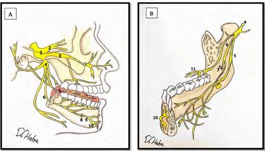
The interior alveolar nerve (IAN) is the largest branch of the posterior division of mandibular nerve, which is a branch of the fifth cranial nerve, the trigeminal nerve (Figure 1).[1,2] Injury to the IAN can result from pathology or from iatrogenic causes while performing oral-related treatments. The injury can lead to temporary or permanent neurosensory disturbance of the innervated sites by anesthesia, hypoesthesia and/or neurogenic discomfort.[3] In addition, this damage can be applied either directly to the nerve, or indirectly through compression. Dental procedures that may result in IAN injury include: nerve block injections,[4] oral surgery in the posterior mandible,[5] dental implant placement,[6,7] orthodontic treatment[8] and endodontic treatment.[9,10]

This paper aims to review the iatrogenic indirect compression phenomenon of the IAN following surgical placement of a dental implant and to report a clinical case of an indirectly compressed IAN with temporary loss of sensation following placement of a dental implant in the posterior mandible.
Case Report
Patient Information
A healthy, mentally stable 27-year-old female presented to our dental implant clinic for replacement of missing tooth #31 (lower right second molar), which was extracted five weeks previously.
Clinical Findings
The clinical examination revealed good vertical and mesio-distal prosthetic space, absence of periodontal pockets around adjacent tooth #30 (>3mm), and a wide zone of keratinized mucosa buccal to the missing tooth site (>5mm). The patient had normal mouth opening, with no signs or symptoms of parafunctional habits. She had good oral hygiene, with a plaque index=0 and gingival index=1.
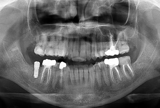
Three-dimensional radiograph cone beam computed tomography (CBCT) revealed sufficient bone volume for surgical placement of dental implant with average of 15 mm height of bone superior to the inferior alveolar canal and an average of 10 mm width of alveolar ridge (Figure 2). A prosthetically driven implant, sized 4 mm x 10 mm was planned considering safety margin of >3 mm of bone superior to inferior alveolar nerve canal.
Timeline
On the day of surgery, the patient signed surgical consent forms. Local anesthesia infiltration buccal and lingual to the site was administered (2% lidocaine with 1:80000 epinephrine). Aided by an acrylic prosthetic stent, a 3i bonelevel implant size 4 mm x 10 mm, with an average distance of 5 mm superior to the inferior alveolar canal was inserted.
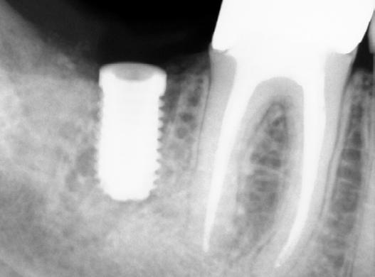
Pre-, peri- and postoperative periapical radiographs were taken for implant alignment and safe drilling (Figures 3-5). An insertion torque of 40 Ncm with good primary stability was achieved.
One-stage implant surgery was performed, with a healing abutment fixed over the fixture, and the flap was sutured using 4/0 Vicryl with multiple-interrupted suturing technique.
Postoperative instructions were given to the patient, along with prescribed analgesics for three days (ibuprofen 400mg; TID), antibiotic for seven days (Augmentin 625mg amoxicillin/Clavulanic acid), TID and chlorhexidine mouthwash 0.2% (10ml, BID) for 14 days. The patient left the clinic in good condition without any pain or bleeding.
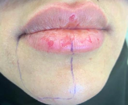
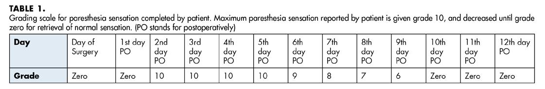
On the second day following surgery, the patient reported numbness and a burning sensation in her ipsilateral lower lip, part of her chin and the buccal mucosa. The numbness and burning continued until it disappeared totally by day 10 postoperatively (Figure 6) (Table 1).
Paresthesia grading was reported and tracked by asking the patient to grade on a scale of zero to 10, where 10 is the maximum paresthesia sensation (second day following surgery) and zero is complete return of sensation (day 11 postoperatively) (Table 1).
Diagnostic Assessment
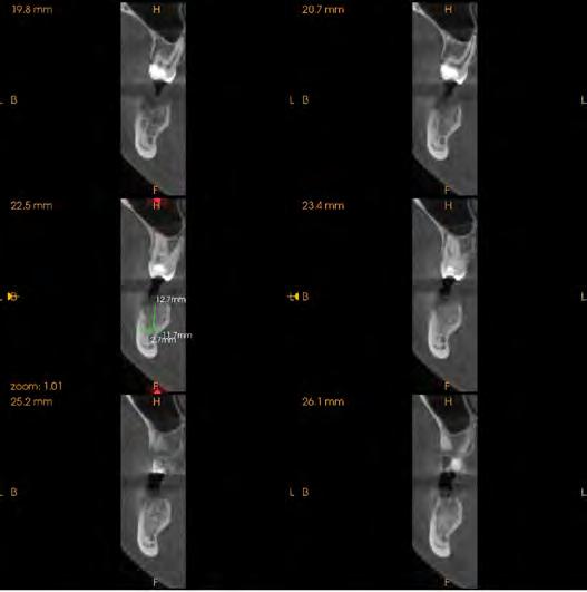
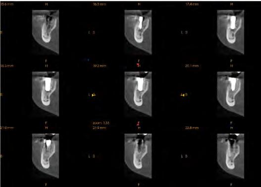
A CBCT was taken postoperatively to assure that there was no direct trauma to the nerve. A distance between the fixture and the inferior alveolar canal was confirmed to be a minimum of 5 mm, with no direct trauma to the inferior alveolar tissues (Figure 7).
A periodontal probe UNC-15 (sharp-right angled) was applied to lower lip, chin and oral mucosa without breaching, followed by asking the patient to compare sensations to that produced by a blunt region of the probe. Areas of sensation disturbance in the skin and lip were marked by a medical skin marker (Figure 6).
Therapeutic Intervention
In addition to the mentioned routine postoperative instructions, medications and intervention were restricted to follow-up and careful communication with the patient with reporting signs and symptoms.
Follow-up and Outcomes
The implant was loaded successfully with a full ceramic prosthetic crown three months after surgery. The patient was followed for six months after loading with no abnormality in sensation reported.
Discussion
One of the complications faced by the clinician and the patient following implant placement in posterior mandible is direct or indirect injury to the IAN. The injury leads to sensory disturbances that provide an unpleasant experience for both the doctor and the patient.[11]
The incidence of altered sensation was reported in literature as short-term (10 days after implant placement) in 13% of cases, while long-term (one year after implant placement) is reported in 3% of cases.[12] For the patients who initially reported altered sensation, 80% of them would return to normal sensation within six months after surgery, and 91% of them would return to normal sensation one year after surgery.[12]
As a protocol in our dental center, cone beam computed tomography (CBCT) is the diagnostic image used for preplanning of our case (Figure 2). CBCT was used again to confirm the absence of direct trauma of the IAN after the patient started feeling the symptoms (Figure 7). As compared to a panoramic radiograph for planning a surgical procedure, Nathalia et al., in a recent systematic review and meta-analysis, found that both a panoramic radiograph and CBCT offer similar ability to reduce the risk of temporary paresthesia of the IAN after oral surgery, specifically, third-molar surgery.[13]
The patient was anesthetized using 1.8 ml local infiltration of 2% lidocaine with 1:80000 epinephrine. Injections were given buccal and lingual to the surgical site, and the patient reported no pain during the surgical procedure. Some authors suggest using infiltration as an injection technique since it would allow a painful stimulus and enable the patient to warn the surgeon of close proximity to the IAN.[14-16] However, in a recent randomized controlled clinical trial comparing IAN block with infiltration, the study concluded that when using mandibular infiltration, and getting closer to the canal, the feeling of pain is not increased and it is, therefore, not recommended to use the presence of pain as a warning to avoid IAN injuries.[17]
It is recommended that after placement of posterior implants near the IAN canal, to check whether the patient experienced a return of normal sensation. This can be six hours postoperatively, after the local anesthesia effect wears off.[11] The presence of persistent numbness/paresthesia in the lower lip and chin on the same side would be the first sign that direct or indirect damage to IAN has occurred.
Indirect trauma to the IAN can result from development of a hematoma and edema[11] from drilling at the surgical site or by indirect compression from the implant body.[18]
Park et al., in 2012, reported two cases of indirect trauma by compression to the IAN. Their explanation for this phenomenon is, since the diameter of the drill is smaller than that of the dental implant, the size difference makes for a tight fit of implant and the bone hole to increase the initial stability of the implant. This will lead to compressing the cancellous bone.[18] In cases where the IAN courses close to the dental implant and is not protected by the nerve canal, it can also lead to indirect nerve injury.[18]
Implant surgical placement can be performed the same day as extraction to limit additional surgeries (immediate approach), four to eight weeks after extraction to allow for soft-tissue healing (early approach), or after >12 weeks from extraction to allow for soft- and hard-tissue healing (delayed approach).[19] In our present case, an early approach of implant placement was used, where the pa-
tient had extraction of her lower right second molar five weeks prior to implant surgery. The site was less mineralized on radiographs compared to the rest of her alveolar bone in the posterior mandible. This may give an additional explanation of how indirect compression and edema led to temporary paresthesia of the ipsilateral IAN. Based on this explanation, we recommend careful evaluation and clear communication with the patient about the possibility of temporary paresthesia of the IAN when an implant is placed in the posterior mandible following a recent extraction. However, it is currently impossible to suggest how long we should wait after extraction, prior to performing implant surgery, to avoid temporary paresthesia of the IAN.
In the literature, there are several tests used to objectively measure sensation disturbance; they all produced similar results.[11,20] In the reported case, we used the sharp blunt discrimination method (SBD), where a sharp right-angle dental probe (UNC #15) was applied to the lower lip, chin skin and oral mucosa area without breaching the tissue. This was followed by asking the patient to compare the sensation produced by the sharp end of the probe to that produced by a blunt region of the probe. The area of sensation disturbance in the skin and lip was marked by a medical skin marker (Figure 6).
Since the patient reported a dramatic improvement of symptoms day-by-day following the surgery, our intervention was restricted to follow-up, careful communication with the patient, and reporting signs and symptoms. This improvement may be explained by body adaptive mechanisms and by the local, gradual absorption of the possible hematoma that formed at the surgical site.
Generally, early management is of extreme importance, and the injury is more likely to be persistent when there is an increased duration between injury and intervention with the patient.[21] Furthermore, late diagnosis and ignorance of intervention may lead to development of tunnel syndrome, where an increased pressure on peripheral nerves might occur, followed by impaired neural microcirculation and focal demyelination.[22]
In some cases, intervention to treat the effects of trauma to the IAN may include medications (i.e., steroidal anti-inflammatory drugs, dexamethasone, tricyclic antidepressants, pregabalin, topical 5% lidocaine patches [Versatis], benzocaine topical local anesthesia [LA] cream, and LA with Botox injections), cognitive behavioral therapy, removal of the implant and IAN canal decompression.[18,23,24]
Evaluation of risk factors is important to avoid direct and indirect injury to the IAN. Risk factors can be classified into general, intraoperative and postoperative,[25] and may include: high patient expectations, pre-existing altered sensation, improper selection of site for implant placement or implant size, anatomical- and radiological-related factors, female patients, increased age, protrusion through the lingual or buccal plate, perforation of the IAN canal, extensive bleeding, inexperienced surgeons, recent extraction, less corticated canal, repeated IAN blocks, the interval between the time of injury, and diagnosis and treatment.[11,18,25-27] Clear communication between dentist and patient, careful evaluation of risk factors and planned treatment are critical for successful treatment and avoidance of direct and indirect injury to the IAN during implant placement.[12]
Conclusion
In general, if there is no infection, no inflammation of clinical significance, and an absence of clinical and radiographic pathology, then a wait-and-see policy should be adopted. In the case at hand, the following points may be considered for a favorable outcome:
1. Monitoring radiographically on a monthly basis is recommended for six months.
2. The patient should be reassured that paresthesia is possible, but it is a resolvable and manageable clinical situation.
3. If paresthesia persists, alternate measures should be adopted, i.e., removal of the implant and/or referral to a neurologist.
Queries about this article can be sent to Dr. Alsalman at wesamcom@hotmail.com.
REFERENCES
1. Monkhouse S. Cranial Nerves: Functional Anatomy. Cambridge University Press; 1st edition (January 1, 2005). Pp 60-65.
2. Khalil H. A basic review on the inferior alveolar nerve block techniques. Anesth Essays Res [Internet] 2014;8(1):3–8. Available from: http://dx.doi.org/10.4103/0259-1162.128891.
3. Manfuso A, Pansini A, Tewfik K, Copelli C. Inferior alveolar nerve reconstruction in extensive mandibular resection: Technical notes. J Plast Reconstr Aesthet Surg [Internet] 2021;74(3):634–6. Available from: http://dx.doi.org/10.1016/j.bjps.2020.11.040.
4. Moon S, Lee S-J, Kim E, Lee C-Y. Hypoesthesia after IAN block anesthesia with lidocaine: management of mild to moderate nerve injury. Restor Dent Endod [Internet] 2012;37(4):232–5. Available from: http://dx.doi.org/10.5395/rde.2012.37.4.232.
5. Ali AS, Benton JA, Yates JM. Risk of inferior alveolar nerve injury with coronectomy vs surgical extraction of mandibular third molars-A comparison of two techniques and review of the literature. J Oral Rehabil [Internet] 2018;45(3):250–7. Available from: http://dx.doi.org/10.1111/joor.12589.
6. Lin M-H, Mau L-P, Cochran DL, Shieh Y-S, Huang P-H, Huang R-Y. Risk assessment of inferior alveolar nerve injury for immediate implant placement in the posterior mandible: a virtual implant placement study. J Dent [Internet] 2014;42(3):263–70. Available from: http://dx.doi.org/10.1016/j.jdent.2013.12.014.
7. Juodzbalys G, Wang H-L, Sabalys G, Sidlauskas A, Galindo-Moreno P. Inferior alveolar nerve injury associated with implant surgery. Clin Oral Implants Res [Internet] 2013;24(2):183–90. Available from: http://dx.doi.org/10.1111/j.1600-0501.2011.02314.x.
8. AlAli AM, AlAnzi TH. Inferior alveolar nerve damage secondary to orthodontic treatment: A systematic scoping review. Int J Risk Saf Med [Internet] 2021;32(3):175–91. Available from: http://dx.doi.org/10.3233/JRS-200098.
9. Bianchi B, Ferri A, Varazzani A, Bergonzani M, Sesenna E. Microsurgical decompression of inferior alveolar nerve after endodontic treatment complications. J Craniofac Surg [Internet] 2017;28(5):1365–8. Available from: http://dx.doi.org/10.1097/SCS.0000000000003672.
10. Biglioli F, Kutanovaite O, Autelitano L, Lozza A, Moneghini L, Bulfamante G, et al. Surgical treatment of painful inferior alveolar nerve injuries following endodontic treatment: a consecutive case series of seven patients. Oral Maxillofac Surg [Internet] 2017;21(4):461–6. Available from: http://dx.doi.org/10.1007/s10006-017-0656-8.
11. Shavit I, Juodzbalys G. Inferior alveolar nerve injuries following implant placement - importance of early diagnosis and treatment: a systematic review. J Oral Maxillofac Res [Internet] 2014;5(4):e2. Available from: http://dx.doi.org/10.5037/jomr.2014.5402.
12. Lin C-S, Wu S-Y, Huang H-Y, Lai Y-L. Systematic review and meta-analysis on incidence of altered sensation of mandibular implant surgery. PLoS One [Internet] 2016;11(4):e0154082. Available from: http://dx.doi.org/10.1371/journal.pone.0154082.
13. Del Lhano NC, Ribeiro RA, Martins CC, Assis NMSP, Devito KL. Panoramic versus CBCT used to reduce inferior alveolar nerve paresthesia after third molar extractions: a systematic review and meta-analysis. Dentomaxillofac Radiol [Internet] 2020;49(4):20190265. Available from: http://dx.doi.org/10.1259/dmfr.20190265.
14. Heller AA, Shankland WE. Alternative to the inferior alveolar nerve block anesthesia when placing mandibular dental implants posterior to the mental foramen. J Oral Implantol 2001;27:127–33.
15. Alhassani AA, Alghamdi AS. Inferior alveolar nerve injury in implant dentistry: diagnosis, causes, prevention, and management. J Oral Implantol 2010;36:401–7.
16. Scher ELC. Risk management when operating in the posterior mandible. Implant Dent [Internet] 2002;11(1):67–72. Available from: http://dx.doi.org/10.1097/00008505-200201000-00016.
17. Garcia-Blanco M, Gualtieri A-F, Puia S-A. A randomized controlled trial comparing nerve block and mandibular infiltration techniques in posterior mandible implant surgeries. J Clin Exp Dent [Internet] 2018;10(10):e1003–10. Available from: http://dx.doi.org/10.4317/jced.54330.
18. Park Y-T, Kim S-G, Moon S-Y. Indirect compressive injury to the inferior alveolar nerve caused by dental implant placement. J Oral Maxillofac Surg [Internet] 2012;70(4):e258-9. Available from: http://dx.doi.org/10.1016/j.joms.2011.11.025.
19. Hämmerle CHF, Chen ST, Wilson TG Jr. Consensus statements and recommended clinical procedures regarding the placement of implants in extraction sockets. Int J Oral Maxillofac Implants 2004;19 Suppl:26–8.
20. Khawaja N, Renton T. Case studies on implant removal influencing the resolution of inferior alveolar nerve injury. Br Dent J [Internet] 2009;206(7):365–70. Available from: http://dx.doi. org/10.1038/sj.bdj.2009.258.
21. Renton T, Yilmaz Z. Managing iatrogenic trigeminal nerve injury: a case series and review of the literature. Int J Oral Maxillofac Surg [Internet] 2012;41(5):629–37. Available from: http:// dx.doi.org/10.1016/j.ijom.2011.11.002.
22. Schmid AB, Bland JDP, Bhat MA, Bennett DLH. The relationship of nerve fibre pathology to sensory function in entrapment neuropathy. Brain [Internet] 2014;137(12):3186–99. Available from: http://dx.doi.org/10.1093/brain/awu288.
23. Khawaja N, Renton T. Case studies on implant removal influencing the resolution of inferior alveolar nerve injury. Br Dent J [Internet] 2009;206(7):365–70. Available from: http://dx.doi. org/10.1038/sj.bdj.2009.258.
24. Renton T, Dawood A, Shah A, Searson L, Yilmaz Z. Post-implant neuropathy of the trigeminal nerve. A case series. Br Dent J [Internet] 2012;212(11):E17. Available from: http://dx.doi. org/10.1038/sj.bdj.2012.497.
25. Juodzbalys G, Wang H-L, Sabalys G. Injury of the inferior alveolar nerve during implant placement: A literature review. J Oral Maxillofac Res [Internet] 2011;2(1):e1. Available from: http://dx.doi.org/10.5037/jomr.2011.2101.
26. Renton T, Janjua H, Gallagher JE, Dalgleish M, Yilmaz Z. UK dentists’ experience of iatrogenic trigeminal nerve injuries in relation to routine dental procedures: why, when and how often? Br Dent J [Internet] 2013;214(12):633–42. Available from: http://dx.doi.org/10.1038/sj.bdj.2013.583.
27. Juodzbalys G, Wang H-L, Sabalys G, Sidlauskas A, Galindo-Moreno P. Inferior alveolar nerve injury associated with implant surgery. Clin Oral Implants Res [Internet] 2013;24(2):183–90. Available from: http://dx.doi.org/10.1111/j.1600-0501.2011.02314.x.

Wesam Talal Alsalman, D.D.S., M.D.S., Haifa Alharbi, B.D.S., and Marwan Abood, B.D.S., J.B.P., are members of the Department of Periodontics and Dental Implantology, Qassim Regional Dental Center, Buraydah, Saudi Arabia.
Saad Mulhi Alharbi, B.D.S., S.B.E., and Heba Alkhair Allah, B.D.S., are members of the Department of Endodontics, Qassim Regional Dental Center, Buraydah, Saudi Arabia.
Abeer Hani Albattah, D.D.S., M.D.S., is a member of the Department of Pediatric Dentistry, Qassim Regional Dental Center, Buraydah, Saudi, Arabia.
Khalid Almas, B.D.S., M.Sc., FDRCS, FRACDS, DDPH, RCS, is a member of preventive dental science, Faculty of Dentistry, Imam Abdulrahman Bin Faisal University, Dammam, Saudi Arabia.