9 minute read
Peripheral Ameloblastoma


Peripheral Ameloblastoma
Case Report and Literature Review
Anna Cappell Cooper, B.A.; Daria Vasilyeva, D.D.S.; Khanh Trinh, D.M.D.; Elizabeth Philipone, D.M.D.
ABSTRACT
Peripheral ameloblastoma is a benign, painless and slow-growing odontogenic tumor, which commonly affects gingival soft tissues or edentulous alveolar areas. Peripheral ameloblastoma is typically found during the 5th to 7th decades of life, with a mean patient age of 52 years and a male predilection. Histopathology features are consistent with those of conventional ameloblastoma. The treatment of choice is conservative surgical excision with minimal disease-free margins and appropriate follow-up.
Peripheral/extraosseous ameloblastoma is a rare and benign odontogenic tumor that affects gingival soft tissues or edentulous areas.[1] The term was first introduced in 2011 by Kuru; and fewer than 200 cases had been reported before 2014. According to the most recent World Health Organization (WHO) classification in 2022, there are four subtypes of ameloblastoma: solid/multicystic; unicystic; desmoplastic; and peripheral/extraosseous.[2] It is believed to be the rarest subtype of ameloblastomas, with a prevalence of 1% to 10%.
Patients typically present between the ages of 40 and 60; and the lesion exhibits a male predilection. Clinically, it is less aggressive than conventional ameloblastomas, typically indolent and painless, although it may rarely cause mild erosion of nearby cortical bone.
Herein we present a case of peripheral ameloblastoma in the right retromolar pad in a 59-year-old male patient.
Case Report
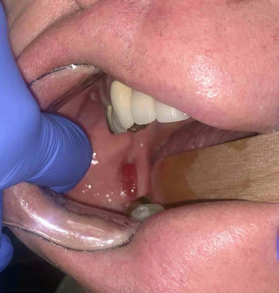
A 59-year-old male in apparent good health was referred to an oral and maxillofacial surgeon by his dentist for evaluation of a lesion of the right retromolar pad. The patient was not aware of the lesion prior to the visit and did not indicate experiencing any associated pain or bleeding. Review of his medical history was noncontributory. Intraoral examination revealed a smooth, well-demarcated red swelling of the soft tissue distal to right mandibular molars (Figure 1). No radiographic changes were noted.
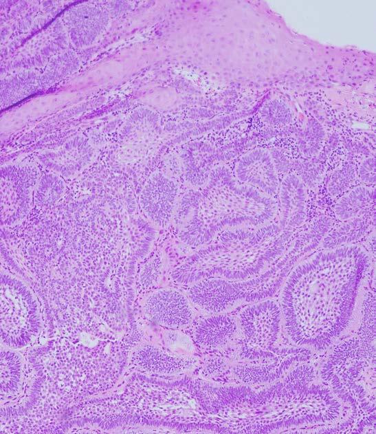
A biopsy of the soft-tissue lesion was performed to establish the diagnosis. On microscopic examination, the lesion consisted of abundant islands and cords of odontogenic epithelium in continuity with overlying surface epithelium. The peripheral cells of these islands and cords were tall and columnar, with reversely polarized hyperchromatic nuclei. The central cores were composed of loosely arranged angular cells resembling stellate reticulum, as well as large polygonal squamous cells with abundant eosinophilic cytoplasm (Figure 2).
Discussion
Peripheral ameloblastoma is a rare, benign, epithelial odontogenic tumor that affects soft tissues of the gingiva or edentulous alveolar areas.[3] In the most recent WHO classification, ameloblastoma was classified into: conventional (formerly, solid/multicystic); unicystic; peripheral (extraosseous); and a newly added adenoid ameloblastoma.[4]
Peripheral ameloblastomas account for 1% to 10% of all ameloblastomas and are nearly twice as common in males as in females.[3] Lesions more commonly occur in the mandible, particularly in the soft tissues of the retromolar pad and on the lingual aspect of the gingiva, with a maxilla-tomandible occurrence ratio of 1:2.4. When present in the maxilla, the lesions commonly occur on the soft, palatal tissue of the maxillary tuberosity.
Although most patients present between the ages of 40 and 60, the age of presentation can range from 9 to 92 years.[4] All types of ameloblastomas are more prevalent in Asian and African-Caribbean populations, with individuals of African descent typically presenting at a younger age.[5]
Although the etiology of peripheral ameloblastoma is not certain, some probable sources of the lesion include remnants of the dental lamina (glands of Serres), odontogenic remnants of the vestibular lamina, and pluripotent cells in the basal cell layer of the mucosal epithelium.[6] Newer data point to gene mutations as pathogenic factors in the development of all types of ameloblastomas. Genetic mutations that lead to dysregulation of the MAP kinase pathway, including those in BRAF and somatic mutations in NRAS and SMO, have been detected in cases of peripheral ameloblastoma. The most common mutation among them is BRAF p.V600E7.
Clinically, peripheral ameloblastoma appears as a solid or cystic lesion with a surface that is either smooth, granular or papillary. The tumor rarely exceeds 4 cm in size and is asymptomatic.[1] Given its extraosseous nature, most cases do not present with radiographic changes. Some, however, exhibit peripheral and superficial bone erosion due to pressure resorption, known as the cupping effect or saucerization.[3] In order to make a diagnosis of peripheral ameloblastoma, radiographs must be taken to rule out intraosseous ameloblastoma that perforated cortical bone, and histologic evaluation is necessary to establish the diagnosis.[8]

Similar to conventional ameloblastoma, peripheral ameloblastomas most often fall into follicular and plexiform microscopic patterns,[1] with either classic and/or acanthomatous cell morphology.[1,9] Microscopically, follicular ameloblastoma consists of islands of odontogenic epithelium within a fibrous connective tissue stroma. The peripheral cells of the islands are usually cubic or columnar in shape, exhibiting reversed polarity and hyperchromatic nuclei, while the cores of the islands consist of loose angular cells resembling stellate reticulum (classic cellular morphology), or large polygonal squamous cells with abundant eosinophilic cytoplasm (acanthomatous cellular morphology).[3]
The second most common plexiform pattern of ameloblastoma is characterized by interlacing and anastomosing strands of epithelial cells with similar palisading and nuclear polarization of basal cells. Fifty percent of peripheral ameloblastomas show continuity with the overlying epithelium.[8]
Peripheral ameloblastoma may raise histologic suspicion for intraoral basal cell carcinoma (IOBCC). Albeit extremely rare, IOBCC has been reported on the gingiva. Therefore, when diagnosing peripheral ameloblastoma, it is important to be aware of some histologic features that are specific to IOBCC, such as its origination from surface epithelium, presence of scattered mitotic figures and apoptotic cells, the presence of mucoid ground substance and infiltration of the tumor throughout the connective tissue.[10]
More recently, immunohistochemical examination has been used to aid in the diagnosis and to differentiate between peripheral ameloblastoma and BCC, particularly the expression of BER-EP4 in BCC4. Peripheral ameloblastomas show positive reactivity to immunostaining of amelogenin, calretinin and cytokeratins 5, 14 and 19, with CK13 preferentially expressed in stellate reticulum-like cells, CK14 in peripheral cells and CK19 in all cells.[1]
The benign, slow-growing nature of peripheral ameloblastoma, coupled with a high recurrence rate, render the issue of preferred treatment controversial.[11] The current treatment of choice is conservative supraperiosteal surgical excision with disease-free margins.[6] The extent of the excision and decision to include healthy margins is dictated by histopathological features, tumor location, size and patient factors, such as age and one’s individual anatomy.[5]
Surgery can be supplemented with curettage, cryotherapy, electrocautery or tissue fixation, which does not seem to play a role in potential recurrence.[12] Long-term follow-up of at least 10 years is necessary, given a relatively high recurrence rate of up to 19%, although the recurrence is mostly attributed to incomplete excision rather than disease behavior.[1]

Conclusion
The gingiva is a site that can be affected by a variety of oral pathologies, and chairside diagnosis presents a challenge for the clinician. A biopsy is often necessary in rendering a definitive diagnosis for gingival lesions.
The authors of this paper report no conflicts of interest nor did their study receive commercial funding. Queries about this article can be sent to Ms. Cooper at Ac4746@cumc.columbia.edu.
REFERENCES
1. Vezhavendhan N, Vidyalakshmi S, Muthukumaran R, Santhadevy A, Sivaramakrishnan M, Gayathri C. Peripheral ameloblastoma of the gingiva. Autopsy and Case Reports 2020;10(1). https://doi.org/10.4322/acr.2019.127.
2. Vered M, Wright JM. Update from the 5th Edition of the World Health Organization Classification of Head and Neck Tumors: Odontogenic and Maxillofacial Bone Tumours. Head and Neck Pathol 2022;16:63–75. https://doi.org/10.1007/s12105-021-01404-7.
3. Assis EM, Gomes HE, Sousa FE, Brener S, Leal RM, Alencar Souza PE, Rebello Horta MC. Recurrent peripheral ameloblastoma in an elderly patient: A case report. Gerodontology 2019;36(1):78–81. https://doi.org/10.1111/ger.12377.
4. Kaneko T, Nakamura S, Kawano R, Horie N, Shimoyama T. Peripheral ameloblastoma of the mandible: A case report. Journal of Oral and Maxillofacial Surgery, Medicine, and Patholog 2016;28(6):565–568. https://doi.org/10.1016/j.ajoms.2016.01.008.
5. Anpalagan A, Tzortzis A, Twigg J, Wotherspoon R, Chengot P, Kanatas A. Current practice in the management of peripheral ameloblastoma: a structured review. The British Journal Oral & Maxillofacial Surgery 2021;59(1): e1–e8. https://doi.org/10.1016/j.bjoms.2020.08.084.
6. Beena VT, Choudhary K, Heera R, Rajeev R, Sivakumar R, Vidhyadharan K. Peripheral ameloblastoma: A case report and review of literature. Case reports in dentistry 2012. https:// doi.org/10.1155/2012/571509.
7. Guimarães LM, Coura BP, Gomez RS, Gomes CC. The molecular pathology of odontogenic tumors: Expanding the spectrum of MAPK pathway driven tumors. Frontiers in Oral Health, 2 2021, https://doi.org/10.3389/froh.2021.740788.
8. Philipsen HP, Reichart PA, Nikai H, Takata T, Kudo Y. Peripheral ameloblastoma: biological profile based on 160 cases from the literature. Oral Oncology 2001;37(1):17–27. https://doi. org/10.1016/s1368-8375(00)00064-6.
9. Hertog D, Bloemena E, Aartman IH, van-der-Waal I. Histopathology of ameloblastoma of the jaws; some critical observations based on a 40-year single institution experience. Medicina Oral, Patologia Oral y Cirugia Bucal 2012;17(1): e76–e82. https://doi.org/10.4317/medoral.18006.
10. Woods TR, Cohen DM, Islam MN, Kratochvil FJ, Stewart J C, Reeder SL, Bhattacharyya I. Intraoral basal cell carcinoma, a rare neoplasm: report of three new cases with literature review. Head and Neck Pathology 2014;8(3):339–348. https://doi.org/10.1007/s12105-013-0505-5.
11. Cadavid AM, Araujo JP, Coutinho-Camillo CM, Bologna S, Junior CA, Lourenço SV. Ameloblastomas: Current aspects of the new WHO classification in an analysis of 136 cases. Surgical and Experimental Pathology 2019;2(17). https://doi.org/10.1186/s42047-019-0041-z.
12. Borrello R, Bettio E, Bacci C, Valente M, Sivolella S, Mazzoleni S, Berengo M. (2016). A conservative approach to a peripheral ameloblastoma. Case Reports in Dentistry 2016, 8254571. https://doi.org/10.1155/2016/8254571.
Anna Cappell Cooper, B.A., is a dental student, Class of 2024, College of Dental Medicine, Columbia University Medical Center, New York, NY.
Daria Vasilyeva, D.D.S., is a resident, Division of Oral and Maxillofacial Pathology, Columbia University Medical Center, New York, NY.
Khanh Trinh, D.M.D., is a resident, Division of Oral and Maxillofacial Pathology, Columbia University Medical Center, New York, NY.
Elizabeth Philipone, D.M.D., is associate professor, Division of Oral and Maxillofacial Pathology, Columbia University Medical Center, New York, NY.


