
9 minute read
Mouth Ulcers in Children


Temitope T. Omolehinwa, B.D.S., DScD; Eric T. Stoopler, D.M.D., FDSRCS, FDSRCPS
ABSTRACT
In this article, we discuss mouth ulcers, which are common in the pediatric population, though rendering a specific diagnosis may present a challenge. Evaluation of patients with mouth ulcers includes obtaining a thorough medical history and conducting an appropriate physical examination. Establishing a differential diagnosis will aid in determining if additional diagnostic studies are warranted, as well as initiation of appropriate management strategies. This article reviews the pertinent clinical aspects of mouth ulcers in children in order to provide optimal patient care.
A mouth ulcer can be defined as any open sore caused by a breach in the epithelial lining of the oral mucous membrane(s);[1] it may, occasionally, involve tissues around the mouth. In children, this can be concerning due to possible decreased nutritional intake[2] associated with discomfort and pain related to the ulcers. Mouth ulcers may be of infectious or non-infectious origins. Recurrent aphthous ulcers and traumatic ulcers are examples of two commonly reported non-infectious conditions that cause oral ulcers in children, with a prevalence of 0.9% to 10.8% and 0.09% to 22.15%, respectively.[3] Herpes simplex virus (HSV) infection, also commonly reported in children and caused by a DNA virus, has a prevalence of 4.52% to 9.28%.[4]
Most mouth ulcers have similar clinical appearances, making diagnosis challenging for the untrained eye.[5] A systematic approach, which includes a thorough medical history, review of systems, careful and detailed physical examination and, possibly, biopsy, as discussed in detail below, are important in arriving at an accurate diagnosis.
Evaluation of Mouth Ulcer(s) in Pediatric Population
Medical History
The role of a detailed history, including history of present illness, cannot be overemphasized in establishing the etiology of mouth ulcers. One of the factors to consider is age of onset of these ulcers. For example, primary herpetic gingivostomatitis is common between the ages of 6 months to 5 years, while recurrent herpetic lesions are found in children older than age 5. On the other hand, recurrent aphthous ulcers have an onset of between 10 to 19 years of age.[5,6]
Other important specific questions to ask patients/caregivers are listed below (adapted from Stoopler ET 2014).[7,8]
• Is this the first time you are having ulcers, or have you had ulcers like these before?
• How long has/have the ulcer(s) been present in your mouth?
• Did the ulcers appear suddenly, or did you notice them over time?
• Have you had any ulcers in the past? If you have had ulcers in the past, how long did each episode last? Was there a tingling sensation before onset of lesions?
• Is anyone around you sick—cold, rashes, other contagious illnesses?
• Any history of trauma in the mouth or around the mouth?

Figure 1. Differential diagnosis of pediatric ulcers.10,15
Review of Systems
Positive and negative responses to review-of-system questions will provide the clinician with information regarding the child’s overall well-being and may provide information suggestive of a systemic disorder as a cause of mouth ulcers. Systemic conditions are likely to present with signs and symptoms in other parts of the body. For example, patients with hand, foot and mouth disease have lesions on their hands and feet in addition to the mouth, while patients with Behcet’s disease are likely to present with ocular and genital lesions.
Questions to ask on review of systems include:
• Was there any generalized body pain, weakness, fever, diarrhea, vomiting, etc., prior to ulcer onset?
• Do you have any problems with swallowing?
• Is there any weight loss, reduced urine production or unusually dry diapers in infants?
• Do you have ulcers or lesions anywhere else on your body— skin, eyes, genital or nose?
• What type of toothpaste do you use? Does it contain sodium lauryl sulfate (SLS)? Do you use cinnamon-flavored mouth products (toothpaste, mouthwash), gums or candies? Do you eat a lot of food flavored with cinnamon? Do you use any tooth-whitening products?
Physical Examination
It is important to conduct a general physical examination in addition to a focused clinical examination of both extraoral and intraoral anatomical structures. The list below is a guide to detailed physical examination in a child with mouth ulcers.
1. Record vital signs and check for constitutional symptoms, including fevers and signs of dehydration.
2. Thorough head and neck exam—check for lymphadenopathy, lesions on face, scalp, etc.
3. Check for number of ulcers in the mouth—single or multiple?
4. Are ulcers on keratinized surfaces (gingiva, dorsum of the tongue), non-keratinized surface (buccal mucosa, labial mucosa, floor of the mouth, ventral tongue, soft palate) or both?
5. What is the size (pinpoint, <1cm, >1cm) and shape (well-defined/not well-defined margins, circumscribed, etc.) of each lesion?
6. Are the ulcers deep or shallow?
7. Any surrounding discoloration around the lesions—none, red/inflamed, whitish/keratinized?
8. Any other visible lesions outside of the oral cavity—perioral, skin, eyes, scalp, nose?
Further Investigation
This will be based on pertinent findings from the medical history and physical examination and is especially warranted if the history, review-of-systems responses and physical examination do not point to any specific diagnosis or if the clinician is unsure what the diagnosis is. It may include one or a combination of the following:
1. Viral testing if viral etiology is suspected [viral culture, Tzanck smear, direct fluorescence assay (DFA), polymerase chain reaction (PCR)]. This is especially important in non-specific ulcers that appear to have a viral origin. They usually will present with an erythematous base. Viral testing can be carried out on ulcers that are persistent or of unknown duration. DFA and PCR are the tests of choice for fast and reliable results. This is especially accurate in testing for herpes simplex and varicella zoster (VZV) viruses.9
2. Laboratory studies (to rule out systemic causes, if suspected).
2i. Complete blood count (CBC) [anemia], iron, folate, B12, ferritin levels [vitamin, nutritional deficiencies]. Anemia has a prevalence of 43% especially in children below 4 years of age.[10,11] It is recommended to carry out a CBC when a systemic cause of oral ulcers is presumed.
2ii. Erythrocyte sedimentation rate (ESR), antinuclear antibody (ANA), C-reactive protein (CRP), rheumatoid factor [connective tissue disorders, such as lupus erythematosus]. Positive results are noted in patients with autoimmune or inflammatory conditions. It is important to refer patients with positive results to an oral medicine specialist, dermatologist (if skin lesions are present) or rheumatologist if an autoimmune process is suspected.
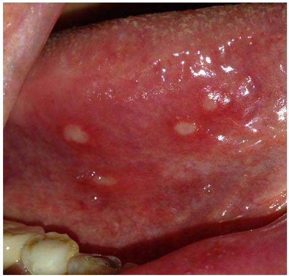
Figure 2. Multiple aphthous ulcers affecting right tongue. Note erythema surrounding each ulcer.
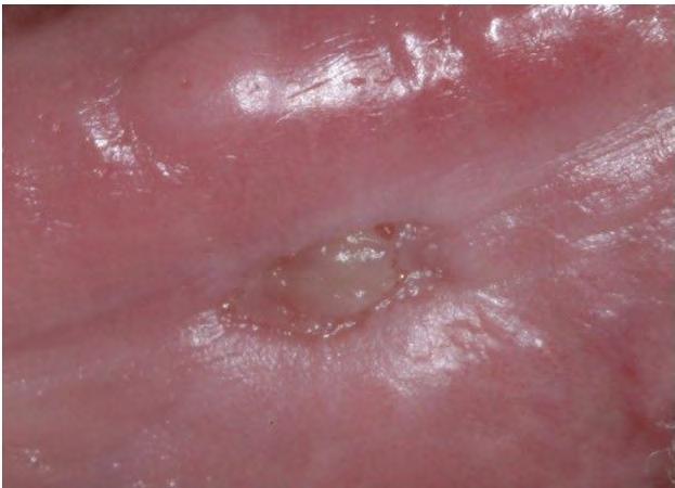
Figure 3. Traumatic ulcer affecting right tongue.
3. Specialist consult if level of care is beyond the scope of the practitioner. May include oral medicine specialist for all oral ulcers with nonspecific presentation; gastroenterologist when conditions like Coeliac or Crohn’s disease is suspected; ophthalmologist—especially in patients with bullous pemphigoid; dermatologist when skin lesions are present, etc. Organ-specific evaluation, such as laboratory assessment, imaging, and/or tissue analysis, may be conducted if indicated.
4. Biopsy for hematoxylin and eosin (H&E) staining and/or direct immunofluorescence (DIF) if: 1) the ulcer persists for longer than two to four weeks; 2) diagnosis is not clear based on clinical examination only; and/or 3) the patient is unresponsive to treatment.
Differential Diagnosis of Mouth Ulcers
Differential diagnosis of pediatric mouth ulcers can be grouped based on number of ulcers present (single v. multiple) (Figure 1).
Viral etiology for herpetic gingivostomatitis is herpes simplex virus, while herpangina, and hand, foot, and mouth disease are both caused by the Coxsackie virus.
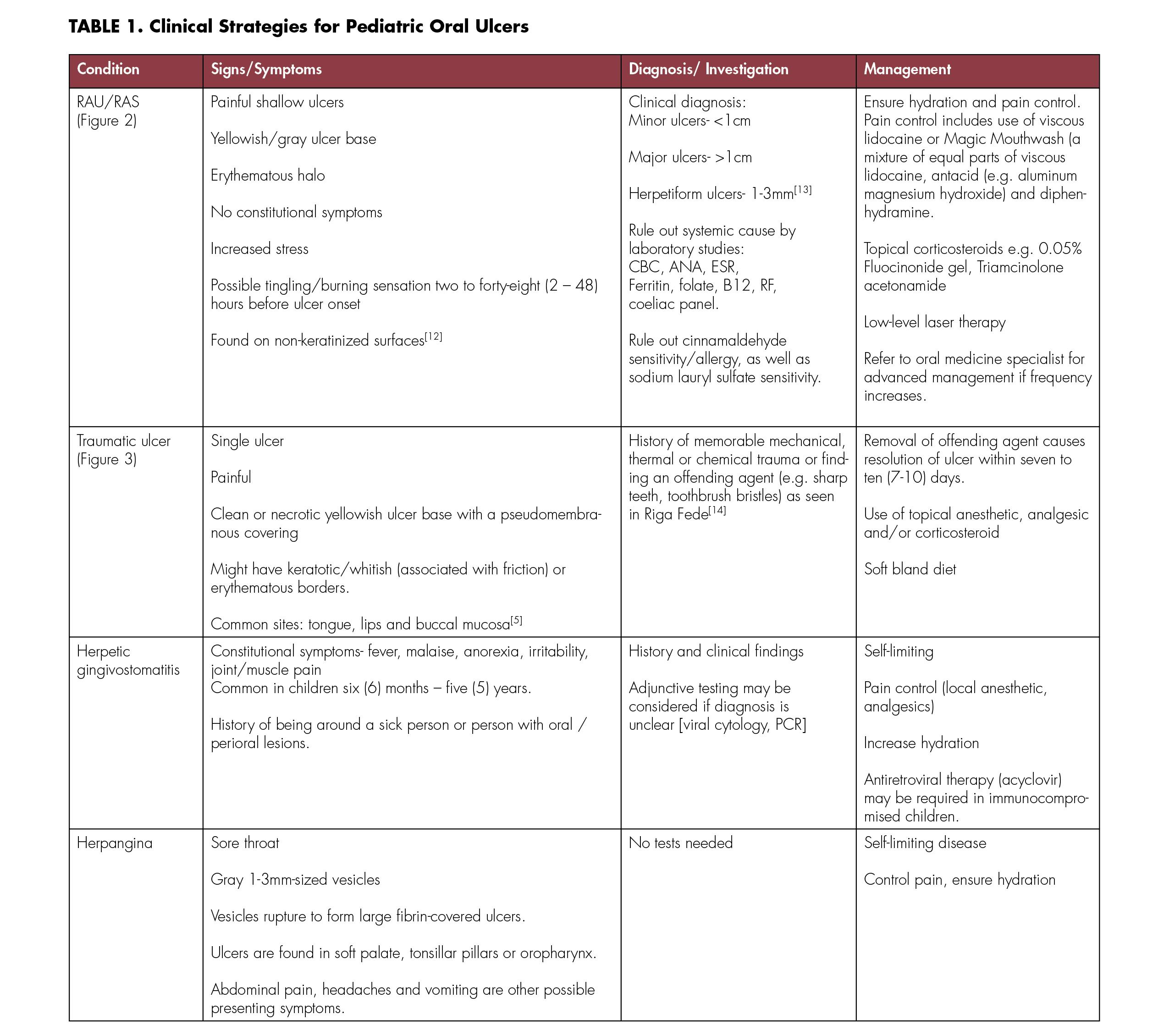
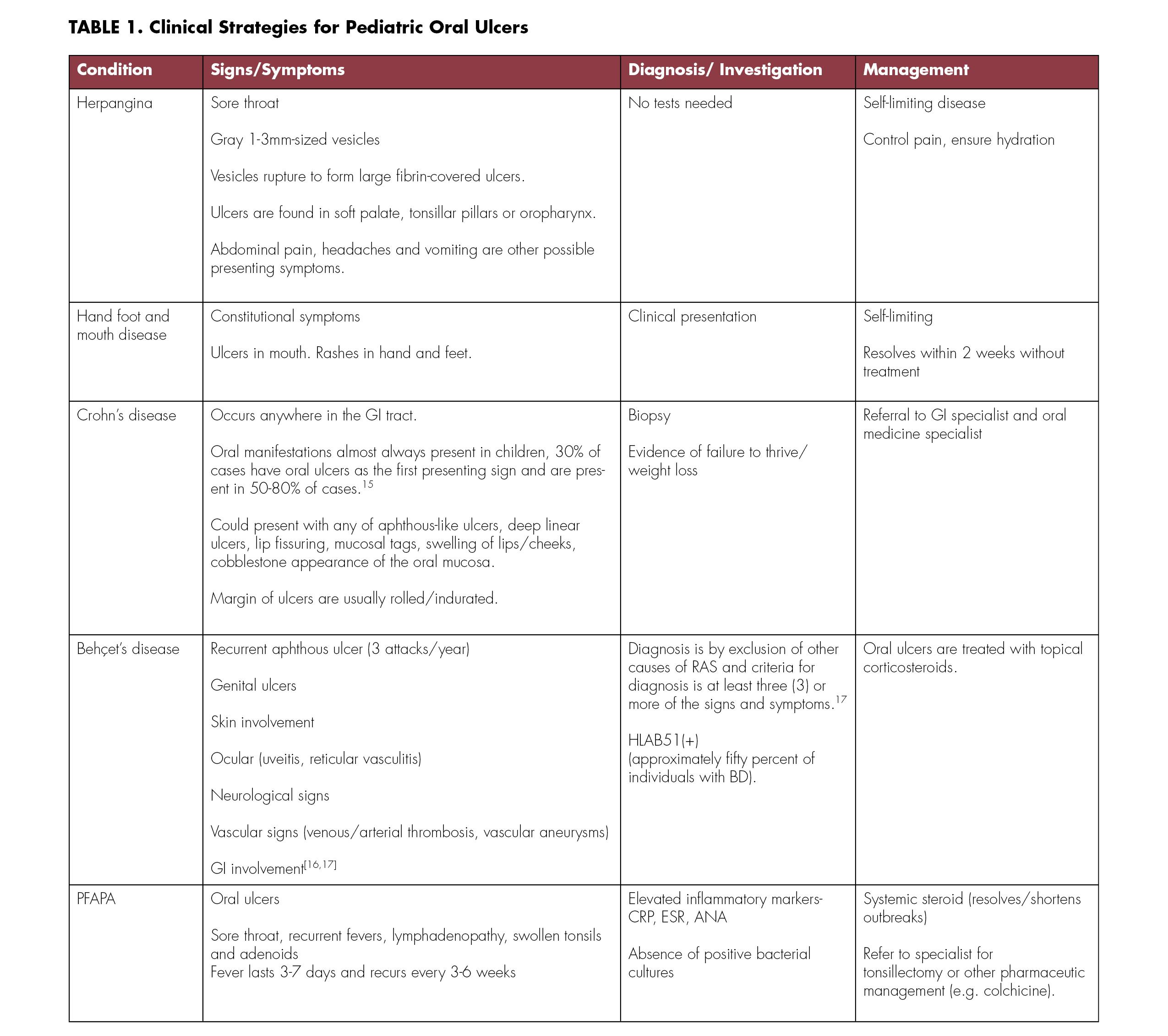
General Management
In managing mouth ulcers in children, it is important to ensure continuous hydration and maintenance of adequate nutrition. This can be achieved by oral intake or, occasionally, by intravenous infusions. Since pain is commonly associated with mouth ulcers, use of analgesics and/or topical anesthetic agents often provide temporary symptom relief. It is important to monitor the child to avoid additional mouth trauma when using topical anesthetics.
After specific diagnosis has been confirmed and treatment initiated (Table 1), follow up within four weeks is recommended, if possible, to ensure adequate response to treatment. Children with chronic conditions, such as inflammatory bowel disease, often require long-term follow up by the appropriate specialists. Pediatric patients with complete resolution of mouth ulcers attributed to a non-chronic etiology may be re-evaluated on an as-needed basis.
The authors declare they have no financial/economic/professional interest in their study. Queries about this article can be sent to Dr. Omolehinwa at omote@upenn.edu.
REFERENCES
1. Regezi JA, Sciubba JJ, Jordan RCK. Mucosal (surface) lesions. In: Oral Pathology: Clinical Pathologic Correlations. 3251 Riverport Lane, St. Louis, Missouri: Elsevier; 2017.
2. Lee Y, Wang T, Chen S, Lin H, Tsai M. Management of viral oral ulcers in children using Chinese herbal medicine: A report of two cases. Complement Ther Med 2017;32:61-65.
3. Rioboo-Crespo MR, Planells-del PP, Rioboo-García R. Oral mucosal diseases in children. Med Oral Patol Oral Cir Bucal (Oral Medicine and Oral Pathology). 2005;10:376-378.4.
4. Majorana A, Bardellini E, Flocchini P, Amadori F, Conti G, Campus G. Oral mucosal lesions in children from 0 to 12 years old: Ten years’ experience. OOOOE 2010;110(1):e13-e18.
5. Mortazavi H, Safi Y, Baharvand M, Rahmani S. Diagnostic features of common oral ulcerative lesions: An updated decision tree. Int J Dent 2016;7278925.
6. Le Doare K, Hullah E, Challacombe S, Menson E. Fifteen-minute consultation: A structured approach to the management of recurrent oral ulceration in a child. Arch Dis Child- Education & Practice Edition 2014;99(3):82-86.
7. Stoopler ET, AlZamel G. How to manage a pediatric patient with oral ulcers. J Canad Dent A 2014;80:e9.
8. Benoit S, Hamm H. Differential diagnosis of oral mucosal erosions and ulcers in children. Dermatologist 2015;66:258-258.9.
9. Chayavichitsilp P, Buckwalter V J, Krakowski AC, Friedlander SF. Herpes simplex. Pediatr Rev 2009;30(4):119-130.
10. Ezzati M, Lopez A, Rodgers A, Vander Hoorn S, Murray C. Selected major risk factors and global and regional burden of disease. Lancet 2002;360:1347-1360.
11. Siegel E, Stoltzfus R, Khatry S, LeClerq S, Katz J, Tielsch J. Epidemiology of anemia among 4 to 17-month-old children living in south central Nepal. Eur J Clin Nutr 2005;60:228.
12. Akintoye SO, Greenberg MS. Recurrent aphthous stomatitis. Dent Clin North Am 2014;58(2):281-297.
13. Preeti L, Magesh KT, Rajkumar K, Karthik R. Recurrent apthous stomatitis. J Oral and Maxillofac Pathol 2011;15(3):252-256.
14. Slayton RL. Treatment alternatives for sublingual traumatic ulceration (Riga-Fede disease). Pediatr Dent 2000;22(5):413-414.
15. Eckel A, Lee D, Deutsch G, Maxin A, Oda D. Oral manifestations as the first presenting sign of Crohn’s disease in a pediatric patient. J Clini Exp Dent 2017;9(7):e934-e938.
16. Johnson EF, Hawkins DM, Gifford LK, Smidt AC. Recurrent oral and genital ulcers in an infant: Neonatal presentation of pediatric Behçet disease. Pediatr Dermatol 2015;32(5):714-717.
17. Kone-Paut I. Bechet’s disease in children, an overview. Pediatr Rheumatol 2016;14(10).
Temitope T. Omolehinwa, B.D.S., DScD, is assistant professor of oral medicine, Department of Oral Medicine, Penn Dental Medicine, University of Pennsylvania, Philadelphia, PA.

Dr. Omolehinwa
Eric T. Stoopler, D.M.D., FDSRCS, FDSRCPS, is professor of oral medicine, Department of Oral Medicine, Penn Dental Medicine, University of Pennsylvania, Philadelphia, PA.

Dr. Stoopler


