
14 minute read
Arteriovenous Malformation Presenting as a Disfigured and Throbbing Face


Arteriovenous Malformation Presenting as a Disfigured and Throbbing Face
Case Report
Abrar Shamim, D.D.S., M.A.; Steven Halepas, D.M.D., M.D.; Alfonso Salcines, D.M.D., M.D.; Sidney B. Eisig, D.D.S.
ABSTRACT
Maxillofacial arteriovenous malformations (AVMs) are a rare condition in which arteries and veins are in continuity without a capillary bed in between, which poses a significant risk of severe bleeding. Intraosseous AVMs can lead to cortical bone expansion and facial deformity. Dental procedures may result in life-threatening bleeding risk if this condition is not recognized. This report describes a patient experiencing pain in the lower right side of the mouth, along with a taste of blood in the mornings and a tingling sensation in the chin and lower lip. The patient had a history of uncontrolled bleeding following a dental extraction at an adjacent site. Examination demonstrated a non-fluctuant facial enlargement, with pulsation on extraoral palpation of the mandible and buccal and lingual vestibules intraorally. CT angiogram confirmed an intraosseous AVM with multiple feeder vessels. The patient underwent preoperative embolization to minimize intraoperative bleeding followed by a hemimandibulectomy and a fibula free-flap reconstruction.
Arteriovenous malformations (AVMs) are a rare pathologic entity, with an estimated prevalence of less than 1/100,000 person years, of which approximately half occur in the head/neck region.[1,2] The AVM is a condition in which the arterial supply anastomoses directly with the venous system without a capillary bed separating them. In the absence of a high-resistance capillary network, blood flows briskly from the arterial to the venous system. The area of this lesion is, therefore, considered a high-risk area for potentially life-threatening hemorrhage. In the absence of a capillary network supplying the associated region, this can also result in reduced nutrient and oxygen supply to the affected area.
AVMs are most often congenita; however, they may be acquired in the setting of trauma or hormonal changes, such as pregnancy.[3] The most common presenting symptom is bleeding in three-quarters of patients, which may manifest as gingival bleeding, acute oral hemorrhage or iatrogenic causes (i.e., following dental extraction). These lesions may also cause disfigurement due to changes in soft tissue or bony architecture. While not a malignant entity, these lesions display a progressive nature. This is because the vein must adapt to high arterial pressures without the protection of a capillary bed, resulting in reactive muscular hyperplasia of the vessel walls, leading to ongoing arterialization and growth of the vessel.[4]
Dental procedures in the context of an underlying AVM can result in uncontrollable hemorrhage; therefore, these lesions must remain on the differential when appropriate physical exam findings and history of bleeding from the jaws are present.


Case Report
Clinical Presentation
A 23-year-old male patient presented to a dental emergency clinic with a complaint of pain and swelling on the right side of his face and neck that he noticed three to four months prior. The patient reports persistent pain from the right mandibular area, coupled with a tingling sensation around his right chin to corner of lip for the last few months. Additionally, he reports experiencing a taste of blood in his mouth upon waking on some mornings. The patient had a tooth extracted from the lower right side one year ago, complicated by bleeding from the extraction site which required hospital admission and a blood transfusion. The patient reports that he now notices a tooth adjacent to this area that is quite loose, and he thinks it requires extraction since it does not permit mastication on his right side. The patient’s medical, psychosocial and family histories are noncontributory.
Clinical Findings
On exam, a non-fluctuant swelling in the right extraoral buccal region with fullness of the right inferior border of the mandible and submandibular triangle is detected. The inferior border is palpable; the sublingual region is not swollen intraorally; and the patient has no difficulty breathing or swallowing secretions. The lateral cortex of the mandible demonstrates bony expansion palpable extraorally.
On intraoral examination, bony expansion is palpable in the right buccal vestibule, in addition to a Grade II mobility of the first molar (tooth #30) adjacent to the healed extraction site of a second molar (tooth #31). The gingiva in the area appears hyperemic. There is a palpable thrill, which can be felt extraorally in the site of bony expansion, in addition to intraorally in the right buccal vestibule. Extraoral auscultation with a stethoscope demonstrates an audible murmur/thrill.
Teeth #27-#30 are vital to electric pulp testing and hypothermia. Hematologic workup demonstrates no abnormalities.
Imaging
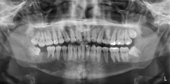
The panoramic radiograph (Figure 1) demonstrates an irregularly eroded inferior mandibular cortex compared to the left. There is a multilocular radiolucency of the right mandibular body superimposed on submandibular gland fossa region. There is unilateral widening of the right mandibular canal and an enlarged mental foramen. Tooth #30 demonstrates root resorption, although no clear odontogenic pathosis is apparent.
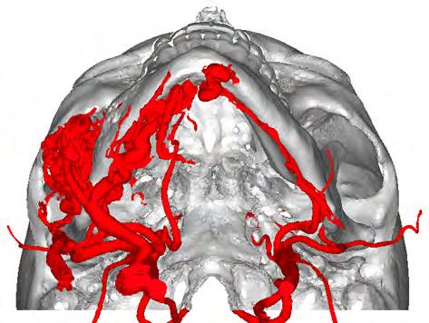
On CT angiogram, there is a facial arteriovenous malformation (AVM) centered within the right mandible and masseter muscle extending to portions of the right submandibular region/floor of mouth. There is a dilated vascular network within the mandible with filling defects suggestive of partial thrombosis in parts of the lesion (Figure 2). Feeding arteries are predominantly from the right external carotid artery (ECA), namely, the facial and lingual branches. There is also supply from left ECA branches. Draining veins are mostly through the internal and external jugular veins and pterygoid plexus.
Diagnosis
Based on the physical examination findings indicating a high-flow arteriovenous malformation and confirmation of the diagnosis through a CT angiogram, dental treatment should be avoided due to risk of severe and potentially fatal hemorrhage. This patient required hospital admission, followed by comprehensive treatment planning, which in this case included embolization of the feeding vessels and resection, with subsequent microvascular fibular free-flap reconstruction.
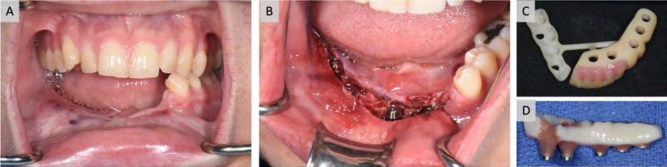
Five endosseous dental implants were placed at the time of the reconstructive surgery (observed in the postoperative panoramic radiograph, Figure 3). Our multidisciplinary team discouraged the option of embolization alone due to the presence of multiple arteries feeding the lesion, which significantly increases the likelihood of recurrence. The patient underwent Stage II implant exposure five months after his free-flap reconstruction and received an unloaded provisional prosthesis at that time (Figure 4).
Discussion
This case underscores the significance of recognizing an important diagnostic entity when confronted with facial swelling, a presentation that is frequently linked to odontogenic infections and abscess formation. Failure to identify vascular malformations within the jaws can have severe consequences, including life-threatening hemorrhage. Patients with a history of uncontrollable bleeding after dental extractions should be approached with caution. It is essential to exercise prudence before initiating any interventions. The clinician must meticulously assess potential factors contributing to uncontrolled bleeding from a tooth socket. Possible reasons for bleeding after an extraction include medications, hematologic abnormalities and vascular anomalies. Vascular anomalies are classified according to the blood vessels from which they are derived. This includes capillary, lymphatic, venous, arteriovenous or combined malformations when there are two or more blood vessel derivatives within a single lesion.[7] Any vascular malformation with an arterial component is a fast-flow lesion, whereas all others are classified as slow-flow. The pathogenesis of these lesions is unclear, although it is speculated that they are a result of embryologic disturbances in the late stages of differentiation and angiogenesis.[6,8] Efforts to identify genes and pathways associated with specific vascular lesions are underway.[9] AVMs most often become symptomatic at the age of 20 to 40; the average age of presentation is between 19 and 22 years old.[5] In the largest known case series, AVMs of the jaws occurred at a male:female ratio of approximately 4:3.2
However, others have found higher prevalence among females or no gender predilection.[1,6] Maxillofacial AVM is significantly more common in the mandible over the maxilla, but it may also occur simultaneously in both jaws. In the mandible, the most common location (95%) is the posterior body inferior to the molar dentition, which was the case for the patient we describe here.
These lesions commonly present with hemorrhage after various oral surgeries, although they may also bleed spontaneously.[10,11] In this case, the patient noted that on some mornings he woke up tasting blood in his mouth and could also sometimes see blood on his teeth, which resulted in significant distress. In addition to various forms of bleeding, less common presenting symptoms include toothache, loose teeth, facial swelling and pain.[2] There may also be mucous membrane or cutaneous pigment changes.[2]
The non-fluctuant nature of this patient’s apparent facial “swelling” was the first indicator that an odontogenic abscess is not the underlying pathology. Swelling related to dental infection presents as a fluctuant, soft, compressible or indurated swelling secondary to cellulitis or abscess formation. Odontogenic infections may demonstrate jelly-to board-like fluctuant swelling (depending on the stage of infection, i.e., edema, cellulitis, abscess), often with a clear odontogenic offender. Additionally, a nonvital tooth in the context of radiolucency in the maxilla or mandible is often associated with odontogenic source of bone destruction and periapical pathology (granuloma or cyst); however, with nonodontogenic pathologic entities, the vitality of teeth is often preserved, as was the case for the patient described in this report.[12]
Odontogenic abscesses may also represent true medical emergencies, especially in the setting of a nonpalpable inferior mandibular border, raised tongue, difficulty swallowing secretions or difficulty breathing. Ludwig’s angina is a concern, in which an odontogenic infection disseminates simultaneously into the sublingual, submandibular and submental spaces, resulting in airway compromise. If ecchymosis is present, then the clinician must be concerned about a bleeding diathesis; however, there were no hematologic abnormalities present in this patient.
Other nonodontogenic cysts and tumors can be considered in the context of facial asymmetry and a multilocular radiolucency on panoramic radiograph, such as myxoma, neurofibroma, eosinophilic granuloma, aneurysmal bone cyst, ameloblastoma and central giant cell granuloma. However, the physical exam findings, notably, the palpable thrill and murmur heard on auscultation on this patient indicated a vascular malformation.

On orthopantomography (OPG, panoramic radiograph) maxillofacial AVMs will typically present as a multilocular radiolucency and may also demonstrate enlargement of the mental foramen and widening of the inferior alveolar canal. The patient in this report presented with a chief complaint of pain in addition to paresthesia in the extraoral lip and chin area, indicating involvement of the inferior alveolar and mental nerves. Dental root resorption may be present, often affecting the molars and premolars and resulting in tooth mobility.[6,13-15] Root resorption may occur secondary to disturbed blood supply due to the absence of normal vascular anatomy and capillary bed. Bony expansion of the buccal, lingual or inferior border cortices with erosion may be seen.[10,16]
Some reports have demonstrated other radiographic appearances, including pin-point punched out lytic radiolucencies, honeycombing or soap bubble appearance.[15] Incisional biopsy is contraindicated in the context of suspected vascular lesions.
A common approach to treatment involves embolization to decrease inflow to the malformation, followed by surgical resection and reconstruction within days to weeks. Other treatment options include sclerotherapy,[17] pharmacotherapy[18] and laser therapy (CM), which may be preferable for the treatment of vascular malformations approximating the skin surface due to limited laser penetration depth.[19] Smaller lesions with uncomplicated feeder vasculature may be amenable to embolization without further surgical management. Surgical resection is used primarily in lesions that may be resected completely or are bulky/have multiple feeder vessels for which embolization would lead to unfavorable prognosis.
Conclusion
This case emphasizes the importance of appropriate workup for patients who present to hospital emergency departments or dental clinics with facial swelling. A history of uncontrollable bleeding following dental procedure or tooth extraction is a critical component that should cause clinicians to work up the patient appropriately for hematologic abnormalities and vascular malformations.
The authors have no disclosures or conflicts of interest to report. This research did not receive any specific grant from funding agencies in the public, commercial or not-for-profit sectors. The authors thank Drs. Grace Mandigo and Sean Levine from the Department of Neurosurgery and Drs. Salvatore Caruana and Scott Troob from the Department of Otolaryngology-Head and Neck Surgery, as well as all the support staff at New York-Presbyterian/Columbia University Irving Medical Center for their assistance in the management of this rare condition. Queries about this article can be sent to Dr. Shamim at ashamim@mgh.harvard.edu.
REFERENCES
1. Rosen R J, Nassiri N, Drury JE. Interventional management of high-flow vascular malformations. Techniques in Vascular and Interventional Radiology 2013;16:22–38.
2. Li X, et al. Clinical and imaging features of intraosseous arteriovenous malformations in jaws: a 15-year experience of single centre. Sci Rep 2020;10: 12046.
3. Pandhare MN, Jyoti DB, Mandale MS, Suresh RB. Acquired arteriovenous malformation of lip occurring as an occupational hazard: a case report with review of literature. J Oral Maxillofac Pathol 2018;22:287.
4. Della Rosa N, Bertozzi N, Adani R. Vascular malformations and their unpredictable evolution: a true challenge for physicians. Acta Biomed 2020;91: e2020067.
5. Deepa D, David D, Lidiya, DA. Management of arterio venous vascular malformation masquerading as a mucocele using sclerotherapy-review of literature and a case report. in Int J Pharmaceutical Science Invention 2013.
6. Noreau G, Landry PP, Morais D. Arteriovenous malformation of the mandible: review of literature and case history. J Can Dent Assoc 2001;67:646–651.
7. Kunimoto K, Yamamoto Y, Jinnin, M. ISSVA Classification of Vascular Anomalies and Molecular Biology. Int J Mol Sci 2022;23:2358.
8. Starke RM, McCarthy D, Komotar RJ, Connolly E S Somatic KRAS mutation found in sporadic arteriovenous malformations. Neurosurgery 2018;83:E14–E15.
9. Queisser A, Seront E, Boon LM, Vikkula M. Genetic basis and therapies for vascular anomalies. Circulation Research 2021;129:155–173.
10. Ferrés-Amat E, et al. Gingival bleeding of a high-flow mandibular arteriovenous malformation in a child with 8-year follow-up. Case Reports in Pediatrics 2015; e745718.
11. Hendriks T, Pollaers K, Phillips T, Kuthubutheen J. Tongue arteriovenous malformation with oral haemorrhage treated by embolisation. BMJ Case Reports CP 2020;13:e235366.
12. Malik N. Cysts of the “oro-maxillofacial region.” in Oral and Maxillofacial Surgery for the Clinician (eds. Bonanthaya, Panneerselvam E, Manuel S, Kumar VV, Rai A) 549–575 (Springer Nature, 2021). doi:10.1007/978-981-15-1346-6_27.
13. Menon S, Roy Chowdhury S, Mohan C. Arteriovenous malformation in mandible. Med J Armed Forces India 2005;61:295–296.
14. Raymond SB, Kaban LB, Rabinov JD. Spontaneous regression of a mandibular arteriovenous malformation. Oral and Maxillofacial Surgery Cases 2015;1:15–18.
15. Shailaja SR, Manika, Manjula M, Kumar LV. Arteriovenous malformation of the mandible and parotid gland. Dentomaxillofacial Radiology 2023;41:609–614.
16. Nabeel AK, et al. Great radiologic imitators: arteriovenous malformation of mandible—a case series. Contemp Clin Dent 2018;9:502–508.
17. Mishra M, Singh G, Gaur A, Tandon S, Singh A. Role of sclerotherapy in management of vascular malformation in the maxillofacial region: our experience. Natl J Maxillofac Surg 2017;8:64–69.
18. Léauté-Labrèze C. Medical management of vascular anomalies of the head and neck. J Oral Pathology Medicine 2022;51:837–843.
19. Rosenberg TL, Richter GT. Lasers in the treatment of vascular anomalies. Curr Otorhinolaryngol Rep 2014;2:265–272.

Abrar Shamim, D.D.S., is an oral and maxillofacial surgery resident at Massachusetts General Hospital/ Harvard Medical School, Boston, MA.

Steven Halepas, D.M.D., M.D., is a graduate of the Oral and Maxillofacial Surgery Residency Program at New York-Presbyterian/Columbia University Irving Medical Center, New York, NY.

Alfonso Salcines, D.M.D., M.D., is a graduate of the Oral and Maxillofacial Surgery Residency Program at New York-Presbyterian/Columbia University Irving Medical Center, New York, NY.

Sidney B. Eisig, D.D.S., FACS, is chair of the Division of Oral and Maxillofacial Surgery, New YorkPresbyterian/Columbia University Irving Medical Center, New York, NY.


