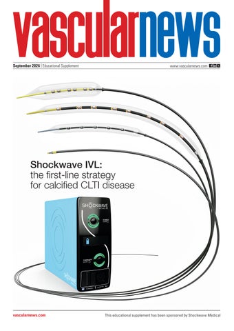
September 2026 | Educational Supplement
www.vascularnews.com
Shockwave IVL: the first-line strategy for calcified CLTI disease
vascularnews.com
This educational supplement has been sponsored by Shockwave Medical
Introduction
The role of Shockwave IVL in calcified CLTI disease treatment
squeezing and other forces that modify superficial and deep calcium—followed by a low-amplitude negative peak pressure, which minimises soft tissue damage from tensile stress. Shockwave IVL’s published acoustic waveform is key to achieving predictably safe and effective outcomes, and it differentiates Shockwave IVL from other Raghuram Lakshminarayan (Hull, UK) provides a detailed overview of how calcium modification technologies. This is Shockwave intravascular lithotripsy (IVL) is poised to meet the increasingly documented in a robust and growing body prevalent challenge of chronic limb-threatening ischaemia (CLTI). of clinical evidence representing >25,000 patients and >600 peer-reviewed journals.8 Single-arm studies and real-world registries demonstrate that CLTI REPRESENTS THE SEVERE calcification.6 Shockwave IVL is effective end of the vascular spectrum of peripheral The need for dedicated calcium across all calcium morphologies. arterial disease (PAD). Patients present with modification that disrupts The key to this effectiveness is rest pain, ulcers or gangrene resulting from calcified plaque before definitive the catheter and emitter design.9 reduced perfusion and significant arterial therapy has therefore become Shockwave IVL’s catheter design disease.1 The incidence of CLTI is increasing, recognised as a prerequisite consists of a series of emitters reflecting the global trends of increasing for durable outcomes in PAD with two 180-degree-spaced advanced age, hypertension, diabetes, and and CLTI. Shockwave IVL has spark gaps per emitter. Energy end-stage renal disease.2 Recent studies emerged as a distinct modality travels from the generator and demonstrate a growing burden of CLTI with that is complementary to other is delivered at the spark gaps Raghuram nearly half a million patients diagnosed with tools, addressing the problem launching two initial shockwaves Lakshminarayan the disease over a two-year period.3 Patients of calcium in endovascular (one per spark gap), which merge with untreated CLTI have high mortality and treatment.7 to create a spherical pressure field coverage 1 amputation rates. around each emitter. The ultrasonic acoustic Underlying this demographic reality is a Shockwave IVL’s mechanism of action output of the spherical shockwave field physiological one—the presence of vascular IVL is the energy-based production of produced is distributed circumferentially calcification. Vascular calcification does not ultrasonic acoustic pressure waves for the around the balloon edge, lacking dead zones passively form with age; it involves active modification, fracture and fragmentation or areas with an acoustic output less than mineralisation of the arterial wall driven of vascular calcification. Shockwave IVL’s the threshold for calcium modification. by metabolic and inflammatory signals. therapeutic payload for calcium modification Shockwave IVL’s emitters are designed Calcium deposition occurs in both the intima are shockwaves, which are a specialised form to prioritise device delivery and crossing (typically as part of atherosclerotic plaque) of ultrasonic acoustic pressure waves tuned to performance while maintaining clinical and the media, where it thickens and stiffens prioritise safety without tradeoffs for efficacy. efficacy across all types of calcium.10 the vessel wall. The resulting calcium Shockwave’s acoustic pressure waveform Shockwave IVL devices consist of burden fundamentally alters the mechanical consists of a very fast transition of highstrategically placed and tandem-firing properties of the artery and complicates amplitude positive peak pressure—which is emitters that ensure a consistent acoustic endovascular management.4 responsible for creating compressive, shear, output along the length of the device to In patients with CLTI, most arterial territories show calcification.5 This systemic calcification burden is strongly associated with adverse clinical outcomes including impaired revascularisation success, restenosis, and major adverse limb events.4 The presence of heavy arterial calcification is a challenge for conventional endovascular techniques. Standard balloon angioplasty might not produce adequate lumen gain in calcified lesions. High-pressure inflations intended to overcome this resistance frequently result in traumatic dissection, often necessitating bailout stenting, which is a less desirable outcome in vessels that may require future reintervention. Adjunct technologies like drug-coated 1 balloons (DCBs) or stents fail to deposit the drug evenly into the vessel wall due to All rights reserved. Published by BIBA Medical, London T:+44 (0)20 7736 8788, publishing@bibamedical.com. The opinions expressed in this supplement are solely those of Shockwave Medical and the featured physicians and may not reflect the views of Vascular News.
2 SPL 83000 Rev B
September 2026
Shockwave IVL
disrupt calcium regardless of where it is located along the catheter. While the area of highest acoustic output is adjacent to the emitters, the acoustic output of Shockwave E8 is measured to be therapeutic along the balloon edge and clinically relevant up to 6mm away from the emitter, allowing for the modification of superficial, deep and medial calcification.* One pulse from a Shockwave IVL device activates two emitters in tandem producing two spherical shockwaves simultaneously per pulse. The benefits of Shockwave IVL’s tandem-firing energy delivery are twofold. First, shock-shock interaction occurs when the simultaneously produced shockwaves intersect between the tandem-firing emitters at the balloon edge. This interaction facilitates a more homogenous acoustic pressure field across the balloon as compared to independentfiring emitters.† Secondly, tandem-firing emitters maximise treatment capability. Due to its tandemfiring emitter, Shockwave E8’s 400 available pulses per catheter provide physicians 800 shockwaves for modification fracture and fragmentation of calcification. Moreover, these 800 shockwaves provide a consistent acoustic output from the initial to last shockwaves available, with 98–102% wave power consistency.**†† The physics of this interaction are central to its efficacy and safety. Calcium, being substantially denser than the surrounding soft tissue, preferentially absorbs the compressive and tensile forces carried by the shockwaves. The high-amplitude positive pressure peak generates compressive stress that initiates fracture planes within calcified deposits; the subsequent negative pressure component generates tensile stress, which promotes
crack propagation, yet has a low amplitude to minimise soft tissue damage. Critically, Shockwave IVL is effective for both intimal and medial calcium, which is a significant advantage over modalities that act primarily at the luminal surface. The consequence of this calcium modification is a change in vessel compliance that has the potential to improve patient outcomes by avoiding high-pressure balloon dilatation and facilitating complete lumen expansion in what would have previously been considered an undilatable calcified lesion.11–14
“The presence of heavy arterial calcification is a challenge for conventional endovascular techniques” Shockwave Peripheral IVL portfolio The Shockwave Peripheral IVL portfolio encompasses a range of devices, all sharing Shockwave IVL’s unique mechanism of action, and tailored to address arterial calcifications within the whole peripheral vasculature. The Shockwave L6 is sized for largerdiameter arteries, offering calcium modification at a scale and profile appropriate for iliac and common femoral interventions. Shockwave L6 is specifically designed to address challenging calcium in large vessels. Featuring a six-emitter configuration for uniform energy delivery, it offers a good solution to effectively treat those focal, tough lesions often seen in iliac arteries. Shockwave M5+ is a versatile device for
above-the-knee (ATK) and proximal belowthe-knee (BTK) treatment. The wide range of sizes make it a sound solution for a variety of patients with calcified PAD. Equipped with five emitters, Shockwave M5+ delivers controlled calcium modification across the superficial femoral artery and popliteal segment. Its design balances effective lesion preparation with trackability, making it suitable for integration into femoropopliteal DCB or stenting workflows. The portfolio also includes Shockwave E8, engineered to extend capabilities when treating long calcified lesions below the knee. Its tapered tip and hydrophilic coating make it the most deliverable catheter of the Shockwave Peripheral IVL portfolio, and the workhorse tool for BTK treatment. An extended working length of 150cm enables the catheter to reach the distal tibial and peroneal vessels—territories critical to wound healing and limb salvage in CLTI. The E8 delivers up to 400 pulses at two per second, offering high treatment density per deployment. Lastly, for those niche cases with focal, tough BTK or below-the-ankle (BTA) calcifications, Shockwave Javelin offers a solution to modify calcium and enable further treatment. This is a forward-firing IVL platform designed for lesions that cannot be crossed by conventional wire-first techniques. It is most used in the BTA segment. Clinical evidence The clinical evidence for Shockwave has been building over the last five years. The DISRUPT PAD III trial is the pivotal randomised controlled trial (RCT) Continued on page 4
4 2
Figure 1. Acoustic pressure waveform Figure 2. Spherical pressure field coverage around each emitter Figure 3. Homogenous acoustic pressure field Figure 4. Consistent acoustic output
3
September 2026
SPL 83000 Rev B 3
Case report Continued from page 3
establishing the superiority of Shockwave IVL over conventional balloon angioplasty for calcium modification in femoropopliteal disease. The study enrolled 306 patients with moderately to severely calcified femoropopliteal lesions (Rutherford class 2–4) and randomised them 1:1 to treatment with IVL (n=153) or standard percutaneous transluminal angioplasty (PTA; n=153) as the primary lesion preparation step before DCB treatment or stenting.15 Shockwave IVL was superior to PTA on the primary endpoint of procedural success, defined as residual stenosis of less than 30% without flow-limiting dissection following the randomised treatment (IVL or PTA), prior to definitive therapy. This superiority was driven by meaningful reductions in the key complications that limit PTA outcomes in calcified disease. The rate of arterial dissection was significantly reduced with Shockwave IVL for both Grade C and Grade D dissections (3.5% vs. 15.1%; p=0.03). The need for bailout stenting was reduced by 75% relative risk in the IVL arm, with stent implantation rates of 4.6% vs. 18.3% in the PTA group (p=0.001). This finding has important implications for long-term management: patients who avoid index stenting preserve vessel anatomy for future reintervention, which is a critical consideration in calcified PAD where repeated procedures over time may be necessary. The durability of these procedural benefits was confirmed at 12 months. Primary patency, defined as freedom from clinically driven target-lesion revascularisation (CDTLR), was significantly greater in the IVL arm at one year: 80.8% vs. 70.9%.15 This was sustained in the second year, 74.4% vs. 57.7%. The DISRUPT PAD III RCT therefore provided level-one evidence proving that Shockwave IVL offers a safer and more predictable procedure than PTA while providing durable longer-term patency with significantly reduced need for provisional stenting. The DISRUPT BTK II study was a prospective, multicentre postmarket study evaluating Shockwave IVL for the treatment of calcified BTK arterial disease. Its patient population was deliberately designed to reflect the real-world complexity of BTK intervention: 80% of the 250 enrolled patients had CLTI, forming a dedicated 200-patient CLTI cohort that provides the most relevant data for this indication.16 The CLTI cohort was characterised by the high-risk features typical of this population: 73% presented with active wounds, 77% had
4 SPL 83000 Rev B
diabetes mellitus, 30.6% had chronic total occlusions (CTOs), and 85.8% had moderateto-severe arterial calcification. These are not the carefully selected patients of early feasibility studies but represent the complex, comorbid individuals who present daily in the vascular unit and in whom durable revascularisation is most difficult to achieve. One-year results, presented at VIVA 2025, demonstrated meaningful clinical durability in this challenging cohort.17 Freedom from major target-limb amputation across the overall study population was 94.8% at 12 months. Freedom from CD-TLR was 84.5% at one year, reflecting sustained patency without the need for early repeat intervention, while durable patency was confirmed in 67.1% of patients.17 Perhaps the most clinically meaningful finding was the transformation in CLTI severity classification. At baseline, 80% of patients were classified within CLTI Rutherford categories; by 12 months, this proportion had fallen to 25.1%, representing a reduction of more than 50 percentage points in CLTI-class disease.17 Quality-of-life data corroborated this improvement: VascuQoL-6 scores in the CLTI cohort improved significantly from a baseline mean of 11.9 to 17.2 at 12 months (p<0.001), confirming patient-reported benefit.17 These results are particularly notable in the context of the DISRUPT BTK II population. Prior studies of BTK revascularisation have typically involved less complex anatomy and lower rates of CLTI; the durability demonstrated in DISRUPT BTK II, in a population with 80% CLTI prevalence, high rates of diabetes, wound burden, and CTOs, establishes a meaningful clinical benchmark for IVL-supported BTK intervention. Conclusion An ageing population and progression of arterial calcification has led to an increasing incidence of complex PAD and CLTI.4 As calcium burden increases with age and comorbidity, so does the imperative for revascularisation tools that can reliably address calcified lesions without compromising safety or durability. Shockwave IVL has established a good evidence base across the full spectrum of peripheral arterial territory, from large inflow vessels through the femoropopliteal segment to the distal BTK runoff critical to limb salvage. The Shockwave Peripheral IVL portfolio provides operators with a dedicated toolset matched to the anatomical and technical demands of each vascular territory. The DISRUPT PAD III RCT and the DISRUPT BTK II one-year CLTI cohort results together
provide a robust clinical foundation including level-one evidence of superiority to PTA for vessel preparation in femoropopliteal disease, and demonstration of durable limb salvage in the most complex BTK CLTI patients. As the burden of CLTI continues to grow, Shockwave IVL occupies an increasingly central role in the armamentarium of endovascular strategies for treatment of CLTI. References: 1. Abu Dabrh A et al. The natural history of untreated severe or critical limb ischemia. J Vasc Surg. 2015; 62:1642–1651.e3 2. Stabley JN, Towler DA. Arterial calcification in diabetes mellitus: preclinical models and translational implications. Arteriosclerosis, Thrombosis, and Vascular Biology. 2017;37(2):205–217. 3. Kwong M et al. Updated estimates for the burden of chronic limb-threatening ischemia in the Medicare population. J Vasc Surg. 2023;77(6):1760–1775. 4. Dong Y et al. Lower limb arterial calcification and its clinical relevance with peripheral arterial disease. Front Cardiovasc Med. 2023 Nov 24;10:1271100. doi: 10.3389/fcvm.2023.1271100. PMID: 38075978; PMCID: PMC10710292. 5. Alexopoulos N et al. Systemic arterial calcium burden in patients with chronic limb-threatening ischemia. J Cardiovasc Comput Tomogr. 2023;17(4):248–254. 6. Marlevi D, Edelman ER. Vascular lesion-specific drug delivery systems: JACC state-of-the-art review. J Am Coll Cardiol. 2021 May 18;77(19):2413–2431. 7. Saratzis A et al. Use of vessel preparation in endovascular peripheral arterial disease (PAD) interventions: a global qualitative analysis. J Endovasc Ther. 2026 Feb 19:15266028261424732. 8. Kereiakes D et al. Principles of intravascular lithotripsy for calcific plaque modification. JACC. June 2021:1275–1292. 9. Madhavan MV et al. Efficacy and safety of intravascular lithotripsy for the treatment of peripheral arterial disease: an individual patient-level pooled data analysis. Catheter Cardiovasc Interv. 2020 Apr 1;95(5):959–968. 10. Adams G et al. Intravascular lithotripsy for treatment of calcified lower extremity arterial stenosis: initial analysis of the Disrupt PAD III study. J Endovasc Ther. 2020 June;27(3):473–480. 11. Riley R et al. Percutaneous Impella mechanical circulatory support delivery using intravascular lithotripsy. JACC: Case Reports. 2020;2(2):250–254. 12. Armstrong E et al. Intravascular lithotripsy for treatment of calcified, stenotic iliac arteries: a cohort analysis from the Disrupt PAD III study. CRM. Epub 13. Rosseel L et al. Intravascular iliac artery lithotripsy to enable transfemoral thoracic endovascular aortic repair. Catheter Cardiovasc Interv. 2019;1–4. DOI: 10.1002/ccd.28379. 14. DiMario C et al. A prospective registry of intravascular lithotripsy-enabled vascular access for transfemoral transcatheter aortic valve replacement. JACC: CI. 2019:12(5);502–504. 15. Tepe G et al. Intravascular lithotripsy for peripheral artery calcification: mid-term outcomes from the randomised DISRUPT PAD III trial. J Soc Cardiovasc Angiogr Interv. 2022;1(6):100325. 16. Chandra V et al. Thirty-day outcomes from the Disrupt PAD BTK II study of the Shockwave intravascular lithotripsy system for treatment of calcified below-the-knee peripheral arterial disease. J Vasc Surg. 2025 Mar;81(3):710–719.e2. 17. Armstrong E. One-year outcomes from the Disrupt PAD BTK II study: treatment of patients with calcified below-the-knee lesions with a peripheral intravascular lithotripsy system. VIVA 2025. * Based on longitudinal acoustic output scans. IEC standards 62127 & 61846. † Based on internal comparative longitudinal scans of Shockwave IVL devices. IEC standards 62127 & 61846. ** Based on acoustic output measurements of Shockwave E8 by hydrophone. IEC standards 62127 & 61846. †† Wave power consistency is a percent of the average acoustic output of shockwaves per cycle / average acoustic output of total shockwaves
Raghuram Lakshminarayan is a consultant vascular radiologist at Hull University Teaching Hospitals NHS Trust in Hull, UK, and a paid consultant of Shockwave Medical. Disclaimer: The views expressed in this article are those of the physician and may not reflect the views of Shockwave Medical.
September 2026
Shockwave IVL
Redefining inflow revascularisation in CLTI with Shockwave L6 Sharing the details of a recent case, Michel Bosiers (Bern, Switzerland) argues that Shockwave intravascular lithotripsy (IVL) is playing an increasingly central role in the treatment of calcified iliac inflow disease. CHRONIC LIMB-THREATENING ischaemia (CLTI) remains one of the most demanding conditions in vascular medicine, and adequate iliac inflow is a prerequisite for any successful revascularisation.1 When significant calcification is present, that inflow is jeopardised not only by the stenosis/ occlusion itself, but by the mechanical properties of the vessel wall, and those properties fundamentally alter how we should approach intervention.2 Heavily calcified iliac arteries present four compounding challenges. First, arterial rigidity reduces compliance, translating into inadequate balloon expansion and significant elastic recoil after conventional angioplasty.3 Second, attempts to overcome that rigidity with high-pressure balloons dramatically increase the risk of dissection or perforation—complications that are particularly consequential in the frail CLTI patient.4 Third, severe calcification impairs device trackability, making it genuinely difficult to deliver balloons and stents to target lesions.5 Fourth, and perhaps most critically for durability, residual calcium prevents full stent expansion, and underexpansion is a well-recognised driver of in-stent restenosis and repeat intervention.6 Taken together, calcified iliac disease is not simply a technical inconvenience; it directly compromises inflow restoration and, ultimately, limb salvage.1 Shockwave IVL addresses these challenges through a mechanism that is fundamentally different from conventional vessel preparation. By delivering ultrasonic acoustic pressure waves capable of modifying both intimal and medial calcium at low pressures, Shockwave IVL improves vessel compliance without the shear forces associated with high-pressure techniques.4,7,8 The result is a more predictable procedure with fewer bailout requirements, which is an important consideration in complex patients with CLTI.4 Critically, this mechanism enables IVL to function not merely as an adjunct to stenting, but as a definitive standalone therapy in appropriately selected cases.4 When calcium is adequately fractured and compliance restored, stenting may be unnecessary, in selected cases, eliminating the additional
September 2026
risks of a permanent implant and preserving future access.4
Treatment was delivered at very low (2) atmospheres across the full length of the CIA. No adjunctive stenting was required. Additional treatment of the CFA and SFA was performed. Immediate duplex assessment of the CFA confirmed triphasic flow, and pressure gradient resolution was complete.
Case report A 63-year-old male with CLTI was referred for revascularisation prior to hallux valgus surgery, with wound healing contingent on restored perfusion. His cardiovascular risk profile was substantial: arterial hypertension, Outcomes diabetes, hyperlipidaemia, prior smoking, At three months, ABI had normalised to 1.14, carotid disease, antiphospholipid with triphasic flow confirmed syndrome, and anticoagulation throughout the popliteal artery. with phenprocoumon Wound healing following (Marcoumar, KohlPharma). hallux valgus surgery was Pre-procedural ankle brachial uncomplicated. At 18 months, index (ABI) was 0.4 on the index the patient maintains an ABI of limb, consistent with severe 1.14, with triphasic flow in the haemodynamic compromise. CFA, SFA, and popliteal artery, Computed tomography representing durable patency (CT) angiography revealed a without an iliac stent. severely stenosed (TASC B) Michel Bosiers common iliac artery (CIA) with Clinical implications extensive calcification (Peripheral This case illustrates several arterial calcium scoring system [PACSS] 3) principles that are shaping practice in throughout the iliac territory. The left CIA calcified iliac disease. Shockwave IVL measured 11mm proximally, narrowing to enables safe, effective vessel preparation 9.4mm distally, with severe circumferential in patients where conventional angioplasty carries elevated procedural risk.8 Its balloonbased workflow integrates seamlessly into standard endovascular practice, with catheter oversizing of approximately 10% as a key technical point. And in highly selected cases where, after IVL treatment, the duplex waveform improves (e.g. from monophasic/ blunted to biphasic/triphasic at the level of the CFA) and/or the translesional gradient calcium burden—the characteristic pressure at rest decreases to <10mmHg, ‘cauliflower’ morphology that signals a standalone Shockwave IVL may represent particularly challenging substrate. Calcified the optimal endpoint, avoiding stent lesions were also evident in the common implantation.4 Further studies are of course femoral artery (CFA) and the superficial mandatory before robust conclusions can femoral artery (SFA). be drawn. Sizing of the Shockwave IVL catheter For CLTI patients with calcified iliac followed the principle of 10% oversizing inflow disease, Shockwave IVL is no longer and matching the largest vessel diameter: a niche adjunct. Experts support its role a 12mm Shockwave L6 catheter was selected as a first-line vessel preparation strategy, for the 11mm and the 9.4mm CIA. This and selected iliac cases argue for its use as sizing strategy is a key technical principle definitive therapy in its own right.4,10 where undersizing reduces acoustic energy delivered to the vessel wall, while References: 1. Conte MS et al. Global vascular guidelines on the appropriate oversizing ensures circumferential contact and maximises Continued on page 6 calcium modification efficacy.9
“For CLTI patients with calcified iliac inflow disease, Shockwave IVL is no longer a niche adjunct”
SPL 83000 Rev B 5
Case report Continued from page 5 management of chronic limb-threatening ischemia. Eur J Vasc Endovasc Surg. 2019 Jul;58(1S):S1–S109.e33. doi: 10.1016/j. ejvs.2019.05.006. Epub 2019 Jun 8. 2. Dong Y et al. Lower limb arterial calcification and its clinical relevance with peripheral arterial disease. Front Cardiovasc Med. 2023 Nov 24;10:1271100. doi: 10.3389/fcvm.2023.1271100. PMID: 38075978; PMCID: PMC10710292. 3. Gardiner G et al. Quantification of elastic recoil after balloon angioplasty in the iliac arteries. J Vasc Interv Radiol. 2001 Dec;12(12): 1389–1393. doi: 10.1016/s1051-0443(07)616947. 4. Fazzini S et al. Mid-term outcomes of Shockwave intravascular lithotripsy in the IVLIAC registry for the treatment of calcified iliac occlusive disease. J Vasc Surg. 2025;82(4):1366–1374.
5. Tepe G et al. Intravascular lithotripsy for peripheral artery calcification 30-day outcomes from the randomized Disrupt PAD III trial. JACC Cardiovasc Interv. 2021 Jun 28;14(12):1352–1361. doi: 10.1016/j.jcin.2021.04.010. 6. Dubosq-Lebaz M et al. Advances in intravascular lithotripsy: mechanisms, devices, and clinical applications. Expert Rev Med Devices. 2025 Mar;22(3):209–218. 7. Adams G et al. Intravascular lithotripsy for treatment of calcified lower extremity arterial stenosis: initial analysis of the Disrupt PAD III study. J Endovasc Ther. 2020 Jun;27(3):473– 480. doi: 10.1177/1526602820914598. Epub 2020 Apr 3. 8. Armstrong E et al. Intravascular lithotripsy for treatment of calcified, stenotic iliac arteries: a cohort analysis from the Disrupt PAD III study. Cardiovasc Revasc Med. 2020 Oct;21(10):1262–1268. doi: 10.1016/j.carrev.2020.02.026. Epub 2020 Mar 2. 9. Brodmann M et al. Primary outcomes and mechanism of action of intravascular lithotripsy in calcified, femoropopliteal lesions: results of Disrupt PAD II. Catheter Cardiovasc Interv. 2019 Feb 1;93(2):335–342. doi: 10.1002/ccd.27943. Epub
2018 Nov 25. 10. Saratzis A et al. Use of vessel preparation in endovascular peripheral arterial disease (PAD) interventions: a global qualitative analysis. J Endovasc Ther. 2026 Feb 19:15266028261424732. doi: 10.1177/15266028261424732. Online ahead of print.
Michel Bosiers is a senior consultant at University Hospital Bern in Bern, Switzerland, and a paid consultant of Shockwave Medical. Disclaimer: The views expressed in this article are those of the physician and may not reflect the views of Shockwave Medical.
Case images: No stent needed for iliac inflow disease
1
4
5
8
9
2
3
6
7
Figure 1. Foot at baseline Figure 2. Baseline CT scan Figure 3. Baseline angiogram Figures 4–7. Treatment with Shockwave L6 12mm Figure 8. Final angiogram Figure 9. Duplex scan of the CFA at 18-month follow-up Figure 10. Duplex scan of the popliteal artery at 18-month follow-up
10
Scan code to link to corresponding video interview
6 SPL 83000 Rev B
September 2026
Shockwave IVL
Optimising femoropopliteal treatment with Shockwave E8 in a patient with CLTI Leyla Schweiger (Graz, Austria) shares a case in which Shockwave intravascular lithotripsy (IVL) enabled effective lesion preparation, supported subsequent treatment, and contributed to wound healing in a patient with heavily calcified femoropopliteal disease. HEAVILY CALCIFIED femoropopliteal disease remains one of the most demanding scenarios in endovascular intervention. In these lesions, the challenge is not only to cross the target segment, but also to prepare the vessel adequately before definitive therapy. Conventional balloon angioplasty in the presence of marked calcium burden may be limited by underexpansion, recoil and dissection, resulting in a less predictable procedural outcome.1 Randomised data from DISRUPT PAD III showed that IVL-based vessel preparation in calcified femoropopliteal arteries was associated with higher procedural success, fewer flow-limiting dissections and less need for stenting compared with conventional angioplastybased preparation.2 Observational data further suggest that Shockwave IVL combined with drug-coated balloon (DCB) angioplasty can achieve promising 12-month patency and freedom from target-lesion revascularisation in severely calcified femoropopliteal lesions.3 This concept is especially relevant in chronic limb-threatening ischaemia (CLTI), where the procedural endpoint should not be angiographic success alone. Contemporary CLTI guidance emphasises evidence-based revascularisation with the aim of restoring perfusion sufficiently to support wound healing, limb salvage and preservation of function. Treatment planning should integrate patient risk, limb severity and anatomic complexity.4,5 Case report A 78-year-old woman presented with Rutherford class 5 CLTI of the right lower limb, with ulceration involving the lower leg and heel region. Her comorbidity profile included bilateral peripheral arterial disease, coronary artery disease, persistent atrial fibrillation on apixaban, renal insufficiency, arterial hypertension, iron deficiency anaemia and chronic venous disease. Baseline wound documentation demonstrated a relevant tissue defect with fibrinous slough and surrounding inflammatory change, consistent with advanced limb-threatening ischaemia. Angiography revealed a long, severely calcified lesion involving the distal superficial
September 2026
femoral artery and extending luminal result, with no significant into the popliteal artery. Lesion recoil and no flow-limiting length was approximately 13cm, dissection. From a technical with subtotal stenosis and severe perspective, this case supports a calcification on angiography. In treatment strategy in which IVL such lesions, calcium burden is is used early in severely calcified clinically relevant because severe femoropopliteal disease to femoropopliteal calcification facilitate definitive therapy while has been associated with poorer minimising vessel injury. outcomes in patients undergoing Most importantly, the Leyla intervention for ischaemic angiographic result translated Schweiger tissue loss.6 In addition, high into clinical benefit. At followcalcification burden has been up, the wound had healed with associated with inferior outcomes after DCB restoration of skin integrity and resolution angioplasty in de novo femoropopliteal of the previously open ulcer. In CLTI, this disease, underlining the importance of remains the outcome that matters most. adequate lesion preparation.7 Current guidelines make clear that The lesion was approached antegradely revascularisation in CLTI should be judged and crossed using an Advantage 0.014 according to limb-based clinical benefit, not guidewire supported by a Navicross catheter angiography alone.4,5 (both Terumo Interventional Systems). Following successful lesion crossing, vessel Clinical implications preparation was performed with a Shockwave This case illustrates the value of Shockwave E8 5/80mm catheter. The reference vessel E8 as part of a vessel-preparation strategy in diameter was 4.5mm, and a 5mm Shockwave heavily calcified femoropopliteal disease in a patient with Rutherford 5 CLTI. In a long, severely calcified lesion where conventional angioplasty alone would likely have been less predictable, Shockwave IVL enabled effective lesion preparation, supported subsequent DCB treatment and contributed to wound healing at follow-up. Practical lessons from this case include calcium modification, careful sizing to achieve wall apposition and E8 catheter was selected to achieve consistent attention to the clinical endpoint of approximately 10% oversizing for adequate limb salvage. wall apposition. A total of 400 pulses were delivered over 10 cycles at 2–4atm. References 1. Lichtenberg M et al. Recoil, dissection, and restenosis in Progressive balloon expansion during pulse below-the-knee (BTK) arteries following standard balloon delivery indicated calcium modification. angioplasty. Vasa. 2025 Nov 18. doi: 10.1024/0301-1526/ a001254. Epub ahead of print. PMID: 41250599. This intra-procedural response is one 2. Tepe G et al; Disrupt PAD III investigators. Intravascular lithotripsy for peripheral artery calcification: 30-day of the practical advantages of Shockwave outcomes from the randomized Disrupt PAD III trial. JACC IVL in calcified peripheral disease: calcium Cardiovasc Interv. 2021;14(12):1352–1361. doi: 10.1016/j. jcin.2021.04.009. PMID: 34167675. modification can be achieved at low balloon 3. Stavroulakis K et al. Intravascular lithotripsy and drug-coated pressure, allowing lesion preparation without balloon angioplasty for severely calcified femoropopliteal arterial disease. J Endovasc Ther. 2023;30(1):106–113. doi: the same degree of barotrauma typically 10.1177/15266028221075563. PMID: 35130782. associated with aggressive high-pressure 4. Conte MS; GVG writing group. Global vascular guidelines on the management of chronic limb-threatening ischemia. 2,3 dilatation. Following IVL, the lesion was J Vasc Surg. 2019;69(6S):3S–125S.e40. doi: 10.1016/j. jvs.2019.02.016. PMID: 31159978. treated with a DCB.
“The wound had healed with restoration of skin integrity and resolution of the previously open ulcer”
Outcomes Final angiography demonstrated an excellent
5. Gornik HL et al. 2024 AHA/ACC peripheral artery disease guideline. Circulation. 2024;150(2):e131–e290. doi: 10.1161/
Continued on page 8
SPL 83000 Rev B 7
Case report 7. Mori S et al. Impact of calcification on clinical outcomes after drug-coated balloon angioplasty for superficial femoral artery disease: assessment using the peripheral artery calcification scoring system. Catheter Cardiovasc Interv. 2023;102(5):1028–1037. doi: 10.1002/ccd.30726. PMID: 36883957.
Continued from page 7 CIR.0000000000001251. PMID: 38752899. 6. Lee HY et al. The effect of severe femoropopliteal arterial calcification on the treatment outcome of femoropopliteal intervention in patients with ischemic tissue loss. Vasc Specialist Int. 2020;36(2):96–104. doi: 10.5758/vsi.200005. PMID: 32611842.
Leyla Schweiger is an internal medicine fellow at the Medical University of Graz in Graz, Austria, and a paid consultant of Shockwave Medical. Disclaimer: The views expressed in this article are those of the physician and may not reflect the views of Shockwave Medical.
Case images: Rutherford class 5 CLTI to wound healing
1
4
5
2
6
3
7
8
12
9
Figure 1. Baseline wound Figure 2. Baseline angiography Figure 3. Crossing Figure 4–8. Treatment with Shockwave E8 5mm Figure 9. Post-IVL angiography Figure 10. Adjunctive drug-coated balloon Figure 11. Final angiographic result Figure 12. Wound healing at follow-up
10
11
Scan code to link to corresponding video interview
8 SPL 83000 Rev B
September 2026
Shockwave IVL
Reframing treatment possibilities in below-the-knee disease Pablo Del Canto (Gijón, Spain) presents the case of a patient with severely calcified below-the-knee (BTK) disease. The Shockwave E8, he shares, was key to a good result in a challenging clinical scenario. Challenges of BTK disease BTK disease represents a particularly complex and challenging area within peripheral arterial disease, introducing a higher level of difficulty in treatment planning and execution due to both patientrelated and lesion-specific factors. Patients treated in the BTK space often present with advanced disease, frequently including chronic limb-threatening ischemia (CLTI) and a high prevalence of comorbidities such as chronic kidney disease and diabetes, which is associated with an increased burden of deep arterial calcification.1 Lesion characteristics further complicate treatment, as BTK arteries commonly exhibit long, diffuse lesions, combined with a higher prevalence of both superficial and deep calcium.2–4 In particular, medial arterial calcification is more frequently observed in BTK vessels and has been identified as a potential predictor of worse clinical outcomes, including an increased risk of amputation.5,6 These anatomical features translate into significant procedural challenges leading to complications and suboptimal outcomes with conventional endovascular approaches. BTK interventions are often limited by poor vessel expansion and acute recoil that can hinder acute gain and reduce patency.7 Additionally, the presence of calcium contributes to a higher incidence of dissections during angioplasty, frequently necessitating bailout stenting.8 Altogether, the combination of complex patient profiles, heavily calcified and anatomically challenging lesions, and the inherent limitations of standard therapies underscores the need for more effective and reliable vessel preparation strategies in the BTK segment to improve both immediate and long-term clinical outcomes. Case report An 85-year-old male patient presented to our department with a foot infection on his left lower limb that had been evolving for several weeks and showing significant worsening over the past 48 hours. The patient’s medical history included diabetes mellitus, hypertension, dyslipidaemia, and chronic kidney disease.
September 2026
tibial artery (PT) and a patent, Initial presentation and albeit diseased, peroneal artery. clinical evaluation Following the diagnostic workup, The patient was admitted because an endovascular strategy was of a septic clinical presentation chosen to treat the ATA in an accompanied by fever and attempt to restore direct in-line increased inflammatory markers, flow to the area of the lesion, with a white blood cell count which will also require a minor of 16,000 cells/mm3, C-reactive amputation. protein (CRP) 301mg/mL and a clear deterioration of renal Revascularisation procedure: function, with an estimated Pablo Del Canto calcification and glomerular filtration rate (eGFR) Shockwave E8 of 25 at the time of admission. At An antegrade ultrasound-guided femoral physical examination the patient presented puncture was performed. A short 6Fr sheath with femoral and popliteal pulses but no palpable pedal pulses. There was an infection was advanced and systemic heparinisation of the hallux with wet gangrene and cellulitis, was administered. The popliteal CTO was crossed with a 0.014 guidewire, which was together with an ischaemic lesion along the inner border of the foot at the level of the first also advanced into the ATA down to the foot. Before vessel preparation, we attempted to metatarsal head. A bedside ankle–brachial confirm intraluminal position but were unable index (ABI) was non-compressible, likely to cross the occlusions with any device, due to arterial calcification. including a 0.014-inch microcatheter or 1mm balloons. At this point, we decided to perform Imaging an ultrasound-guided retrograde puncture of Diagnostic angiography revealed severe the DP to externalise the wire. Then, using a calcific arterial disease, with dense Badform technique we were able to advance calcification throughout all the tibial vessels. a 1mm balloon, which allowed confirmation The popliteal artery demonstrated a heavily of intraluminal position and predilatation of calcified chronic total occlusion (CTO) at the lesions. the level of its bifurcation. The origin and In view of the severe calcification of proximal third of the anterior tibial artery the lesions and the associated difficulty in (ATA) were patent, but showed diffuse advancing devices across them, the use of the narrowing along its course, with complete Shockwave E8 catheter for vessel preparation was deemed appropriate. The device was chosen due to its favourable crossing profile and balloon length, which would allow treatment of the entire vessel segment using a single catheter. The 3.5mm Shockwave E8 catheter was advanced down to the distal ATA and activated, delivering 400 pulses at low inflation pressures of 2–4atm. This improved compliance and enabled subsequent balloon expansion without complications. The procedure continued with a 3mm nonocclusion at the mid segment and distal compliant balloon angioplasty to maximise reconstitution in the distal segment, which luminal gain. also exhibited moderate and severe calcified stenosis. The distal runoff to the foot via the Results and follow-up dorsalis pedis (DP) appeared acceptable. There was severe disease of the tibioperoneal Continued on page 10 trunk with total occlusion of the posterior
“This case demonstrates the typical severe BTK calcification commonly seen in patients with diabetes mellitus and chronic kidney disease”
SPL 83000 Rev B 9
Case report Case images: Severe BTK disease to full ambulation
1
2
3
12 8
9
Continued from page 9
The subsequent angiography demonstrated excellent luminal gain with no residual stenosis or dissections, and no recoil after 15 minutes. The procedure was finally completed with a 3mm drug-coated balloon along the entire ATA with direct in-line flow to the foot on completion angiography. Forty-eight hours after the procedure, a minor amputation of the first toe was performed. The patient remained admitted on intravenous antibiotic therapy for 10 days and was then discharged. At follow-up in the outpatient clinic, complete healing of the amputation site and
10 SPL 83000 Rev B
10
the medial border lesion was observed at one month after the index procedure, with the patient recovering full ambulatory function. Discussion This case demonstrates the typical severe BTK calcification commonly seen in patients with diabetes mellitus and chronic kidney disease. The calcification involves not only the intimal layer but also extends into the medial layer. This pattern of calcification represents a significant therapeutic challenge, predisposing to a high incidence of recoil and residual stenosis. In addition, when more aggressive dilation is pursued, the risk of
13
14
11
dissection or vessel rupture due to marked barotrauma can be increased.8 The Shockwave intravascular lithotripsy (IVL) mechanism of action has represented a paradigm shift in the management of calcified lesions and, consequently, in the treatment of patients with CLTI. The Shockwave E8 catheter further extends its application to more distal vascular segments, which are particularly prone to calcification. In particular, its improved crossing profile, increased pushability, and flexibility facilitate navigation through small-calibre, highly tortuous vessels, enabling access to the most distal segments. Secondly, its mechanism of action enables treatment of both superficial
September 2026
Shockwave IVL
4
5
6
7
Figure 1. Wound at baseline Figures 2–3. Baseline angiograms Figures 4. Retrograde puncture Figure 5. Predilation with 1mm plain balloon angioplasty Figures 6–10. Treatment with Shockwave E8 3.5mm Figures 11–13. Final angiograms Figure 14. Wound at 30 days post-revascularisation 12
13
and deep calcium in a highly safe manner. Calcium modification is performed at very low inflation pressures (2–4 atm), thereby minimising barotrauma and reducing the risk of complications such as dissection or vessel rupture. Finally, the longer balloon length and increased number of pulses enable treatment of the entire distal vessel segment with a single catheter, thereby reducing procedural complexity, procedure time, and costs. References 1. Stabley JN, Towler DA. Arterial calcification in diabetes mellitus: preclinical models and translational implications. Arteriosclerosis, Thrombosis, and Vascular Biology. 2017;37(2):205–217. 2. Li J et al. Below-the-knee endovascular revascularization: a position statement. JACC: Cardiovascular Interventions.
September 2026
14
2024;17(5):589–607. 3. Soor GS et al. Peripheral vascular disease: who gets it and why? A histomorphological analysis of 261 arterial segments from 58 cases. Pathology. 2008;40(4):385–391. 4. Kim SH, Jung SL. Endovascular recanalization for infrapopliteal artery disease in chronic limb-threatening ischemia: a practical review. Vascular Specialist International. 2025 Dec 26;41:39. doi: 10.5758/vsi.250076. 5. DiBartolomeo AD et al. Medial arterial calcification score is associated with increased risk of major limb amputation. Journal of Vascular Surgery. 2023;78(5):1286–1291. 6. Losurdo JA et al. Medial artery calcification: the silent killer of the leg, American College of Cardiology expert analysis, 2021. 7. Baumann F et al. Early recoil after balloon angioplasty of tibial artery obstructions in patients with critical limb ischemia. Journal of Endovascular Therapy. 2014;21(1):44–51. 8. Fitzgerald PJ et al. Contribution of localized calcium deposits to dissection after angioplasty: an observational study using intravascular ultrasound. Circulation. 1992;86(1):64–70.
Pablo Del Canto is a vascular and endovascular surgeon at the Hospital Universitario de Cabueñes in Gijón, Spain, and a paid consultant of Shockwave Medical. Disclaimer: The views expressed in this article are those of the physician and may not reflect the views of Shockwave Medical.
Scan code to link to corresponding video interview
SPL 83000 Rev B 11
Shockwave Peripheral IVL
Establishing a new benchmark for patients with CLTI Results from the DISRUPT BTK II study show that in a majority of patients with CLTI, Shockwave™ IVL plays a critical role in laying the foundation for successful wound healing through safe and effective revascularization.
9/10 2/3
patients were free from major amputation1 patients achieved wound healing or improvement*
Explore Shockwave IVL for calcified BTK lesions
*Mean wound follow up time: 157 days 1. Armstrong E, One-Year Outcomes From the Disrupt PAD BTK II Study: Treatment of Patients With Calcified Below-the-Knee Lesions With a Peripheral Intravascular Lithotripsy System, VIVA 2025 Prior to use, please reference the Instructions for Use for the information on indications, contraindications, warnings, precautions, and adverse events. www.shockwavemedical.com/IFU Product availability may vary by country. ©2026 Shockwave Medical Inc. All rights reserved. SPL 83000 Rev. B.