9 minute read
Labeling Tumors with Quantum Dots Nachiket Girish


Labeling tumors with quantum dots
Advertisement
BY NACHIKET GIRISH
The prefix “nano-,” the word “quantum”—both are overused tropes in science fiction. Perhaps writers guess that they instantly make anything they are attached to sound futuristic and cuttingedge.
While in the movies these scientificsounding prefixes are used to describe portals, time machines, and other fantastical objects, the real world is often even weirder than fantasy, and lends surprising plausibility to the existence of those cliches. Perhaps no single entity provides greater evidence of this weirdness than the baffling world of quantum dots.
DISCOVERING CONFINEMENT IN COLLOIDS
Louis Brus, born in Cleveland in 1943, might have been destined to be a chemical physicist. He attended Rice University on a Navy scholarship, where during his freshman year, a major in chemical physics was offered for the first time ever—an opportunity he pounced upon. After his graduation, he was commissioned to the Navy, but was given a special dispensation to go to graduate school instead. Later, having obtained a PhD in chemical physics
from Columbia University, Brus joined Bell Labs, one of the premier research centers in the world. 1
It was here in 1983 that Brus made the accidental observation which would become his most significant work. Brus was working on the redox reactions of organic molecules, reactions taking place on the surfaces of semiconductors. A crucial factor in these reactions was the semiconductor’s band gap, the energy the electrons need to begin conduction and moving charge. After testing various models, Brus realized that the organic reactions would occur faster if the molecules were suspended in a colloidal solution (a mixture of evenly dispersed particles suspended in a medium) so that they could bind to the semiconductor surface from all directions.
Upon performing the experiment in the colloid, however, Brus noticed something odd—the band gap of the semiconductor particles in the colloid seemed to be shrinking with time! Upon some more analysis, Brus was able to link this effect to an increase in the particle size inside the colloid over time. Particles would agglomerate and naturally get larger inside a colloid, and this increase in size was causing a decreasing band gap and, consequently, increased conductivity of
the semiconductor. Brus had just observed a system whose energetic properties depended on its physical size—a system which we now know as a quantum dot. “We realized,” says Brus in his autobiography, “that this was (and is) an excellent research problem.” In 2006, Brus, along with the other fathers of quantum dots, scientists Alexei Ekimov and Alexander Efros, won the R. Wood Prize of the Optical Society of America. He probably would not have dreamed, however, that his excellent research problem would, among other things, find applications in cancer research and renewable energy. 2
BUT FIRST—WHAT ARE QUANTUM DOTS?
A quantum dot at the most fundamental level is just a particle bouncing around in a box. That’s it. Apart from size, there are few other special conditions to their existence. This, however, is the puzzling, unintuitive beauty of quantum mechanics—just going down a few scales of size takes us to a whole new world. In this case, it is a world where we can make that box emit different colors of light just by squeezing it. To understand the particle in a box, we need to understand the basic tenets of quantum mechanics.
The development of quantum mechanics was first motivated by the observation that particles often behave surprisingly like waves, and vice versa. The particle in a box model can be understood if we consider the particle to not actually be a particle, but a wave reflecting back and forth in a box. In most cases, a wave bouncing around inside a box would be a confusing mess—think of water sloshing around in a bathtub. There is one special condition, however, which brings order to this chaos: if the waves have wavelengths such that the crest of the incoming wave overlaps exactly with the crest of the reflected wave, they add up quite neatly to form a stable configuration known as a standing wave.
Now, our box can only manage to exactly overlap incoming and reflected waves if it is large enough to fit a whole crest, or an integer number of whole crests exactly within it (Fig. 1). And since the wavelength of the particle depends on its energy, for a given size of a box we thus have a set of special energies for which the associated wavelength (and an integer number of them) matches the size of the box. The particle inside the box can only ever possess one of these special energies as its energy value, no other. 3
If a particle gains some energy, it will emit the same energy, in the form of a definite wavelength of light. And since we know that for different sizes of the box we have different corresponding sets of special energies, the upshot of this becomes that for
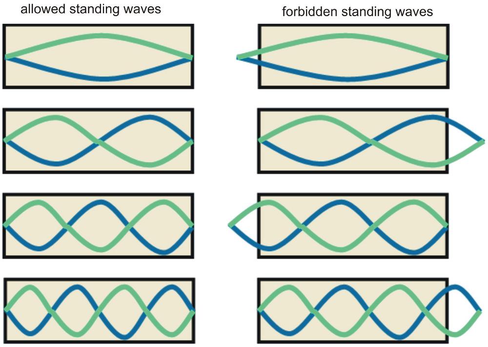
Figure 1: Notice that all the allowed standing waves fit perfectly inside the box, while all the forbidden ones have a partial crest sticking out.
different sizes of the box, we can make the particle emit light of different wavelengths. This is the fundamental property which makes quantum dots so fascinating—by tuning the sizes of particle boxes, we can control their color! 4,5
BIO-INDICATOR DOTS
The discovery that nanoparticles show quantum confinement (the technical term for the effect discussed above) has led to a wide spectrum of applications. Probably the most fascinating is the use of quantum dots for biomedical imaging.
Biomedical imaging is the technique of looking inside the human body without cutting it open. It’s not as exotic as the name might suggest—the X-ray and CT scan are both types of biomedical imaging techniques.
Biomedical imaging is extremely useful for targeted uses, such as when we need to spot tumors among healthy tissue. The name of the game is to find a molecule that has an affinity to the tumor cells and attach to it a label dye, a compound which gives off radiation we can detect. Once released into the body through one of several possible delivery systems, these labeled molecules reach tumor cells and bind to them, thus indirectly attaching to them the radiation-emitting label dye. Doctors can then analyze the radiation being emitted to get a real-time display of where the tumor cells are in the body. 6
Organic label dyes have traditionally been used for quite some time now; however, most of these dyes emit optical radiation, i.e., visible light, which gets easily blocked/absorbed by the tissues surrounding them.
This is where quantum dots come in. Since they offer dyes with emission ranges in the Near Infrared (NIR) range, which
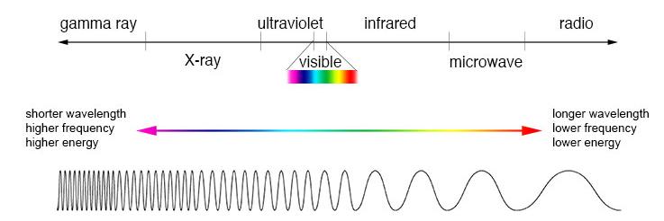
is sparsely blocked by tissue, they are transmitted through living cells, enabling easy detection by instruments (Fig. 2). 7 Moreover, since the emission wavelength of quantum dots is so closely linked to their size, by controlling the size of the quantum dots we can very finely tune the emission wavelength to our desired value. They also fluoresce for several hours and do not disperse away from the tumor into the body as quickly as conventional label dyes, so that surgeons have much more time to carry out operations. 8 Of course, this technology is still relatively new, and there are still challenges to overcome. Universal protocols involving the use of quantum dots have still not been developed, making their present use challenging. Moreover, their relatively large size compared to conventional label dyes raises potential complications in accessing cellular targets. Nevertheless, their myriad advantages mean that we can expect their use to only grow in the future. 9
AN EXCITING TIME AHEAD
There are simply far too many exciting possibilities for quantum dots to cover in a few pages. They are revolutionizing solar panel technology, they are being used to develop effective indoor farming, and they are at the center of Samsung’s fight for dominance against LG in the television industry—an industry where quantumdot-enabled technologies such as emissive quantum dot LED displays and MicroLED promise to bring in a new era of display technology. 10 What we can certainly be sure of, however, is that the future of quantum dots holds nothing but exciting possibilities.
Acknowledgements: I would like to thank my physics professor Matthias Reinsch for reviewing the physics of this article. 1. 2. 3. 4. 5. 6. Davis, T. (2005). Biography of Louis E. Brus. Proceedings of the National Academy of Sciences, 102(5), 1277–1279. https://doi.org/10.1073/ pnas.0409555102 Brus, L.E. (2008). Scientific autobiography of Louis Brus. The Kavli http://kavliprize.org/prizes-andlaureates/laureates/louis-e-brus French, A. P., & Taylor, E. F. (1978). An introduction to quantum physics. Norton. Jacak, L., Hawrylak, P., & Wójs, A. (2013). Quantum dots. SpringerVerlag Berlin Heidelberg. https://doi. org/10.1007/978-3-642-72002-4 Bajwa, N., Mehra, N. K., Jain, K., & Jain, N. K. (2016). Pharmaceutical and biomedical applications of quantum dots. Artificial Cells, Nanomedicine, and Biotechnology, 44(3), 758–768. https://doi.org/10.3109/21691401.201 5.1052468 McCann, T. E., Kosaka, N., Choyke, P. L., & Kobayashi, H. (2012). The use of fluorescent proteins for developing cancer-specific target imaging probes. In Hoffman R. M. (Ed.), In Vivo 7. 8. 9. 10. 1. 2. 3.
Figure 2: Light of higher wavelength, as shown here, has lower energy and is closer to the red end of the spectrum.
Shorter wavelength (and hence higher frequency) light has more energy and is more towards the blue end of the
“The name of the game is to find a molecule that has an affinity to the tumor cells and attach to it a label dye, a compound which gives off radiation we can detect. ”
REFERENCES
spectrum. Cellular Imaging Using Fluorescent Proteins (pp. 191-204). Humana Press. https://doi.org/10.1007/978-1-61779- 797-2_13 Matea, C. T., Mocan, T., Tabaran, F., Pop, T., Mosteanu, O., Puia, C., Iancu, C., & Mocan, L. (2017). Quantum dots in imaging, drug delivery and sensor applications. International Journal of Nanomedicine, 12, 5421–5431. https:// doi.org/10.2147/IJN.S138624 Naasani, I. (2016, September 21). The cancer surgeon’s latest tool: Quantum dots. IEEE Spectrum. https://spectrum. ieee.org/biomedical/imaging/thecancer-surgeons-latest-tool-quantumdots Resch-Genger, U., Grabolle, M., Cavaliere-Jaricot, S., Nitschke, R., & Nann, T. (2008). Quantum dots versus organic dyes as fluorescent labels. Nature Methods, 5(9), 763–775. Morrison, G. (2018, February 9). How quantum dots supercharge farming, medicine and solar, too. CNET. https:// www.cnet.com/news/how-quantumdots-supercharge-farming-medicineand-solar-too/
IMAGE REFERENCES Banner: Fehling-Lab. (Oct. 3, 2019). Experiment test tube fluorescence [jpg image]. Pixabay, https://pixabay. com/photos/experiment-test-tubefluorescence-4520604/ Figure 1: CK-12 Foundation. (2010). Standing waves [png image]. https:// commons.wikimedia.org/wiki/ File:Allowed_and_forbidden_ standing_waves.png Figure 2: NASA. (2013). Imagine the Universe [jpg image]. https://imagine. gsfc.nasa.gov/science/toolbox/ emspectrum1.html