4 minute read
AUSTFHU Year book 2022/2023


Peeking into the Art of Creation
Dr. Einat Reichert-Zelinger still remembers the drawer in her childhood bedroom. Her father filled it with newspaper clippings about his uncle, Einat’s grandfather’s brother, cut from the daily paper and preserved ever so carefully. But her great uncle wasn’t an important politician or a world-renowned rockstar; he was a scientist.
Advertisement
By Devora M. Liss
Prof. Israel Reichert had established the field of phytopathology (the study of plant diseases) in Israel and retained recognition throughout his career as its foremost researcher, culminating with being awarded the Israel Prize in 1955.
When Einat decided to study plant protection sciences at Hebrew University, it was just a matter of time until someone asked about her last name. That person was Prof. Jaacov Katan, who had conducted his doctoral research under her grandfather’s supervision. He invited Einat to his lab, where she first encountered microscopy. She developed such good imaging skills that she would often assist Prof. Katan’s doctoral students with their measurements.
Einat continued to her own doctoral studies at a joint program between Oxford University and Oxford Brookes University, under the supervision of the late Prof. Chris Hawes and Dr. Molly Dewey, where she studied a specific fungus that plagues a variety of wheat grown also in Israel. It was here that she
fully understood the immense power of imaging as a research tool. She had access to so many different types of machines: the confocal laser light microscope, the scanning electron microscope (SEM) that can also handle frozen samples, and the transmission electron microscope (TEM). She also mastered the technique of cutting thin sections and antibody staining with gold particles. “I was enchanted by the radical glimpse that microscopy offered into things too small for the naked eye,” Dr. Reichert-Zelinger recalls.
After graduating, Dr. Reichert-Zelinger returned to Hebrew University to conduct postdoctoral research in Dr. Yael Heifetz’s lab. She was studying the connection between nutrition, aging, and fertility – specifically, the biological clock in Drosophila (small fruit flies), along with Prof. Oren Froy from the Department of Biochemistry, Food Science and Nutrition. But she couldn’t escape the microscopes. Einat quickly became responsible for all things microscopy in the lab, including training new students.
When the position opened at the Center for Scientific Imaging in the Core Research Facility, Einat felt in
her heart that it was her destiny. The unit holds and operates more complex machinery than regular labs and has extremely high-end image processing programs. The staff assists researchers and students to plan and execute their experiments most effectively.
Einat is currently working to develop microscopy protocols for researchers from different fields, identifying which microscope is best suited for which measurement, and how to optimize findings by combining measurements from different machines.
This is no small feat – the Smith Faculty alone has researchers from biology, plant sciences, entomology, soil and water, biochemistry, and more – not to mention researchers from other campuses and external companies who hire Hebrew University’s services.
“The new frontier is correlative microscopy – combining different machines, each with its advantages and disadvantages, to glean the best measurements possible for experiments of any and every kind,” Einat explains. “The question becomes one of big data – analyzing your findings. To this end, I’m leading the way towards applying machine learning to microscopy data.”
Einat needed a project on which to develop these protocols, a testing grounds of sort. She has retained a strong affiliation with Dr. Heifetz’s lab and her ongoing research on Drosophila serves that purpose. She reflects, “Microscopes never cease to amaze. Whether it’s me peering through for the umpteenth time or high school students on a field trip getting their first look, you cannot ignore that sense of awe.”
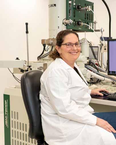
Dr. Reichert-Zelinger next to the Jeol 7800F high esolution scanning electron microscope, funded with the support of the U.S. Agency for International Development (USAID) (Photo: Maxim Dinshtein)
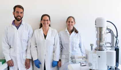
Left to right: Daniel Waiger, Dr. Reichert-Zelinger, and Tali Kossovsky alongside the Jeol IT100 LV scanning electron microscope, funded with the support of the U.S. Agency for International Development (USAID) (Photo: Maxim Dinshtein)
This article originally appeared in the Hebrew University’s Scopus magazine, and is reprinted with permission from the Hebrew University.


