Introduction
Adeno-associated virus (AAV) vectors are widely adopted as delivery vehicles for the treatment of genetic disorders. Of the multiple AAV serotypes, AAV9 has demonstrated tropism towards the central nervous system, retina, inner ear, lung, liver, cardiovascular, and multiple other targets and has been reported as being used in 25% of the programs in clinical development (Advancing AAV production with high throughput screening and transcriptomics Cell & Gene Therapy Insights 2024; 10(6), 821–840).
Serotype independent ligands demonstrate poor binding of AAV9 (Efficient AAV9
Purification Using a Single-Step AAV9 Magnetic Affinity Beads Isolation Int. J. Mol. Sci. 2024, 25(15), 8342;).
The potency of AAV9 is dramatically affected by pH and salt conditions, due to aggregation, depurination, and other structural factors. (Lengler, J., Gavrila, M., Brandis, J. et al. Crucial aspects for maintaining rAAV stability. Sci Rep 14, 27685 (2024). ). Many current affinity ligands require pH of 3 or below for full elution, whereas at pH 4 AAV9 has demonstrated maintenance of physical structure and activity. We determined that a serotype specific AAV9 ligand with a milder elution pH would be beneficial for efficient clinical production of AAV9.
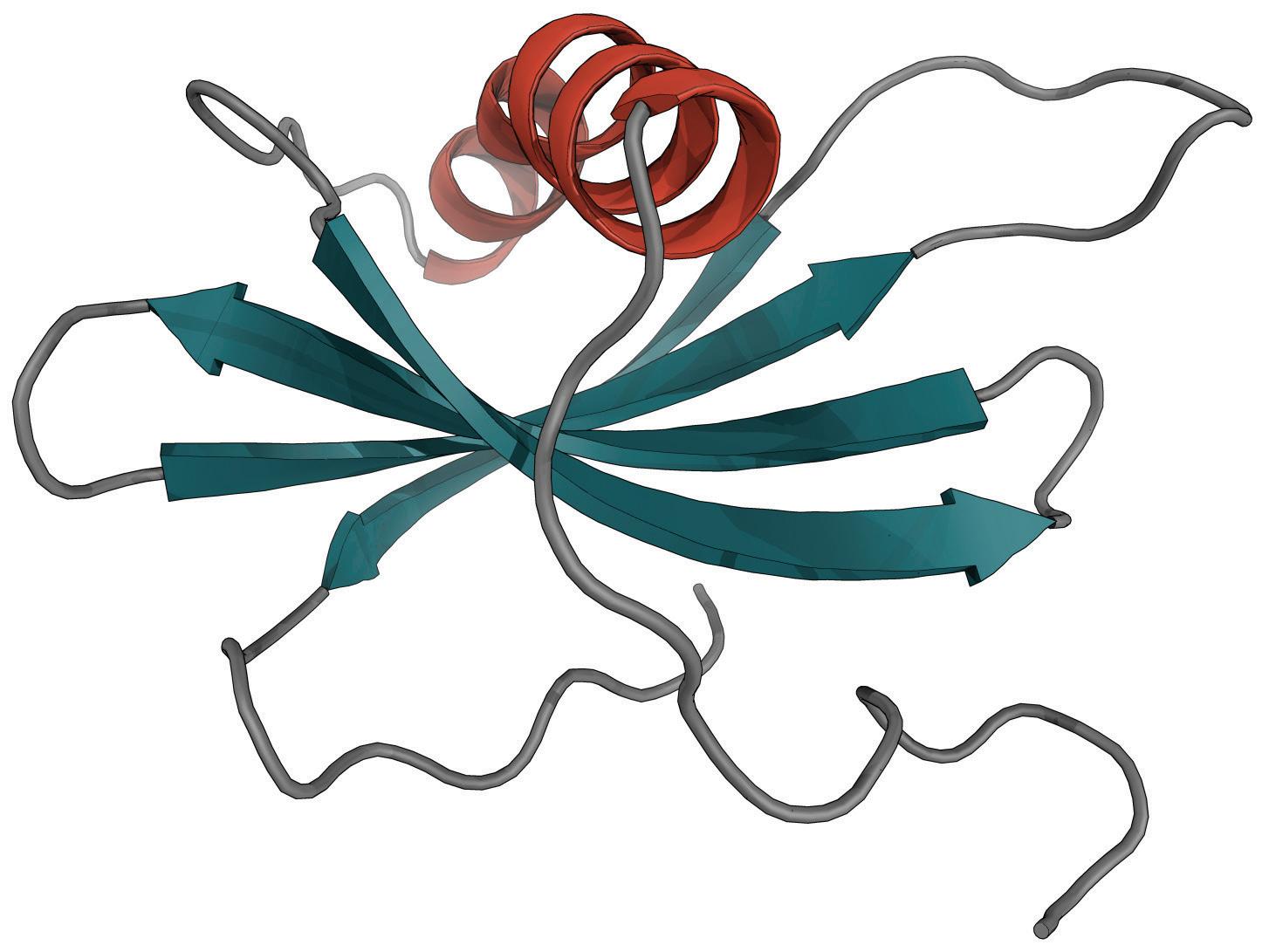
We present a screening process for an Affimer® protein ligand to selectively bind AAV9. Affimer®’s are cystatin-derived single-domain proteins (10.7 kDa), affinity is tested as a function of the inclusion of randomized sequences in loops 2 and 4. The highest performing candidates were immobilized on an agarose bead matrix (PuraBead®) and the resulting adsorbent demonstrated binding at pH 7.5 and elution at pH 4.5 of clarified AAV9 from transiently transfected HEK 293 cells.
Affimer® ligand structure
Primary screening
Phage display was used to generate a library of His-tagged proteins. Screening was performed on a flow cytometry platform using a sandwich assay format. Anti-His antibody–conjugated beads were used to capture Affimer® candidates, which were then incubated with clarified lysate containing recombinant AAV9. Binding was detected using an AlexaFluor-488–labelled anti-AAV9 antibody.
Lead candidate evaluation
Initial screening
Fourteen compounds were identified in the primary screen
Breakthrough capacity Affimer®s immobilized on PuraBead® loaded with AAV9 lysate
Hit validation
Hits from the initial screen were verified using a reverse-sandwich flow cytometry assay. In this format, anti-AAV9 antibody was immobilized, recombinant AAV9 lysate was applied, and binding of Affimer® hit candidates was detected using an AlexaFluor-488 anti-His antibody.
Cross-validation by ELISA
Both assay formats were subsequently repeated using ELISA to cross-validate and strengthen the hit identification dataset.
Seven sequences were selected for scale-up to 1 L expression in E. coli
• Proteins were purified using IMAC followed by SEC, achieving >95% purity as confirmed by capillary electrophoresis
High-throughput binding assessment
• Purified candidates were immobilized on the PuraBead® Edge matrix
Binding performance was evaluated for total AAV9 particle capture in a high-throughput plate-based format
Recovery was quantified using an AAV9-specific ELISA
Column-scale evaluation
• Lead candidates from the plate assay were further assessed in a 1 mL column format



21 sequential column runs were performed. AAV9 lysate was loaded on cycles 1, 11, and 21, corresponding to: post-0, post-10, and post-20 exposures to 0.1 M NaOH.
Chromatography method
• Load: >1 × 10¹⁴ capsids/mL adsorbent (AAV9)
• Equilibration: PBS, 0.2 M NaCl, 0.001% Pluronic F-68, pH 7.5
• Elution 1: 50 mM citrate, 0.3 M NaCl, pH 4.5
• Elution 2: 50 mM citrate, 0.3 M NaCl, pH 3.0
Clean‑in‑place (CIP): 0.1 M NaOH, 15 column volumes at 1 mL/min flow rate (15-minute exposure per cycle)
Analysis: ELISA and SDS-PAGE
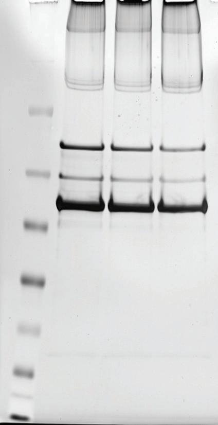
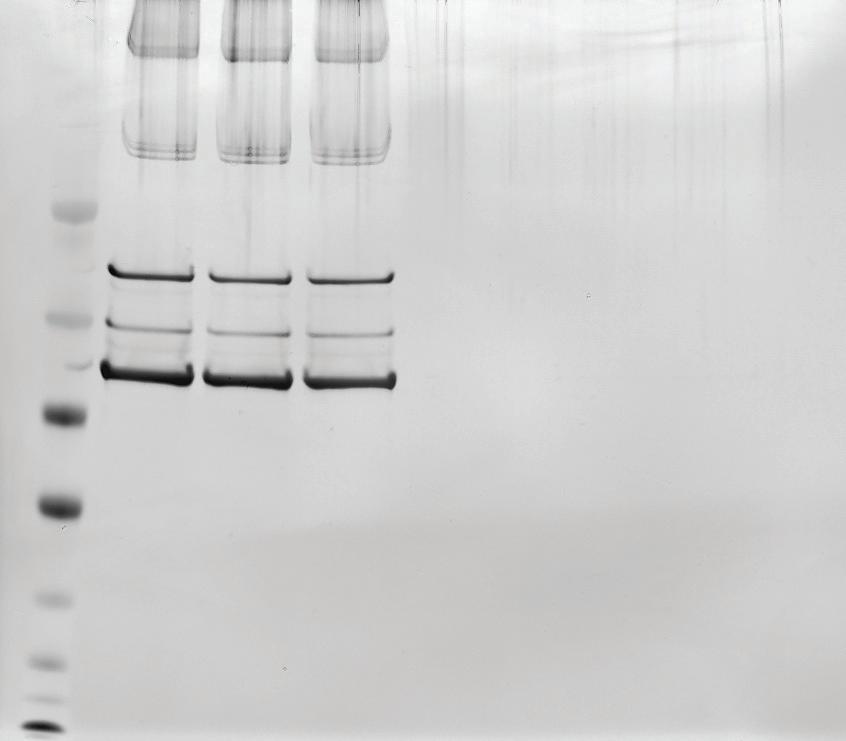
Screening methodology and lead optimization Validation of AAV9 eluate purity following Affimer® 1 purification
Conclusions